A Double Antibody Sandwich ELISA Assay for the Detection of
advertisement
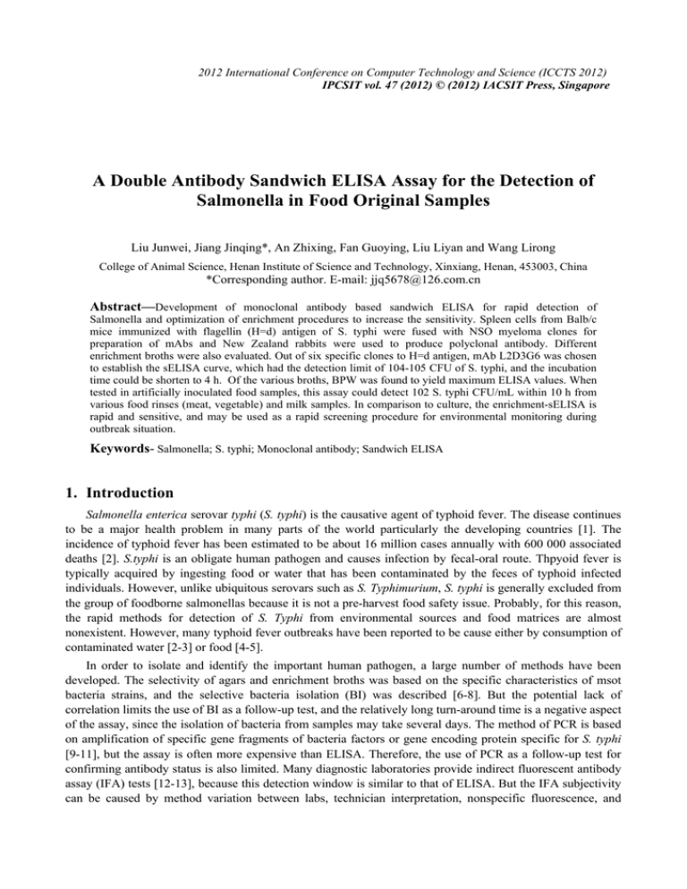
2012 International Conference on Computer Technology and Science (ICCTS 2012) IPCSIT vol. 47 (2012) © (2012) IACSIT Press, Singapore A Double Antibody Sandwich ELISA Assay for the Detection of Salmonella in Food Original Samples Liu Junwei, Jiang Jinqing*, An Zhixing, Fan Guoying, Liu Liyan and Wang Lirong College of Animal Science, Henan Institute of Science and Technology, Xinxiang, Henan, 453003, China *Corresponding author. E-mail: jjq5678@126.com.cn Abstract—Development of monoclonal antibody based sandwich ELISA for rapid detection of Salmonella and optimization of enrichment procedures to increase the sensitivity. Spleen cells from Balb/c mice immunized with flagellin (H=d) antigen of S. typhi were fused with NSO myeloma clones for preparation of mAbs and New Zealand rabbits were used to produce polyclonal antibody. Different enrichment broths were also evaluated. Out of six specific clones to H=d antigen, mAb L2D3G6 was chosen to establish the sELISA curve, which had the detection limit of 104-105 CFU of S. typhi, and the incubation time could be shorten to 4 h. Of the various broths, BPW was found to yield maximum ELISA values. When tested in artificially inoculated food samples, this assay could detect 102 S. typhi CFU/mL within 10 h from various food rinses (meat, vegetable) and milk samples. In comparison to culture, the enrichment-sELISA is rapid and sensitive, and may be used as a rapid screening procedure for environmental monitoring during outbreak situation. Keywords- Salmonella; S. typhi; Monoclonal antibody; Sandwich ELISA 1. Introduction Salmonella enterica serovar typhi (S. typhi) is the causative agent of typhoid fever. The disease continues to be a major health problem in many parts of the world particularly the developing countries [1]. The incidence of typhoid fever has been estimated to be about 16 million cases annually with 600 000 associated deaths [2]. S.typhi is an obligate human pathogen and causes infection by fecal-oral route. Thpyoid fever is typically acquired by ingesting food or water that has been contaminated by the feces of typhoid infected individuals. However, unlike ubiquitous serovars such as S. Typhimurium, S. typhi is generally excluded from the group of foodborne salmonellas because it is not a pre-harvest food safety issue. Probably, for this reason, the rapid methods for detection of S. Typhi from environmental sources and food matrices are almost nonexistent. However, many typhoid fever outbreaks have been reported to be cause either by consumption of contaminated water [2-3] or food [4-5]. In order to isolate and identify the important human pathogen, a large number of methods have been developed. The selectivity of agars and enrichment broths was based on the specific characteristics of msot bacteria strains, and the selective bacteria isolation (BI) was described [6-8]. But the potential lack of correlation limits the use of BI as a follow-up test, and the relatively long turn-around time is a negative aspect of the assay, since the isolation of bacteria from samples may take several days. The method of PCR is based on amplification of specific gene fragments of bacteria factors or gene encoding protein specific for S. typhi [9-11], but the assay is often more expensive than ELISA. Therefore, the use of PCR as a follow-up test for confirming antibody status is also limited. Many diagnostic laboratories provide indirect fluorescent antibody assay (IFA) tests [12-13], because this detection window is similar to that of ELISA. But the IFA subjectivity can be caused by method variation between labs, technician interpretation, nonspecific fluorescence, and increased background staining with some samples. Due to these inherent difference, decreased IFA reproducibility can be expected between laboratories. ELISA methods, available in 96-well format are best suited for screening a large number of environmental samples, and the procedure can also be easily performed by the peripheral laboratories. Therefore, several enzyme immunoassays using polyclonal or monoclonal antibodies (mAbs) have been established for the detection of Salmonella [14-16]. However, ELISAs can have higher detection limits of order of 105 CFU/mL. As the infectious dose of S. typhi can be less than 105 bacteria, it is pertinent to achieve a better sensitivity which possibly can be obtained by including an enrichment step before ELISA. The objective of this study was to produce a monoclonal antibody (mAb) and a polyclonal antibody (pAb) highly specific for S. typhi, and to develop a sandwich ELISA method for the rapid detection of Salmonella in food samples. We have also evaluate various pre-enrichment and enrichment broths for sELISA, examined the efficacy of the enrichment-ELISA in various artificially inoculated food samples. The present study also examined the presence of S. typhi in natural field samples. 2. Materials and Methods 2.1. Bacterial strains and materials S. typhi (strain SKST), a clinical isolate originally obtained from the veterinary clinic in College of Animal Science of Henan Institute of Science and Technology (Xinxiang, China), was used in the present study for mouse immunization, hybridoma production and preparation of flagellin for rabbit antiserum. Other bacterial strains used for characterization of mAbs and pAbs included 14 isolates of S. typhi, 12 strains of 9 other serovars of Salmonella, 3 Proteus spp., 2 Yersinia spp., and 9 other bacteria. FCA and FIA were obtained from Pierce. HAT and HT were obtained from Sigma-Aldrich (USA). RPMI-1640 with L-glutamine was obtained from Gibco. PEG 1500 was from Roche Diagnostics Corporation (Indianapolis, USA). Fetal bovine serum (FBS) was from Hangzhou Sijiqing Biological Engineering Materials Co. Ltd. (Hangzhou, China). TMB, phenacetin, urea peroxide were obtained from Sigma Company. All other solvents and reagents were of analytical grade or higher, unless otherwise stated. New Zealand white rabbits and female Balb/c mice were obtained from the Laboratory Animal Center, Beijing Medical University, China, and raised under strictly controlled conditions in our laboratory chamber. 2.2. Instruments A spectrophotometric microtitre reader, MULTISKAN MK3 (Thermo company, USA), provided with a 450 nm filter, was used for absorbance measurements. A Legend Micro 17 microcentrifuge and GS15R high speed refrigerated centrifuge were supplied by Thermo Company (USA). CO2 incubator from RS-Biotech (Galaxy S+, UK) was used for cell cultivation. SW-CJ-2FD Superclean Bench was purchased from Suzhou purification equipment Co., Ltd (Suzhou, China). 2.3. Media and growth conditions Buffered peptone water (BPW) and brain heart infusion broth (BHIB) were used for pre-enrichment. Selenite F broth (SFB) and Rappaport-vassiliadis broth (RVB) were used as enrichment broths. BHI was used as post-enrichment broth for isolation of S. typhi by culture method. S. typhi bacteria were grown in the broths and incubated at 37 ℃ for maximum of 24 h under shaking conditions (150 rpm). Plates were incubated at 37 ℃ of 24 h. the bacterial counts were determined by pour plate method. 2.4. Preparation and characterizatio of mAbs Flagellin was purified from S. typhi strain SKST for antibody production. A group of 6-8 weeks old female Balb/c mice were injected 5 times with flagellin (60 μg) on day 0, 7, 10, 13, and 15. Mice were bled 3 days after the last injection and the mouse with the highest titre of serum antibodies to flagellin antigen was given 2 booster injections intraperitoneally on consecutive days before use for hybridoma production. The procedures of cell fusion and cloning conditions were described previously by Ko¨hler and Milstein [17] with further modifications by Chen et al [18]. Hybridomas producing antiflagellar antibodies were purified at least twice by limiting dilution. Once established, the clones were expanded in tissue culture flasks and stored in liquid nitrogen for future use. Two mature female Balb/c mice were injected intraperitoneally (i.p.) with 0.5 mL of paraffin 10 days before receiving an i.p. injection of the positive hybridoma cells suspended in RPMI 1640 medium. Ascites fluid was collected 10 days after the injection and then stored at –20 °C. The ascites fluid was precipitated with ammonium sulphate solution and dialyzed against PBS for three days. Protein concentration was determined with ultraviolet spectrophometer by the equation: protein concentration (mg/mL)=1.45OD280nm- 1.74OD260nm, where OD value is the optical density [19]. Isotyping of clones was established using a Bio-Rad isotyping kit according to manufacturer’s instructions. 2.5. Polyclonal antiflagellar antibody Two female New Zealand white rabbits were subcutaneously immunized at four sites in the back with purified flagellin. FCA was employed in the first immunization and FIA was used in the subsequent boost injections. Rabbits were immunized every 14 days with 200 μg of immunogen, and blood samples were taken for identification from the marginal vein of the ear after each immunization (from the third immunization onward). Ten days after the final boost, all rabbits were exsanguinated by heart puncture and the serum was separated from blood cells by storing at 4 ℃ overnight. Then the serum was centrifuged with 5000 rpm for 15 min, and this crude serum was purified with saturated ammonium sulfate (SAS) precipitation method. 2.6. Sandwich ELISA procedure The sandwich ELISA was developed using the pAb as the capture antibody and mAb as detection antibody. Each well of the 96-well microtitre plate was coated with 100 μL of purified pAb (10 μg/mL in CBS) and kept at 4 ℃ overnight. After washing, the remaining binding sites were blocked with 300 μL/well of 6.67% new-born bovine serum in PBS for 1 h at 37 ℃. The blocking agent was removed, and 100 μL of test samples (bacterial cells) were allowed to react with the antibodies at 37 ℃ for 1 h. This was followed by the addition of mAbs (detection antibody, 2 μg/mL in PBS), and the incubation was kept at 37 ℃ for 1 h. Then the plates were washed three times and 100 μL of goat anti-mouse IgG conjugated to horseradish peroxidase (1: 1000 in PBST) was added and the mixture was incubated at 37 ℃ for 30 min. After the wells was washed, 60 μL/well of TMB substrate solution was added, followed by incubation for 15 min at room temperature. The enzymatic reaction was stopped with sulfuric acid (2 M, 100 μL/well) and the yellow plate was spectrophotometrically read in a single wavelength mode at 450 nm. A positive ELISA reaction was established when A450 of a sample was greater than 2.1 times the value of negative controls. 2.7. Effects of selective enrichment broth media S. typhi bacteria was grown in BPW, BHIB, RVB, and SFB at a concentration of ca 101 to 105 CFU/mL and incubated at 37 ℃ for 24 h. Prior to detection by sELISA after 6 h or 24 h, bacteria were inactivated by heating the cultures at 96 ℃ for 10 min. Enumeration of bacteria was accomplished by plate count. 2.8. Artificial inoculation of food samples Meat or chicken carcasses and green leafy vegetables (100 g) from local retail supplier were each rinsed manually with 100 mL of BPW. The rinses were dispensed in 10 mL aliquots and frozen at –20 °C. Each batch of carcass or vegetable rinse was tested for the presence of S. typhi by culture method. Raw milk and curd samples were collected from dairy and local market respectively and tested for the presence of S. typhi by culture method. Meat and chicken carcass rinse, vegetable rinse, milk, and curd free of S. typhi were inoculated with S. typhi strain SKST at concentration of 10-1, 100, 101, and 102 CFU/mL. BPW (90 mL) was added to 10 mL of the inoculated samples and incubated at 37 ℃ for 24 h. Two samples of one mL each were taken at 6 and 24 h of incubation and heated at 96 ℃ for 10 min before testing by sELISA. 2.9. Authentic field tests Food (n=30) and potable water (n=25) samples were evaluated for the presence of S. typhi by enrichmentELISA and conventional culture method. Food samples were obtained from the local market and included milk (n=10), green leafy vegetables (n=5), meat (n=10), and chicken (n=5) samples. The water samples were collected from different parts of the local river, and included samples from public water supply (n=15), open drain (n=10), and groundwater (n=10). When tested by enrichment-ELISA, all food samples were pre-enriched in BPW at 37 ℃ for 24 h at a ratio of 10:90 (v/v). The water sample from public water supply and groundwater (10 mL) was filtered through membrane filter (0.45 μm). The filter was removed and inoculated in BPW (50 mL). The water sample from open drain was centrifuged at 2000 rpm for 15 min to get rid of particulate matter before filtration. The media was incubated at 37 ℃ for 24 h. One milliliter of BPW pre-enriched sample was heated at 96 ℃ for 10 min and tested by ELISA. 3. RESULTS and ANALYSIS 3.1. Production of mAbs and pAbs Six clones produced antibodies that were found to be specific for the flagellin (H=d) antigen of S. typhi and other Salmonella serovars carrying the H=d antigen. Different cross reactions (CR) were observed with other Salmonella strains, while no CR was detected for other bacterial types. Culture supernatant of clone L2D3G6 showing the highest titre of 1:50000 were selected for use in sELISA. Isotyping revealed that this clone belonged to IgG2a class of heavy chain and κ isotype for the light chain. Immunobloting results showed the presence of single 52 kDa band with S. typhi purified flagellin and whole cell sonicated antigen on reaction with mAb clone of L2D3G6 (results not shown). The detection limit of this antibody was 104/mL in pure culture of S. typhi cells by indirect ELISA and 1.0 ng of purified flagellin by dELISA. The antiserum to flagellin was raised in New Zealand white rabbits and was found to have the titre of 1:104 when tested by dELISA. After purification, the titre of the pAb was reduced to 1:2000. 3.2. Sandwich ELISA standard curve The sandwich ELISA method was developed for detecting Salmonella in enrichment cultures of food samples, and the results based on the checkerboard assays are shown in Fig. 1. In this procedure, pAb was used as capture antibody and the specific mAb L2D3G6 was employed as detection antibody. The method was found to have a detection limit of 103-104 S. typhi CFU in pure culture as shown in Fig. 1. Based on the OD values observed in the negative control wells (0.080-0.085), an OD of 0.165 was taken as the cutoff value to determine the positive reaction. 2.0 OD values at 450 nm 1.6 1.2 0.8 0.4 0.0 -3 10 10 -2 -1 0 1 10 10 10 10 3 S. typhi (CFU/mL) X10 2 10 3 10 4 Fig. 1. Sensitivity of double antibody sandwich ELISA method for detecting S. typhi bacteria. The horizontal line represents the cutoff value for sELISA. Data were obtained by averaging six independent curves, each run in triplicate. PAb as capture antibody was prepared in CBS (pH 9.6); purified mAb produced by L2D3G6 as detection antibody was prepared in PBS; GaMIgG-HRP was diluted 1:1000 in incubation buffer. 3.3. Represent curves for Salmonella strains Results of different Salmonella strains tested in field sample using culture and ELISA procedures are presented in Fig. 2. OD450nm 2.0 OD values at 450 nm 1.6 1.2 2.0 1.5 1.0 0.5 0.0 -3 -1 1 3 10 10 10 10 S. typhi 0.8 Hadar Anatum Enteritidis Thompson Typhimurium 0.4 0.0 -3 10 10 -2 -1 0 1 10 10 10 10 3 S. typhi (CFU/mL) x 10 2 10 3 10 4 Fig. 2. Different sandwich ELISA curves for representative Salmonella strains bacteria. The inset indicates the standard curve for S. tyhpi. It can be seen the established sandwich ELISA curve could be used for simultaneously detection of Salmonella Typhimurium (serogroup B), Salmonella Thompson (serogroup C1), Salmonella Hadar (serogroup C2), Salmonella Enteritidis (serogroup D1), and Salmonella Anatum (serogroup E1). 3.4. Effects of enrichment media on sELISA The effects of various selective enrichment media are shown in Table 1. The growth of S. typhi bacteria was poor in SCB and RVB after 6 h of incubation. On most occasions, not even 1 log increase in growth was observed after 6 h and this increase was not-significant. Pure culture grew to 107-108 CFU/mL after 24 h could be observed. When tested by sELISA, S. typhi bacteria grown to a concentration of 105 CFU/mL could only be detected in SCB, but in RVB it could increase to a concentration of 108 CFU/mL. Table 1. Quantification of S. typhi in different enrichment broth by sELISA. SFB RVB Initial 2.6 3.6 4.6 5.6 CFU/mLa 6h 24 h 3.0 8.4 4.2 8.2 4.7 8.7 5.7 8.6 ELISAb 6h 24 h 0.072 0.771 0.074 0.802 0.068 0.853 0.279 0.861 2.6 3.6 4.6 5.6 3.0 4.2 4.7 5.0 0.090 0.086 0.091 0.120 7.6 7.9 8.2 8.1 0.106 0.102 0.108 0.141 Note: a Number represent the mean CFU/mL (log10). b ELISA A450 cut off value is 0.165; values are mean of three readings. 3.5. Field study results Except one water sample (ground water) that showed an OD of 0.225 from 24 h BPW enrichment after 24 h, none of the food or water samples resulted in an OD of more than 0.1 by enrichment-ELISA. All samples tested negative by traditional culture method. 4. Discussion The ELISA method described in this study is a double antibody sandwich assay, in which antiflagellar pAb was used as capture antibody and a mAb L2D3G6 specific to H=d antigen of Salmonella was employed as a detection antibody. The assay described in this study was rapid and took about 4 h to complete with being at least 10 times more sensitive than indirect ELISA. The detection limit of sELISA was 104-105 CFU of S. typhi cells/mL. The possibility of using selective enrichment broth by growing the bacteria in SCB and RVB are also investigated. Generally, poor growth of S. typhi was observed in these media, because the selective broth had agents that could be inhibitory to the target bacteria. In conclusion, the described ELISA procedure, which was a mAb based sELISA preceded by an enrichment culture step in BPW, was a sensitive and rapid method for detection of S. typhi from food or water samples. It offered considerable reduction in detection time over the traditional culture methods for Salmonella. This method can be of valuable in application of environmental protection during the investigation of outbreak situations. 5. References [1] S. Kumar, K. Balakrishna and H. V. Batra, Enrichment-ELISA for detection of Salmonella typhi from food and water samples. Biomed Environ Sci, 2008, 21(2):137-143. [2] D. J. Kopecko, H. Sieber, J. A. Ures, A. Fürer, J. Schlup, U. Knof, A. Collioud, D. Xu, K. Colburn and G. Dietrich, Genetic stability of vaccine strain Salmonella Typhi Ty21a over 25 years. Int J Med Microbiol, 2009, 299(4):233-246. [3] J. H. Mermin, R. Villar, J. Carpenter, L. Roberts, A. Samaridden, L. Gasanova, S. Lomakina, C. Bopp, L. Hutwagner, P. Mead, B. Ross and E. D. Mintz, A massive epidemic of multidrug-resistant typhoid fever in Tajikistan associated with consumption of municipal water. J Infect Dis, 1999, 179(6):1416-1422. [4] A. Valdivieso-Garcia, E. Riche, O. Abubakar, T. E. Waddell and B. W. Brooks, A double antibody sandwich enzyme-linked immunosorbent assay for the detection of Salmonella using biotinylated monoclonal antibodies. J Food Prot, 2001, 64(8):1166-1171. [5] T. R. Coté, H. Convery, D. Robinson, A. Ries, T. Barrett, L. Frank, W. Furlong, J. Horan and D. Dwyer, Typhoid fever in the park: epideminology of an outbreak at a cultural interface. J Community Health, 1995, 20(6):451-458. [6] R. Khakhria, D. Woodward, W. M. Johnson and C. Poppe, Salmonella isolated from humans, animals and other sources in Cannda. Epidemiol Infect, 1997, 119(1):15-23. [7] N. Korsak, J. N. Degeye, G. Etienne, B. China and G. Daube, Comparison of four different methods for Salmonella detection in fecal samples of porcine origin. J Food Prot, 2004, 67(10):2158-2164. [8] W. Xu, G. Zhang, X. Li, S. Zhou, P. Li, Z. Hu and J. Li, Delayed secondary enrichment for the isolation of Salmonella from broiler chickens and their environment. Appl Environ Microbiol, 1980, 40(4):783-786. [9] A. R. Bennett, D. Greenwood, C. Tennant, J. G. Banks and R. P. Betts, Rapid and definitive detection of Salmonella in foods by PCR. Lett Appl Microbiol, 1998, 26(6):437-441. [10] D. H. D'Souza and L. A. Jaykus, Nucleic acid sequence based amplification for the rapid and sensitive detection of Salmonella enterica from foods. J Appl Microbiol, 2003, 95(6):1343-1350. [11] C. Burtscher and S. Wuertz, Evaluation of the use of PCR and reverse transcriptase PCR for detection of pathogenic bacteria in biosolids from anaerobic digestors and aerobic composters. Appl Environ Microbiol, 2003, 69(8):4618-4627. [12] C. Morissette, J. Goulet and G. Lamoureux, Rapid and sensitive sandwich enzyme-linked immunosorbent assay for detection of staphylococcal enterotoxin B in cheese. Appl Environ Microbiol, 1991, 57(3):836-842. [13] A. Nakamura, N. Ishikawa and H. Suzuki, Diagnosis of invasive candidiasis by detection of mannan antigen by using the avidin-biotin enzyme immunoassay. J Clin Microbiol, 1991, 29(11):2363-2367. [14] S. Kerr, H. J. Ball, D. P. Mackie, D. A. Pollock and D. A. Finlay, Diagnostic application of monoclonal antibodies to outer membrane protein for rapid detection of Salmonella. J Appl Bacteriol, 1992, 72(4):302-308. [15] B. W. Blais and A. Martinez-Perez, Detection of group D salmonellae including Salmonella Enteritidis in eggs by polymyxin-based enzyme-linked immunosorbent assay.. J Food Prot, 2008, 71(2):392-396. [16] R. L. Brigmon, S. G. Zan and H. R. Wilson, Detection of Salmonella enteritidis in eggs and chicken with enzymelinked immunosorbent assay. Poult Sci, 1995, 74(7):1232-1236. [17] G. Köhler and C. Milstein, Continuous cultures of fused cells secreting antibody of predefined specificity. J Immunol, 2005, 174(5):2453-2455. [18] Y. Chen, Z. Wang, Z. Wang, S. Tang, Y. Zhu and X. Xiao, Rapid enzyme-linked immunosorbent assay and colloidal gold immunoassay for Kanamycin and Tobramycin in swine tissues. J Agric Food Chem, 2008, 56(9):2944-2952. [19] Z. Wang, Y. Zhu, S. Ding, F. He, R. C. Beier, J. Li, H. Jiang, C. Feng, Y. Wan, S. Zhang, Z. Kai, X. Yang and J. Shen, Development of a monoclonal antibody-based broad-specificity ELISA for fluoroquinolone antibiotics in foods and molecular modeling studies of cross-reactive compounds. Anal Chem, 2007, 79(12):4471-4483.
