Host Susceptibility Is Altered by Light Intensity After Exposure to... Michelle L. Steinauer and Kaitlin M. Bonner*,
advertisement
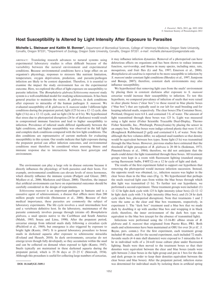
J. Parasitol., 98(5), 2012, pp. 1052–1054 Ó American Society of Parasitologists 2012 Host Susceptibility Is Altered by Light Intensity After Exposure to Parasites Michelle L. Steinauer and Kaitlin M. Bonner*, Department of Biomedical Sciences, College of Veterinary Medicine, Oregon State University, Corvallis, Oregon 97331; *Department of Zoology, Oregon State University, Corvallis, Oregon 97331. e-mail: michelle.steinauer@oregonstate.edu ABSTRACT: Translating research advances to natural systems using experimental laboratory studies is often difficult because of the variability between the natural environment and experimental conditions. Because environmental conditions have a large effect on an organism’s physiology, responses to stressors like nutrient limitation, temperature, oxygen deprivation, predation, and parasite/pathogen infection are likely to be context dependent. Therefore, it is essential to examine the impact the study environment has on the experimental outcome. Here, we explored the effect of light exposure on susceptibility to parasite infection. The Biomphalaria glabrata/Schistosoma mansoni study system is a well-established model for studying schistosomiasis. It has been general practice to maintain the vector, B. glabrata, in dark conditions after exposure to miracidia of the human pathogen S. mansoni. We evaluated susceptibility of B. glabrata to S. mansoni under 3 different light conditions during the prepatent period, light (125 lx) on a 12–12 cycle, dim light (3 lx) on a 12–12 cycle, and no light (24 hr at 0 lx). We hypothesized that stress due to photoperiod disruption (24 hr of darkness) would result in compromised immune function and lead to higher susceptibility to infection. Prevalence of infected snails differed significantly between the light conditions, and higher susceptibility was observed in the full light and complete dark conditions compared with the low light conditions. The dim conditions are representative of current methods for evaluating susceptibility in this system. Our results indicate that light exposure during the prepatent period can affect infection outcomes, and environmental conditions must therefore be considered when assessing fitness and immune response due to interactions between host genotype and environment. The environment can play a large role in disease outcome because it directly influences the physiology of both parasites and their hosts. For example, environmental conditions can elevate levels of stress hormones, which directly influence the immune system (Padgett and Glaser, 2003; Mydlarz et al., 2006; Marketon and Glaser, 2008). Therefore, the impact that artificial environments can have on experimental outcomes should be carefully considered in the design of experiments. Schistosoma mansoni is an important pathogen in humans and is a causative agent of schistosomiasis, a disease that afflicts more than 200 million people world-wide (Steinmann et al., 2006). Because of their medical importance, these parasites are commonly the subject of laboratory experiments. The life cycle involves a snail intermediate host and a vertebrate definitive host. In the laboratory, maintenance of the parasite commonly involves passage through (strains of) Biomphalaria glabrata, a snail species native to the Caribbean and South America (Malek, 1985; Souza and Lima, 1990). After the prepatent period, cercariae emerge from infected snails daily following a circadian cycle (Pitchford et al., 1969), but emergence is also triggered by exposure to bright light (Kuntz, 1947). It is general laboratory procedure to house snails in darkened aquaria after exposure to schistosome miracidia (Eveland and Haseeb, 2010). In dark environments, cercariae do not emerge (even though fully developed), so they accumulate within the snail and can be collected on demand when exposed to light (Kuntz, 1947). Snails typically are maintained in these darkened aquaria during the prepatent period, which is 35–56 days at 23–25 C (Stirewalt, 1954). Although this procedure is useful for collecting large numbers of cercariae, DOI: 10.1645/GE-3109.1 it may influence infection dynamics. Removal of a photoperiod can have deleterious effects on organisms and has been shown to reduce immune function, survivorship, and fitness in many species, including mice, rats, mosquitoes, and fruit flies (Li and Xu, 1997; Emerson et al., 2008). Biomphalaria alexandrina is reported to be more susceptible to infection by S. mansoni under constant light conditions (Shoukry et al., 1997; Jamjoom and Banaja, 2007); therefore, constant dark environments may also influence susceptibility. We hypothesized that removing light cues from the snails’ environment by placing them in constant darkness after exposure to S. mansoni cercariae would increase their susceptibility to infection. To test this hypothesis, we compared prevalence of infection of snails that were raised in clear plastic boxes (‘‘clear box’’) to those reared in blue plastic boxes (‘‘blue box’’) that are typically used in our lab for snail breeding and for keeping infected snails, respectively. The clear boxes (The Container Store, Portland, Oregon) were 6.8 L and contained 3 L of water. The amount of light transmitted through these boxes was 125 lx. Light was measured using a light meter (Fisher Scientific Traceable Dual-Display, Thermo Fisher Scientific, Inc., Pittsburgh, Pennsylvania) by placing the sensor in an empty box. The blue boxes were indigo colored plastic that were 11.4 L (Roughneck Rubbermaid [3 gal]) and contained 6 L of water. Note that although the box volumes differ, we adjusted snail numbers so that density would be equivalent between box types. Some light (3 lx) was transmitted through the blue boxes. However, previous studies have estimated that the threshold of light perception of B. glabrata is 20–80 lx (Sodeman, 1973; Pimentel-Souza et al., 1984); therefore, we did not anticipate that the snails could detect the light transmitted through these boxes. All treatment groups were kept in a room with fluorescent lighting (standard energy saving fluorescent bulbs, F40T12) on a 12 hr cycle of light and dark. The results of the first experiment failed to support our hypothesis that removing the photoperiod would increase infection succession. Instead, the opposite result was obtained, i.e., infection success was higher in the clear boxes than in the blue ones (Fig. 1). We hypothesized that perhaps the snails received light cues from within the blue boxes through which dim light was transmitted (3 lx). To further test our hypothesis, we performed a second experiment. Three treatment groups were included: (1) 12–12 hr light dark cycle with 125 lx light intensity (clear box); (2) 12–12 hr light dark cycle with 3 lx light intensity (blue box); and (3) 24 hr dark cycle (dark box, photoperiod disruption). Note that treatments 1 and 2 were the same as the clear and blue box treatments, respectively, in experiment 1. The ‘‘dark box’’ treatment used a blue box that we made dark by doubling it up with another blue box and wrapping it in black cloth; therefore, the inner environment of the dark box type was equivalent to the blue box (except for the absence of transmitted light). Infections were performed using the 13-16-R1 albino strain of B. glabrata and S. mansoni that originated from Puerto Rico, PR1. Both snails and schistosomes have been maintained at OSU for over 26 yr (C. J. Bayne, pers. comm.). For the first experiment, each treatment group included 48 snails, and for the second experiment, each included 72 snails. Juvenile snails (4–6 mm shell diameter) were exposed to 5 miracidia for 4 hr in individual wells of a 24-well tissue culture plate under fluorescent lighting. Snails were then moved to the treatment boxes so that their densities were equivalent between the clear and blue boxes (snails were split between 2 clear boxes in the light group and put in 1 box for the dim and dark groups in order to keep their densities equivalent between the clear boxes and blue boxes). After the prepatent period, infection status was determined for individual snails by isolating them in artificial spring 1052 RESEARCH NOTES 1053 FIGURE 1. Prevalence of Schistosoma mansoni in Biomphalaria glabrata reared under 3 different lighting conditions during the prepatent period of infection. Error bars indicate 95% confidence intervals following the methodology of Newcombe (1998) as implemented in VassarStats (http://vassarstats.net/). Sample sizes are given on each of the bars. No data (ND) were collected for the dark treatment for the first replicate. water in the wells of 12-well plates for 3 hr under bright light. The wells were then examined with a stereomicroscope for emergent cercariae. Infection status was determined at 5, 6, 7, and 9 wk post-exposure in experiment 1 and 6, 8, and 10 wk post-exposure in experiment 2. Infected snails were removed at each time point. We calculated prevalence by counting the number of infected snails, dividing by the number of snails present at the time of first shedding, and multiplying by 100. The majority of infected snails were discovered at the time of first shedding (74–75% for experiment 1 and 91–95% for experiment 2). Prevalence of infected snails was significantly different between treatment groups, and these differences were consistent across experiments (Fisher’s exact test: experiment 1, P ¼ 0.04; experiment 2, P ¼ 0.0375). Prevalence was highest in the light and dark boxes and lowest in the dim blue boxes (Fig. 1). These results suggest that the light intensity to which a snail is exposed after encountering S. mansoni can influence their susceptibility to infection. Counter to our hypothesis, infection success was higher with the brightest light (125 lx) and no light (0 lx) and was lower when snails were exposed to dim light (3 lx). This would suggest that snails can perceive the difference between a completely dark environment and a dim one (3 lx). An alternative explanation is that box volume may have had an effect on infection success, since the clear boxes contained half as much water as the blue boxes. However, we adjusted the snail numbers so that snail density was equivalent between the 2 treatments. Other factors associated with box size could potentially have influenced the outcome, such as aeration rate, which was not measured, or temperature (although snails were kept in a temperature controlled room typically ranging between 25.5 and 27.2 C). Even though the clear box and the blue box differed in dimensions, the blue boxes and dark boxes were of the same dimensions and were internally identical environments except for the transmitted light. This implies that the difference in prevalence between the 2 treatments was indeed due to difference in light intensity. Therefore, variation in light regimes was the primary difference among the artificial environments. Although light intensity was the primary difference between the treatments, it is also possible that certain wavelengths were filtered out with the blue boxes yielding the difference in infection success. It should also be noted that it is possible that the differences we detected among our FIGURE 2. Prevalence of new infections detected over time in snails reared under different lighting conditions. Note the similar acquisition of new infections detected over time. (A) Experiment 1; (B) Experiment 2. treatment groups reflect delayed development of the parasites in dim light conditions rather than the snails differing in susceptibility, since we monitored infections for 9 or 10 wk in the experiments. However, we do not believe that this is likely because the rates at which infected snails were discovered over the 5 wk intervals declined sharply after the first assessment and were similar among all treatment groups (Fig. 2). If the dim light treatment caused delayed development of the parasites, it would be expected that the number of infected snails would increase over time. Previous studies have shown that although melanic forms of B. glabrata exhibit positive photokinesis (Pimentel-Souza et al., 1984), an albino form (M line) exhibits negative photokinesis (Sodeman and Dowda, 1974). The snails in this study were also an albino form, so it is possible that they also react negatively to light. We hypothesize that stress caused by relatively bright light or constant darkness reduced immune function, allowing greater infectivity of S. mansoni under these circumstances. The relationship between stress and immunity is not fully understood and is particularly poorly understood for Biomphalaria. Stress in vertebrates can be both immunosuppressive and immune-enhancing, depending on the type of stressor and duration (Dhabhar, 2008; Martin, 2009; Ellis et al., 2011). Environmental stressors have been shown to reduce the numbers of hemocytes or phagocytic activity of hemocytes in a variety of mollusks (Lacoste et al., 2001, 2002; Hooper et al., 2007; Ellis et al., 2011) and thus could also reduce the function of hemocytes in B. glabrata, which play a 1054 THE JOURNAL OF PARASITOLOGY, VOL. 98, NO. 5, OCTOBER 2012 large role in the defense against S. mansoni (Bayne et al., 2001; Hahn et al., 2001; Bender et al., 2005). To understand the relationship between light cycle and immune function, stress needs to be assessed in the important snail hosts of the human pathogen S. mansoni. In conclusion, the environment into which an experimental host is placed after exposure to a parasite can influence the infection outcome resulting in over- or under-estimates of infection susceptibility if the artificial habitat does not reflect natural conditions. For this albino form of B. glabrata, both light and absolute dark environments resulted in higher rates of infection than dim environments. These husbandry conditions should be considered when assessing fitness and immunity of hosts because they could be greatly influenced by interactions between host genotype and environment. We acknowledge Michael Blouin and Christopher Bayne for providing resources and helpful discussion regarding this experiment, including the snails and parasites involved in this study. Partial Funding was supplied by NIH grant AI016137 and the General Research Fund at Oregon State University. LITERATURE CITED BAYNE, C. J., U. K. HAHN, AND R. C. BENDER. 2001. Mechanisms of molluscan host resistance and of parasite strategies for survival. Parasitology 123: S159–S167. BENDER, R. C., E. J. BRODERICK, C. P. GOODALL, AND C. J. BAYNE. 2005. Respiratory burst of Biomphalaria glabrata hemocytes: Schistosoma mansoni-resistant snails produce more extracellular H2O2 than susceptible snails. Journal of Parasitology 91: 275–279. DHABHAR, F. S. 2008. Enhancing versus suppressive effects of stress on immune function: Implications for immunoprotection versus immunopathology. Allergy, Asthma, and Clinical Immunology 4: 2–11. ELLIS, R. P., H. PARRY, J. I. SPICER, T. H. HUTCHINSON, R. K. PIPE, AND S. WIDDICOMBE. 2011. Immunological function in marine invertebrates: Responses to environmental perturbation. Fish and Shellfish Immunology 30: 1209–1222. EMERSON, K. J., W. E. BRADSHAW, AND C. M. HOLZAPFEL. 2008. Concordance of the circadian clock with the environment is necessary to maximize fitness in natural populations. Evolution 62: 979–983. EVELAND, L. K., AND M. A. HASEEB. 2010. Laboratory rearing of Biomphalaria glabrata snails and maintenance of larval schistosomes in vivo and in vitro. In Biomphalaria snails and larval trematodes, R. Toledo and B. Fried (eds.). Springer, New York, New York, p. 33–55. HAHN, U. K., R. C. BENDER, AND C. J. BAYNE. 2001. Killing of Schistosoma mansoni sporocysts by hemocytes from resistant Biomphalaria glabrata: Role of reactive oxygen species. Journal of Parasitology 87: 292–299. HOOPER, C., R. DAY, R. SLOCOMBE, J. HANDLINGER, AND K. BENKENDORFF. 2007. Stress and immune responses in abalone: Limitations in current knowledge and investigative methods based on other models. Fish and Shellfish Immunology 22: 363–379. JAMJOOM, M. B., AND A. A. BANAJA. 2007. Comparative studies on the susceptible and non-susceptible Biomphalaria alexandrina in the intermediate snail host of Schistosoma mansoni in Western Saudi Arabia. World Journal of Medical Sciences 2: 108–114. DATE OF PUBLICATION Volume 98, No. 5, was mailed 17 October 2012 KUNTZ, R. E. 1947. Effect of light and temperature on emergence of Schistosoma mansoni cercariae. Transactions of the American Microscopical Society 66: 37–49. LACOSTE, A., F. JALABERT, S. K. MALHAM, A. CUEFF, AND S. A. POULET. 2001. Stress and stress-induced neuroendocrine changes increase the susceptibility of juvenile oysters (Crassostrea gigas) to Vibrio splendidus. Applied and Environmental Microbiology 67: 2304–2309. ———, S. K. MALHAM, F. GELEBART, A. CUEFF, AND S. A. POULET. 2002. Stress-induced immune changes in the oyster Crassostrea gigas. Developmental and Comparative Immunology 26: 1–9. LI, J. C., AND F. XU. 1997. Influences of light-dark shifting on the immune system, tumor growth and life span of rats, mice and fruit flies as well as on the counteraction of melatonin. Biological Signals 6: 77–89. MALEK, E. A. 1985. Snail hosts of schistosomiasis and other snailtransmitted diseases in tropical America: A manual. Scientific publication no. 478, Pan American Health Organization, Washington, D.C., 325 p. MARKETON, J. I. W., AND R. GLASER. 2008. Stress hormones and immune function. Cellular Immunology 252: 16–26. MARTIN, L. B. 2009. Stress and immunity in wild vertebrates: Timing is everything. General and Comparative Endocrinology 163: 70–76. MYDLARZ, L. D., L. E. JONES, AND C. D. HARVELL. 2006. Innate immunity environmental drivers and disease ecology of marine and freshwater invertebrates. Annual Review of Ecology Evolution and Systematics 37: 251–288. NEWCOMBE, R. G. 1998. Two-sided confidence intervals for the single proportion: Comparison of seven methods. Statistics in Medicine 17: 857–872. PADGETT, D. A., AND R. GLASER. 2003. How stress influences the immune response. Trends in Immunology 24: 444–448. PIMENTEL-SOUZA, F., V. T. SCHALL, R. LAUTNER, N. D. C. BARBOSA, M. SCHETTINO, AND N. FERNANDES. 1984. Behavior of Biomphalaria glabrata (Gastropoda, Pulmonata) under different lighting conditions. Canadian Journal of Zoology 62: 2328–2334. PITCHFORD, R. J., A. H. MEYLING, J. MEYLING, AND J. F. DU TOIT. 1969. Cercarial shedding pattern of various schistosome species under outdoor conditions in the Transvaal. Annals of Tropical Medicine and Hygiene 63: 359–371. SHOUKRY, N. M., F. M. EL-ASSAL, G. N. SOLUMAN, AND N. S. MANSOUR. 1997. Susceptibility of three successive snail generations from positive and negative laboratory bred Biomphalaria alexandrina from different localities in Egypt to infection with Schistosoma mansoni from Giza. Journal of the Egyptian Society of Parasitology 27: 317–329. SODEMAN, W. A. 1973. The influence of light on Biomphalaria glabrata. Nautilus 87: 103–106. ——— AND M. C. DOWDA. 1974. Behavioral responses of Biomphalaria glabrata. Physiological Zoology 47: 198–206. SOUZA, C. P., AND L. C. LIMA. 1990. Moluscos de interesse parasitológico do Brasil. Serie de Equistossomose 1. Centro de Pesquisas René Rachou, Fundação Oswaldo Cruz, Belo Horizonte, Brasil, 76 p. STEINMANN, P., J. KEISER, R. BOS, M. TANNER, AND J. UTZINGER. 2006. Schistosomiasis and water resources development: Systematic review, meta-analysis, and estimates of people at risk. Lancet Infectious Diseases 6: 411–425. STIREWALT, M. A. 1954. Effect of snail maintenance temperatures on development of Schistosoma mansoni. Experimental Parasitology 3: 504–516.
