The PET-MRI Brain Image Fusion Using Wavelet Transforms 6

International Journal of Engineering Trends and Technology (IJETT) – Volume 23 Number 6 - May 2015
The PET-MRI Brain Image Fusion Using Wavelet
Transforms
Brundashree .R
#1
, Nita Kakhandaki
#2
, S.B.Kulkarni
#3
, S.F. Rodd
#4
, M. M. Matt
#5
#1
PG Scholar, CSE, SDMCET, Dharwad , Karnataka, India
#2
Asst Professor, CSE, SDMCET, Dharwad, Karnataka, India
#3
Professor, CSE, SDMCET, Dharwad , Karnataka, India
# 4
Associate Professor, dept of CSE, GIT, Belguam, Karnataka, India
#5
Associate Professor of Head, MCA, GIT, Belguam, Karnataka, India
Abstract — In this paper, PET and MRI brain images are fused using wavelet transforms. After wavelet decomposition and gray level fusion, by applying our method good fused result can be obtained, by adjusting structural information in gray matter
(GM) area and patching the spectral information in white matter
(WM) area. For testing and comparison we have used three datasets: normal axial, normal coronal, Alzheimer’s disease brain images. The performance of our fusion method in terms of spectral discrepancy (SD) and average gradient (AG) proved to be better both visually and quantitatively.
Keywords
— PET image, MR image, image fusion, wavelet transform, average gradient (AG), spectral discrepancy (SD).
I.
I NTRODUCTION
There are many medical image imaging methods with different imaging equipments, by which different medical images are produced. The images generated from Magnetic resonance imaging (MRI), positron emission tomography
(PET), Computerized Tomography (CT), are used in the clinical treatments. The image data retrieved by different sensors have limit and difference in the geometry, spectrum, time and space resolutions, so it is hard to use just one type of image data. In order to have the more complete and accurate comprehension and knowledge of the target, one must find a technical method to make use of the different types of image data. So it is important to combine different types of image data. Consider PET and MRI images, the former reveals the biochemical changes in color without anatomical information and latter reveals high-resolution anatomical information in gray intensity. The PET and MRI images are used by the doctors in order to diagnose the brain diseases as they contain important complementary information.
There are many methods for fusing PET and MRI images like principal component analysis (PCA), High-pass filtering
(HPF), Intensity-hue-saturation (IHS) transform based fusion, wavelet transforms (WT) those are generally used. One of the method is HIS substitution method with which rich anatomical structural information can be obtained but color distortion occur [2,3]. Many multiresolution methods have been proposed to get fused result with less color distortion but the detailed structural information was found to be still missing.
In order to regain missing information a new method is proposed where the image fusion based on wavelet transform is done by which good fusion result can be generated by adjusting the structural information in the gray matter (GM) area, and then patching the spectral information in the white matter (WM) area. The performance of our fusion method is better in terms of two metrics spectral discrepancy (SD) and average gradient (AG).
II.
I MAGE FUSION BASED ON WAVELET TRANSFORM
The features of image which are sensitive to human visual system exist in different scales. It is necessary to preserve all the salient features in source images and should not introduce any artifacts or inconsistency that distracts human observer.
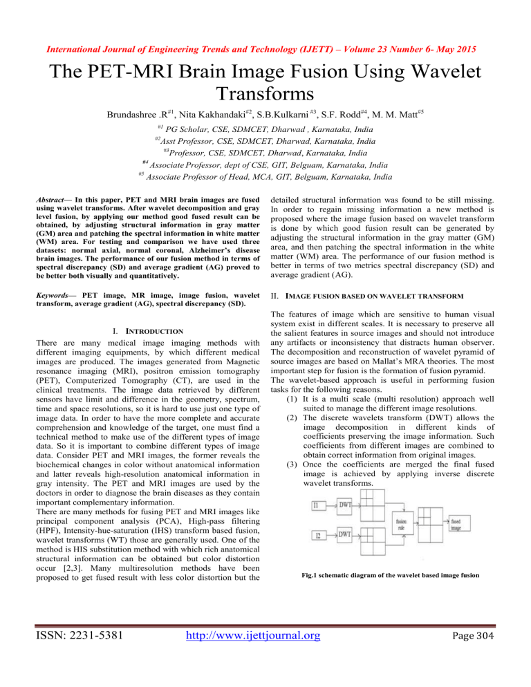
The decomposition and reconstruction of wavelet pyramid of source images are based on Mallat’s MRA theories. The most important step for fusion is the formation of fusion pyramid.
The wavelet-based approach is useful in performing fusion tasks for the following reasons.
(1) It is a multi scale (multi resolution) approach well suited to manage the different image resolutions.
(2) The discrete wavelets transform (DWT) allows the image decomposition in different kinds of coefficients preserving the image information. Such coefficients from different images are combined to obtain correct information from original images.
(3) Once the coefficients are merged the final fused image is achieved by applying inverse discrete wavelet transforms.
Fig.1 schematic diagram of the wavelet based image fusion
ISSN: 2231-5381 http://www.ijettjournal.org
Page 304
International Journal of Engineering Trends and Technology (IJETT) – Volume 23 Number 6 - May 2015
There are many advantages of image fusion wavelet transformation, the high activity region will be lacking some anatomical structural information in the Gray
The image fusion has several advantages in the field of digital image processing. The useful information can be gathered into a single image from multiple images which reduce the time complexity of processing.
The image fusion improves the reliability of the resultant image by grouping the redundant information into a single image.
The capability of the image can be improved by having the complementary information in the fused image. matter (GM) because GM pixel intensity which is near to WM is close to WM pixel intensity. This pixel intensity difference can be adjusted by fuzzy c means algorithm (FCM). FCM is a data clustering technique in which a dataset is grouped into n clusters, where data belongs to a cluster up to certain degree.
To find the lacking structural information the average difference (D avg
) between the I component pixel intensity of original PET image and gray level fused image is calculated.
III.
PROPOSED METHODOLOGY
The Image fusion plays an important role in digital image processing and is used to fuse images captured from multiple sensors to enhance the content of image. The main objective of fusion to find the missing anatomical information in the gray matter (GM) and patching the spectral information in the white matter (WM) area, which is based on wavelet transformation and is different from regular simple DWT fusion method.
(1)
The pixel intensity of high component of fused image( I f,H
) is higher than high component of pet image (I p,H
), therefore D avg is negative. The intensity level for each non-WM pixel in Icomponent fused image can be lowered down using the below formula.
(2)
Where I f,H
(x,y) can be replaced by I p,H
(x,y). The pixel intensity for each non WM pixel can be reduced by adjusting w=50% or w=70%. The low and high activity components are combined to obtain I fused , which represents I component of fused image. The S p
, H p,
and I fused are taken together and inverse HIS transformation is applied. To enhance the contrast of gray scale image, Adapthisteq method is applied on the fused image. The spectral quality of image is measured by spectral discrepancy (SD) of fused image, and spatial resolution of an image is measured by average gradient (AG).
The fusion result is acceptable if SD is smaller and AG is higher [10]. The fusion of PET and MRI images are performed, SD and AG values are tabulated for different values of w=50% and w=70% and comparison of fused result is done.
Fig.2The block diagram of proposed method
IV.
EXPERIMENTAL RESULTS
The fig.2 shows the proposed method. In the first step the PET and MRI images are taken, and HIS transformation is applied such that the PET image is decomposed into intensity, hue and saturation components. In the PET image there is color difference between high and low activity regions, and in order to differentiate between them hue angle is used. The region of interest (ROI) is extracted from the PET and MRI images using Region-mapping technique. The PET image is color image, in which high activity and low activity region gives structural information and spectral information respectively.
The wavelet transformation is applied to high and low activity regions to obtain good color preservation. Then we combine the wavelet co-efficient of PET and MRI image of high and low activity region separately and inverse wavelet transforms is applied. In the obtained fused result after the inverse
Th e image fusion provides an easy way to gather the useful information from multiple images. The test data consist of
PET and MRI brain images where dataset -1, dataset -2, dataset -3 are normal axial, normal coronal and Alzheimer diseases respectively, taken and fused based on our proposed method. The spectral discrepancy of fused image gives spectral quality of that image and average gradient is used to measure the spatial resolution of the image. The different values of SD and AG values are calculated by varying the percentage of w for 50% and 70% in our implementation.
ISSN: 2231-5381 http://www.ijettjournal.org
Page 305
International Journal of Engineering Trends and Technology (IJETT) – Volume 23 Number 6 - May 2015
The fig (d), (e), (f) shows the fusion result for our method when w=50%
Table 1. Performance comparison based on Average Gradient
Average gradient based on Existing method
Fusion Dataset1 Dataset2 Dataset3
(a)
When w=0.5 4.6574 7.7659 6.9527
(b)
When w=0.7 5.168 8.7693 8.0089
Average gradient based on Our method
When w=0.5 4.6574 9.9193 6.9527
When w=0.7 5.168 8.6098 9.4414
Table1. Shows the performance comparison based on w=50% and w=70% for the existing method and our method for average gradient (AG) which is increasing when our proposed method is applied.
Table2. Performance comparison based on Spectral discrepancy
Spectral discrepancy based on existing method
(c)
The fig (a), (b), (c) shows the fusion result for the existing method when w=50%
(d)
(e)
Fusion Dataset1 Dataset2
When w=0.5 5.2418 7.7991
Dataset3
2.5911
When w=0.7 2.4176 7.8178 0.3951
Spectral discrepancy based on our method
When w=0.5 5.2418 5.2768 2.5911
When w=0.7 2.4176 8.3077 0.3951
Table 2 shows the performance comparison based on w=50% and w=70% for the existing method and our method for spectral discrepancy (SD) which is similar for dataset -1and dataset-3 and is decreasing for dataset -2 when our proposed method is applied.
The proposed method is suitable for obtaining fused result with high average gradient (AG) which gives spatial resolution of image but fails in obtaining low spectral discrepancy (SD).
(f)
V .
CONCLUSION
The image fusion helps in having useful information from multiple images in a single image. The fusion of PET and
MRI brain disease images for the proposed method produces the higher average gradient by which the spatial resolution of image is higher. The spectral discrepancy for dataset 1 and
ISSN: 2231-5381 http://www.ijettjournal.org
Page 306
International Journal of Engineering Trends and Technology (IJETT) – Volume 23 Number 6 - May 2015 dataset -3 produces same result as the existing method only dataset -2 is changes to lower values. Hence the spatial resolution of the image is higher when used our proposed method. However different techniques may be developed to improve the spectral quality of the image.
REFERENCES
[1] P.L. Lin, P.W. Huang, C.I. Chen, T.T. Tsai, and C.H.
Chan, “ Brain medical image fusion based on IHS and Log-
Gabor with suitable decomposition scale and orientation for different regions, ” IADIS Multi Conference on Computer
Science and Information Systems 2011CGVCVIP, Rome,
Italy, 2011.
[2] T. Tu, S. Su, H. Shyu, and P.S. Huang, “ A new look at
IHS-like image fusion methods ,” In Information Fusion, vol. 2, no 3, pp. 177-186(10), September 2001 .
[3] A. Wang, H. Sun, and Y. Guan, “ The application of wavelet transform to multimodality medical image fusion, ”
Proceedings of IEEE International Conference on Networking,
Sensing and Control, pp. 270- 274, 2006.
[4] Y. Chibani and A. Houacine, “ Redundant versus orthogonal wavelet decomposition for multisensor image fusion ,” In Pattern Recognition, vol.36, pp.879-887, 2003.
[5] G. Pajares, J. M. D. L. Cruz, “ A wavelet-based images fusion tutorial,” In Pattern Recognition, vol. 37, pp. 1855-
1872, 2004.
[6] H. Zhang, C. Chang, L. Liu, N. Lin, “A novel wavelet medical image fusion method,” International Conference on
Multimedia and Ubiquitous Engineering, pp. 548-553, 2007.
[7] S. Daneshvar and H. Ghassemian, “ MRI and PET image fusion by combining IHS and retina-inspired models ,” In
Information Fusion, vol.11, pp.114-123, 2010.
[8] Maruturi Haribabu , CH.Hima Bindu, Dr.K.Satya Prasad
“ Multimodal Medical Image Fusion of MRI – PET Using
Wavelet Transform ” 2012 International Conference on
Advances in Mobile Network, Communication and Its
Applications.
[9] Dong-Chen He*, Li Wang+ and Massalabi Amani “ A new technique for multi-resolution image fusion .“ Proceedings of
IEEE 2004.
[10]. Po-Whei Huang, Cheng-I Chen, Phen-Lan Lin, Ping
Chen, Li-Pin Hsu, “PET and MRI brain Image fusion using
Wavelet Transform with Structural Information Adjustment and Spectral Information Patching”, International Symposium on Bioelectronics and Bioinformatics, 2014 IEEE.
ISSN: 2231-5381 http://www.ijettjournal.org
Page 307


