J V , Apr. 1996, p. 2586–2592 Vol. 70, No. 4
advertisement
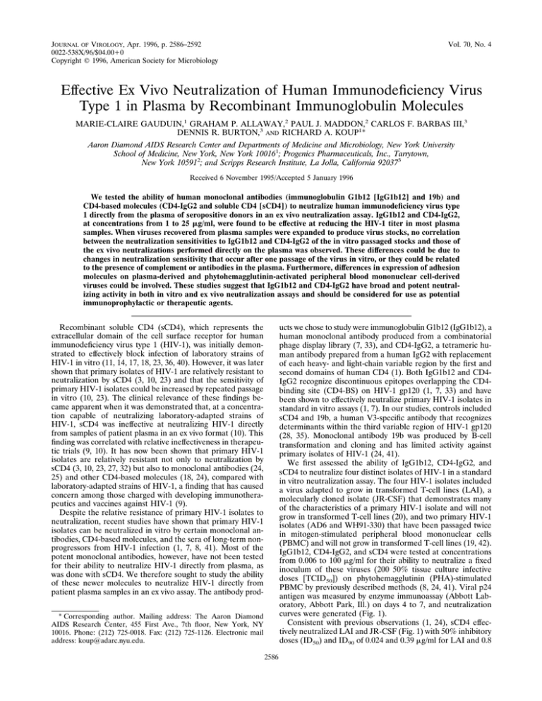
JOURNAL OF VIROLOGY, Apr. 1996, p. 2586–2592 0022-538X/96/$04.0010 Copyright q 1996, American Society for Microbiology Vol. 70, No. 4 Effective Ex Vivo Neutralization of Human Immunodeficiency Virus Type 1 in Plasma by Recombinant Immunoglobulin Molecules MARIE-CLAIRE GAUDUIN,1 GRAHAM P. ALLAWAY,2 PAUL J. MADDON,2 CARLOS F. BARBAS III,3 DENNIS R. BURTON,3 AND RICHARD A. KOUP1* Aaron Diamond AIDS Research Center and Departments of Medicine and Microbiology, New York University School of Medicine, New York, New York 100161; Progenics Pharmaceuticals, Inc., Tarrytown, New York 105912; and Scripps Research Institute, La Jolla, California 920373 Received 6 November 1995/Accepted 5 January 1996 We tested the ability of human monoclonal antibodies (immunoglobulin G1b12 [IgG1b12] and 19b) and CD4-based molecules (CD4-IgG2 and soluble CD4 [sCD4]) to neutralize human immunodeficiency virus type 1 directly from the plasma of seropositive donors in an ex vivo neutralization assay. IgG1b12 and CD4-IgG2, at concentrations from 1 to 25 mg/ml, were found to be effective at reducing the HIV-1 titer in most plasma samples. When viruses recovered from plasma samples were expanded to produce virus stocks, no correlation between the neutralization sensitivities to IgG1b12 and CD4-IgG2 of the in vitro passaged stocks and those of the ex vivo neutralizations performed directly on the plasma was observed. These differences could be due to changes in neutralization sensitivity that occur after one passage of the virus in vitro, or they could be related to the presence of complement or antibodies in the plasma. Furthermore, differences in expression of adhesion molecules on plasma-derived and phytohemagglutinin-activated peripheral blood mononuclear cell-derived viruses could be involved. These studies suggest that IgG1b12 and CD4-IgG2 have broad and potent neutralizing activity in both in vitro and ex vivo neutralization assays and should be considered for use as potential immunoprophylactic or therapeutic agents. ucts we chose to study were immunoglobulin G1b12 (IgG1b12), a human monoclonal antibody produced from a combinatorial phage display library (7, 33), and CD4-IgG2, a tetrameric human antibody prepared from a human IgG2 with replacement of each heavy- and light-chain variable region by the first and second domains of human CD4 (1). Both IgG1b12 and CD4IgG2 recognize discontinuous epitopes overlapping the CD4binding site (CD4-BS) on HIV-1 gp120 (1, 7, 33) and have been shown to effectively neutralize primary HIV-1 isolates in standard in vitro assays (1, 7). In our studies, controls included sCD4 and 19b, a human V3-specific antibody that recognizes determinants within the third variable region of HIV-1 gp120 (28, 35). Monoclonal antibody 19b was produced by B-cell transformation and cloning and has limited activity against primary isolates of HIV-1 (24, 41). We first assessed the ability of IgG1b12, CD4-IgG2, and sCD4 to neutralize four distinct isolates of HIV-1 in a standard in vitro neutralization assay. The four HIV-1 isolates included a virus adapted to grow in transformed T-cell lines (LAI), a molecularly cloned isolate (JR-CSF) that demonstrates many of the characteristics of a primary HIV-1 isolate and will not grow in transformed T-cell lines (20), and two primary HIV-1 isolates (AD6 and WH91-330) that have been passaged twice in mitogen-stimulated peripheral blood mononuclear cells (PBMC) and will not grow in transformed T-cell lines (19, 42). IgG1b12, CD4-IgG2, and sCD4 were tested at concentrations from 0.006 to 100 mg/ml for their ability to neutralize a fixed inoculum of these viruses (200 50% tissue culture infective doses [TCID50]) on phytohemagglutinin (PHA)-stimulated PBMC by previously described methods (8, 24, 41). Viral p24 antigen was measured by enzyme immunoassay (Abbott Laboratory, Abbott Park, Ill.) on days 4 to 7, and neutralization curves were generated (Fig. 1). Consistent with previous observations (1, 24), sCD4 effectively neutralized LAI and JR-CSF (Fig. 1) with 50% inhibitory doses (ID50) and ID90 of 0.024 and 0.39 mg/ml for LAI and 0.8 Recombinant soluble CD4 (sCD4), which represents the extracellular domain of the cell surface receptor for human immunodeficiency virus type 1 (HIV-1), was initially demonstrated to effectively block infection of laboratory strains of HIV-1 in vitro (11, 14, 17, 18, 23, 36, 40). However, it was later shown that primary isolates of HIV-1 are relatively resistant to neutralization by sCD4 (3, 10, 23) and that the sensitivity of primary HIV-1 isolates could be increased by repeated passage in vitro (10, 23). The clinical relevance of these findings became apparent when it was demonstrated that, at a concentration capable of neutralizing laboratory-adapted strains of HIV-1, sCD4 was ineffective at neutralizing HIV-1 directly from samples of patient plasma in an ex vivo format (10). This finding was correlated with relative ineffectiveness in therapeutic trials (9, 10). It has now been shown that primary HIV-1 isolates are relatively resistant not only to neutralization by sCD4 (3, 10, 23, 27, 32) but also to monoclonal antibodies (24, 25) and other CD4-based molecules (18, 24), compared with laboratory-adapted strains of HIV-1, a finding that has caused concern among those charged with developing immunotherapeutics and vaccines against HIV-1 (9). Despite the relative resistance of primary HIV-1 isolates to neutralization, recent studies have shown that primary HIV-1 isolates can be neutralized in vitro by certain monoclonal antibodies, CD4-based molecules, and the sera of long-term nonprogressors from HIV-1 infection (1, 7, 8, 41). Most of the potent monoclonal antibodies, however, have not been tested for their ability to neutralize HIV-1 directly from plasma, as was done with sCD4. We therefore sought to study the ability of these newer molecules to neutralize HIV-1 directly from patient plasma samples in an ex vivo assay. The antibody prod* Corresponding author. Mailing address: The Aaron Diamond AIDS Research Center, 455 First Ave., 7th floor, New York, NY 10016. Phone: (212) 725-0018. Fax: (212) 725-1126. Electronic mail address: koup@adarc.nyu.edu. 2586 VOL. 70, 1996 NOTES 2587 FIG. 1. In vitro neutralization of four HIV-1 isolates by CD4-IgG2, IgG1b12, and sCD4. CD4-IgG2 (h), IgG1b12 (F), and sCD4 (Ç) were tested for neutralization activity in vitro against a standard inoculum (200 TCID50) of HIV-1 isolates, including LAI, JR-CSF, AD6, and WH91-330. Assays were performed on PHA-stimulated PBMC. The virus replication was monitored by measurement of p24 antigen on days 5 to 8. Assays were run in triplicate, and the mean values were plotted as the percentage of maximum p24 output against input concentration (in micrograms per milliliter). Horizontal lines represent 50 and 90% inhibition of p24 output. MAb, monoclonal antibody. and 5.5 mg/ml for JR-CSF, respectively. However, sCD4 was not effective in neutralizing WH91-330 (,20% reduction in p24 production at 100 mg/ml) but was moderately effective in neutralizing the other primary isolate AD6, with an ID50 of 9 mg/ml and an ID90 of 90 mg/ml. Neither LAI nor JR-CSF showed significant differences in neutralization sensitivity to sCD4, IgG1b12, and CD4-IgG2. In contrast, primary isolates AD6 and WH91-330 were more sensitive to neutralization by IgG1b12 and CD4-IgG2 than by sCD4. While there was some loss in neutralization sensitivity to IgG1b12 and CD4-IgG2 when moving from laboratory strains to primary isolates, the greatest loss in sensitivity was to sCD4 (Fig. 1). These results confirm that primary isolates AD6 and WH91-330 are relatively resistant to neutralization, even by IgG1b12 and CD4IgG2, but to a much lesser degree than they are resistant to neutralization by sCD4. We next sought to test the ability of IgG1b12 and CD4-IgG2 to neutralize viruses that had not previously been subjected to passage, and possible selection, in vitro, by using a previously published method (10). All plasma samples used in this study were drawn from HIV-1-infected patients in the New York metropolitan area. Briefly, sCD4, 19b, IgG1b12, or CD4-IgG2 (final concentration, 25 mg/ml) was added to 24-well plates containing serial fivefold dilutions of HIV-1-infected plasma. The mixture was incubated with 2 3 106 PHA-stimulated PBMC from an uninfected donor. After 24 h, the cultures were washed extensively and cultured for 14 days. Viral replication was measured by the expression of p24 antigen in the culture supernatants by using a commercial enzyme immunoassay (Abbott) on days 7 and 14. An end-point titer of infectious HIV-1 in the presence or in the absence of added reagent was calculated (10, 16). A culture was considered positive if the p24 value was above 50 pg/ml. Plasma samples from six donors were used to perform ex vivo neutralization assays. These HIV-1-infected plasma samples were selected on the basis of having an initial infectious titer of at least 250 TCID50/ml and therefore were derived from patients in the later stages of HIV-1 infection. The infectious titer of HIV-1 in the plasma samples in the presence or in the absence of each monoclonal product is shown in Fig. 2. With this assay we were unable to reproducibly measure a fivefold or smaller reduction in infectious titer within any given plasma sample (data not shown), and we therefore defined effective neutralization as a greater-than-fivefold decrease in viral titer in a plasma sample. Both IgG1b12 and CD4-IgG2 neutralized HIV-1 in 5 of 6 plasma samples (Fig. 2). The degree of neutralization ranged from a 25- to a 625-fold reduction in the original infectious titer. In comparison, sCD4 and 19b, when used at the same concentration, neutralized HIV-1 in 0 of 4 and 2 of 6 plasma samples, respectively. Therefore, IgG1b12 and CD4-IgG2 appear to be more effective in neutralizing plasma HIV-1 isolates in ex vivo neutral- 2588 NOTES J. VIROL. FIG. 2. Ex vivo neutralization of viruses from plasma samples by CD4-IgG2, IgG1b12, 19b, and sCD4. Each reagent (25 mg/ml) was incubated with serial dilutions of plasma samples from six HIV-1-infected patients. Viral replication was measured by the expression of p24 antigen in the culture supernatant on days 7 and 14, and an end-point titer was calculated. Each of the six HIV-1-infected patients is represented by a different symbol: Ç, patient no. 2; å, patient no. 3; E, patient no. 8; F, patient no. 9; h, patient no. 13; ■, patient no. 14. MAb, monoclonal antibody. ization assays than are sCD4 and 19b. It is also important to note that viruses within one of the six plasma samples were resistant to neutralization by IgG1b12 but sensitive to CD4IgG2 (patient no. 2), while viruses within another plasma sample (patient no. 14) were resistant to CD4-IgG2 but sensitive to IgG1b12. This result is not unexpected, considering that the two antibody preparations recognize distinct, though overlapping, sites on gp120 and that virus isolates that are resistant to one, the other, or both products have been previously described (41). The ex vivo neutralization assay involves serially diluting patient plasma samples in 24-well tissue culture plates. Therefore, each well contains not only a different infectious inoculum of HIV-1 but also a different concentration of human plasma. To rule out the possibility that these different plasma concentrations could affect the ex vivo neutralization results, we tested the ability of IgG1b12 and CD4-IgG2 to neutralize virus present in four of the previous plasma samples when the plasma was diluted in culture medium or normal human plasma. Under the latter conditions, the concentration of human plasma was kept constant in all wells. As shown in Fig. 3, FIG. 3. Effect of normal human plasma on ex vivo neutralization. CD4-IgG2 and IgG1b12 were each incubated with two HIV-1-infected plasma samples diluted in medium (open symbols) or in normal human plasma (closed symbols). Viral replication was measured by the expression of p24 antigen present in the culture supernatants on days 7 and 14. Each of the four HIV-1-infected patients is represented by a different symbol: Ç, patient no. 3; É, patient no. 13; E, patient no. 2; h, patient no. 14. MAb, monoclonal antibody. the use of plasma as a diluent did not affect the neutralization results by more than fivefold, indicating that high or low concentrations of plasma do not significantly affect the ex vivo neutralization assays. In addition, the results in these assays were similar to those shown in Fig. 1, indicating the reproducibility (within a fivefold difference) of the assays. We next tested IgG1b12 and CD4-IgG2 at 1, 5, and 25 mg/ml in ex vivo assays. Results from seven different plasma samples are shown in Fig. 4. Viruses in some of the plasma samples (patients no. 301, 404, and 17) were sensitive to these antibody products at 1 mg/ml, while viruses in other plasma samples (patients no. 410 and 20) were sensitive only at 25 mg/ml. In general, however, it is remarkable how similar the activities of these two products against viruses in these seven plasma samples were. This would not be apparent, however, if only one concentration of antibody were tested (i.e., 25 mg/ml on plasma from patient no. 20). These results indicate that, in contrast to previous studies with sCD4 (10), effective ex vivo neutralization of virus in most plasma samples can be achieved with between 1 and 25 mg of IgG1b12 or CD4-IgG2 per ml. To determine if passage of virus in plasma through PHAactivated PBMC would alter the neutralization sensitivity, in vitro neutralization assays against virus passaged once in PHAactivated PBMC (P1 isolates) recovered from six HIV-1-infected plasma samples were performed. Both reagents were utilized at graded concentrations from 0.001 to 100 mg/ml. In general, both IgG1b12 and CD4-IgG2 were active against most of the P1 isolates; the ID90 were ,1 mg/ml for virus from patient no. 14, 1 to 10 mg/ml for patients no. 2 and 3, and 10 to 100 mg/ml for patients no. 17 and 20. Only virus from patient no. 9 could not be neutralized 90% or more by 100 mg of IgG1b12 or CD4-IgG2 per ml. No correlation between the neutralization sensitivities of IgG1b12 and CD4-IgG2 in ex vivo assays and those in in vitro neutralization assays performed on P1 isolates from those plasma samples (Fig. 2, 4, and 5) was observed. As shown in Fig. 2, HIV-1 in the plasma from patient no. 2 was potently neutralized ex vivo by CD4-IgG2 (.625-fold at 25 mg/ml) and not by IgG1b12 (at the same concentration). However, the P1 isolate generated from that plasma was comparably neutralized in vitro by both reagents (ID50 and ID90, 0.1 and 3.12 mg/ml, respectively [Fig. 5]). A further example is provided by a plasma sample from patient no. 14 that was not significantly neutralized in the ex vivo assay by CD4-IgG2 but was neutral- VOL. 70, 1996 NOTES 2589 FIG. 4. Ex vivo neutralization at various concentrations of CD4-IgG2 and IgG1b12. CD4-IgG2 (h) and IgG1b12 (F), at 1, 5, and 25 mg/ml, were incubated with serial dilutions of plasma samples from seven HIV-1-infected patients. Viral replication was measured by the expression of p24 antigen in the culture supernatant on days 7 and 14, and an end-point titer was calculated. ized by IgG1b12 (5- and 625-fold reduction in infectivity at 25 mg/ml, respectively [Fig. 2]), yet the corresponding P1 isolate was equally neutralized in vitro by both reagents (.95% neutralization at 25 mg/ml). Therefore, the viruses best neutralized in vitro were not necessarily derived from the plasma samples with the best ex vivo neutralization profiles. Much of this discrepancy may relate to selection of viruses upon passage in PHA-stimulated PBMC. The biphasic nature of many of the in vitro neutralization curves (IgG1b12 curve for patient no. 14, for instance [Fig. 5]) indicates that some of the P1 isolates contain mixtures of viruses with different sensitivities to IgG1b12 and CD4-IgG2. Our results indicate that both human monoclonal antibody IgG1b12 and the CD4-based molecule CD4-IgG2 effectively neutralize HIV-1 directly from the plasma of seropositive donors in an ex vivo neutralization assay. Additionally, IgG1b12 and CD4-IgG2 effectively neutralize T-cell line-adapted strains and primary isolates of HIV-1. However, when viruses recovered from plasma samples were expanded in PBMC to produce virus stocks, the in vitro neutralization sensitivity of those plasma-derived stocks could not be predicted by the previous ex vivo neutralization assays. Both IgG1b12 and CD4-IgG2 interact with the discontinuous CD4-BS on gp120 (1, 4, 33). In order to maintain the ability to bind CD4, certain as yet undefined structural features of this epitopic region remain conserved between virus isolates (29). It is therefore logical that the two broadly reactive neu- tralizing antibody-based products described here interact with this region. Since gp120 exists in multimeric form on the surface of the virus (6, 12, 34), the structural constraints upon the CD4-BS may also be apparent in the multimeric structure of the surface protein. The structural and functional differences in gp120s from different isolates of HIV-1 may not be measurable in assays based upon the monomeric form of this glycoprotein. Indeed, it has been shown that the CD4-BS of primary HIV-1 isolates from diverse geographic clades are conserved (26) and accessible to monoclonal antibodies when measured on gp120 monomers. The CD4-BS as expressed on multimeric gp120-gp41 complexes may therefore be significantly different from the domain as expressed on gp120 monomers (22, 34, 39). Assaying the neutralization sensitivity of unpassaged virus to monoclonal antibodies may be one method for measuring the functional differences in multimeric gp120. Many other antibodies that interact with the CD4-BS on gp120 have failed to demonstrate the broad and potent neutralization of primary isolates of HIV-1 as demonstrated by IgG1b12 and CD4-IgG2 (24). This fact can probably be explained by the fact that most of those products bound well to monomeric gp120 but were not specifically capable of binding to multimeric gp120. It has now been shown that virus neutralization correlates broadly with monoclonal antibody binding to the multimeric form of gp120 (34, 39). IgG1b12 has been shown to bind well to multimeric gp120 (34). This observation 2590 NOTES J. VIROL. FIG. 5. In vitro neutralization of P1 isolates by CD4-IgG2 and IgG1b12. CD4-IgG2 (h) and IgG1b12 (F) were incubated at different concentrations with 200 TCID50 of P1 isolates recovered from plasma samples. The virus replication was monitored by measurement of p24 antigen on days 5 to 8. Assays were run in triplicate, and the mean values were plotted as the percentage of maximum p24 output against input concentration (in micrograms per milliliter). Horizontal lines represent 50 and 90% inhibition of p24 output. MAb, monoclonal antibody. probably explains its broad ability to neutralize primary HIV-1 isolates (7, 41). Two observations make it unlikely that V3-specific antibodies (like 19b) will have the broad and potent neutralizing activity observed with IgG1b12 and CD4-IgG2. First, it is unlikely that a monoclonal product will overcome the amino acid sequence heterogeneity in this region (30). Second, while the V3 region may be well exposed on both oligomeric and monomeric forms of gp120 from T-cell line-adapted viruses, this region is shielded from monoclonal antibodies on the surface of primary isolates of HIV-1 (5). We have demonstrated that there are differences in virus sensitivity to monoclonal products between ex vivo and in vitro neutralization assays. There are several potential explanations for this finding. During the process of expanding a P1 virus stock, selection of minor variants within the original sample may occur (21, 38). In this case, the virus species within the P1 stock will no longer accurately reflect the virus quasispecies present in the initial plasma sample. Since this expansion is performed in the absence of antibody-mediated immune pressure, it is not unreasonable to assume that a neutralizationsensitive virus, which would have been somewhat suppressed in vivo by the antibodies present in plasma, might rapidly grow to be the dominant species in the P1 isolate. Factors other than selection of viral variants through in vitro passage may also be involved in the differences between the results of ex vivo and in vivo neutralization assays. The ex vivo assays are performed in the presence of plasma. The monoclonal products will therefore be affected by prebound antibodies which may compete for epitopes on the viruses. In addition, the plasma samples are not heated, so complement remains active. The ex vivo assay may therefore also measure antibody-dependent complement-mediated neutralization that is not measured in the in vitro assay (37). Finally, since the P1 isolates are expanded in PHA-activated PBMC, they probably express higher levels of adhesion and class II molecules than VOL. 70, 1996 do viruses in plasma (2, 13, 15, 31). This may affect their sensitivity to neutralization. The heterogeneity of gp120 among HIV-1 isolates and the lack of sensitivity of primary isolates of HIV-1 to neutralization have been major obstacles to vaccine development and the use of antibody-based therapeutic or prophylactic strategies (9, 10). The breadth and potency of CD4-IgG2 and IgG1b12 in neutralizing HIV-1 directly from plasma make them good candidates for future studies of HIV-1 prophylaxis in animal studies and in human trials. We thank M. Markowitz for providing patient plasma samples, Y. Cao and Gregory Melcher for providing virus stocks, W. Chen for assistance with graphics, and A. Trkola for critical review of the manuscript. This work was supported by grants from the National Institutes of Health (AI30358 [R.A.K.], AI35522 [R.A.K.], AI33292 [D.R.B.], AI37470 [C.F.B.], and AI36818 [G.P.A.]), the NYU Center for AIDS Research (AI27742), and the Pediatrics AIDS Foundation (555001-1ARI). M.-C.G. was supported by summer internship awards from the Pediatric AIDS Foundation. REFERENCES 1. Allaway, G. P., K. L. David-Bruno, G. A. Beaudry, E. B. Garcia, E. L. Wong, A. M. Ryder, K. W. Hasel, M.-C. Gauduin, R. A. Koup, J. S. McDougal, and P. J. Maddon. 1995. Expression and characterization of CD4-IgG2, a novel heterotetramer that neutralizes primary HIV type 1 isolates. AIDS Res. Hum. Retroviruses 11:533–539. 2. Arthur, L. O., J. W. Bess, R. C. Sowder II, R. E. Benveniste, D. L. Mann, J. C. Chermann, and L. E. Henderson. 1992. Cellular proteins bound to immunodeficiency viruses: implications for pathogenesis and vaccines. Science 258:1935–1938. 3. Ashkenazi, A., D. H. Smith, S. A. Marsters, L. Riddle, T. J. Gregory, D. D. Ho, and D. J. Capon. 1991. Resistance of primary isolates of human immunodeficiency virus type 1 to soluble CD4 is independent of CD4-rgp120 binding affinity. Proc. Natl. Acad. Sci. USA 88:7056–7060. 4. Barbas, C. F., III, D. Hu, N. Dunlop, L. Sawyer, D. Cababa, R. M. Hendry, P. L. Nara, and D. R. Burton. 1993. In vitro evolution of a neutralizing human antibody to human immunodeficiency virus type 1 to enhance affinity and broaden strain cross-reactivity. Proc. Natl. Acad. Sci. USA 91:3809– 3813. 5. Bou-Habib, D. C., G. Roderiquez, T. Oravecz, P. W. Berman, P. Lusso, and M. A. Norcross. 1994. Cryptic nature of envelope V3 region epitopes protects primary monocytotropic human immunodeficiency virus type 1 from antibody neutralization. J. Virol. 68:6006–6013. 6. Broder, C. C., P. L. Earl, D. Long, S. T. Abedon, B. Moss, and R. W. Doms. 1994. Antigenic implications of human immunodeficiency virus type 1 envelope quaternary: oligomer-specific and -sensitive monoclonal antibodies. Proc. Natl. Acad. Sci. USA 91:11699–11703. 7. Burton, R. D., J. Pyati, R. Koduri, S. J. Sharp, G. B. Thornton, P. W. H. I. Parren, L. S. W. Sawyer, R. M. Hendry, N. Dunlop, P. L. Nara, M. Lamacchia, E. Garratty, E. R. Stiehm, Y. J. Bryson, Y. Cao, J. P. Moore, D. D. Ho, and C. F. Barbas III. 1994. Efficient neutralization of primary isolates of HIV-1 by a recombinant human monoclonal antibody. Science 266:1024– 1027. 8. Cao, Y., L. Qin, L. Zhang, J. Safrit, and D. D. Ho. 1995. Virologic and immunologic characterization of long-term survivors of human immunodeficiency virus type 1 infection. New Engl. J. Med. 332:201–208. 9. Cohen, J. 1993. Jitters jeopardize AIDS vaccine trials. Science 262:980–981. 10. Daar, E. S., X. L. Li, T. Moudgil, and D. D. Ho. 1990. High concentrations of recombinant soluble CD4 are required to neutralize primary human immunodeficiency virus type 1 isolates. Proc. Natl. Acad. Sci. USA 87:6574– 6578. 11. Deen, K. C., J. S. McDougal, R. Inacker, G. Folena-Wasserman, J. Arthos, J. Rosenberg, P. J. Maddon, R. Axel, and R. W. Sweet. 1988. A soluble form of CD4 (T4) protein inhibits AIDS virus infection. Nature (London) 331: 82–84. 12. Earl, P. L., R. W. Doms, and B. Moss. 1990. Oligomeric structure of the human immunodeficiency virus. Proc. Natl. Acad. Sci. USA 87:648–652. 13. Etzioni, A. 1994. Adhesion molecules in host defense. Clin. Diagn. Lab. Immunol. 1:1–4. 14. Fisher, R. A., J. M. Bertonis, W. Meier, V. A. Johnson, D. S. Costopoulos, T. Liu, R. Tizard, B. D. Walker, M. S. Hirsch, R. T. Schooley, and R. A. Flavell. 1988. HIV infection is blocked in vitro by recombinant soluble CD4. Nature (London) 331:76–78. 15. Gomez, M. B., and J. E. Hildreth. 1995. Antibody to adhesion molecule LFA-1 enhances plasma neutralization of human immunodeficiency virus type 1. J. Virol. 69:4628–4632. NOTES 2591 16. Ho, D. D., T. Moudgil, and M. Alam. 1989. Quantitation of human immunodeficiency virus type 1 in the blood of infected persons. N. Engl. J. Med. 321:1621–1625. 17. Hussey, R. E., N. E. Richardson, M. Kowalski, N. R. Brown, H.-C. Chang, R. F. Siliciano, T. Dorfman, B. Walker, J. Sodroski, and E. L. Reinherz. 1988. A soluble CD4 protein selectively inhibits HIV replication and syncytium formation. Nature (London) 331:78–81. 18. Klasse, P. J., and J. A. McKeating. 1993. Soluble CD4- and CD4 immunoglobulin-selected HIV-1 variants: a phenotypic characterization. AIDS Res. Hum. Retroviruses 9:595–604. 19. Koup, R. A., J. T. Safrit, Y. Cao, C. A. Andrews, G. McLeod, W. Borkowsky, C. Farthing, and D. D. Ho. 1994. Temporal association of cellular immune responses with the initial control of viremia in primary human immunodeficiency virus type 1 syndrome. J. Virol. 68:4650–4655. 20. Koyanagi, Y., S. Miles, R. T. Mitsuyasu, J. E. Merrill, H. V. Vinters, and I. S. Y. Chen. 1987. Dual infection of the central nervous system by AIDS viruses with distinct cellular tropisms. Science 236:819–822. 21. Meyerhans, A., R. Cheynier, J. Albert, M. Seth, S. Kwok, J. Sninsky, L. Morfeldt-Manson, B. Asjo, and S. Wain-Hobson. 1989. Temporal fluctuations in HIV quasispecies in vivo are not reflected by sequential HIV isolations. Cell 58:901–910. 22. Moore, J. P. 1995. HIV vaccines. Back to primary school. Nature (London) 376:115. 23. Moore, J. P., L. C. Burkly, R. I. Connor, Y. Cao, R. Tizard, D. D. Ho, and R. A. Fisher. 1993. Adaptation of two primary human immunodeficiency virus type 1 isolates to growth in transformed T cell lines correlates with alterations in the responses of their envelope glycoproteins to soluble CD4. AIDS Res. Hum. Retroviruses 9:529–539. 24. Moore, J. P., Y. Cao, L. Qing, Q. J. Sattentau, J. Pyati, R. Koduri, J. Robinson, C. F. Barbas III, D. R. Burton, and D. D. Ho. 1995. Primary isolates of human immunodeficiency virus type 1 are relatively resistant to neutralization by monoclonal antibodies to gp120, and their neutralization is not predicted by studies with monomeric gp120. J. Virol. 69:101– 109. 25. Moore, J. P., and D. D. Ho. 1995. HIV-1 neutralization: the consequences of viral adaptation to growth on transformed T cells. AIDS 9:S117–S136. 26. Moore, J. P., F. E. McCutchan, S.-W. Poon, J. Mascola, J. Liu, Y. Cao, and D. D. Ho. 1994. Exploration of antigenic variation in gp120 from clades A through F of human immunodeficiency virus type 1 by using monoclonal antibodies. J. Virol. 68:8350–8364. 27. Moore, J. P., J. A. McKeating, Y. X. Huang, A. Ashkenazi, and D. D. Ho. 1992. Virions of primary human immunodeficiency virus type 1 isolates resistant to soluble CD4 (sCD4) neutralization differ in sCD4 binding and glycoprotein gp120 retention from sCD4-sensitive isolates. J. Virol. 66:235– 243. 28. Moore, J. P., A. Trkola, B. Korber, L. J. Bouts, J. A. Kessler II, F. E. McCutchan, J. Mascola, D. D. Ho, J. Robinson, and A. J. Conley. 1995. A human monoclonal antibody to a complex epitope in the V3 region of human immunodeficiency virus type 1 has broad reactivity within and outside clade B. J. Virol. 69:122–130. 29. Moore, J. P., R. L. Willey, G. K. Lewis, J. Robinson, and J. Sodroski. 1994. Immunological evidence for interactions between the first, second, and fifth conserved domains of the gp120 surface glycoprotein of human immunodeficiency virus type 1. J. Virol. 68:6836–6847. 30. Myers, G., B. Korber, J. A. Berzofsky, R. F. Smith, and G. N. Pavlakis. 1994. Human retroviruses and AIDS. A compilation and analysis of nucleic acid and amino acid sequences. Los Alamos National Laboratory, Los Alamos, N.Mex. 31. Orentas, P. A., and J. E. Hildreth. 1993. Association of host cell surface adhesion receptors and other membrane proteins with HIV and SIV. AIDS Res. Hum. Retroviruses 9:1157–1165. 32. Orloff, S. L., M. S. Kennedy, A. A. Belperron, P. J. Maddon, and J. S. McDougal. 1993. Two mechanisms of soluble CD4 (sCD4)-mediated inhibition of human immunodeficiency virus type 1 (HIV-1) infectivity and their relation to primary HIV-1 isolates with reduced sensitivity to sCD4. J. Virol. 67:1461–1471. 33. Roben, O., J. P. Moore, M. Thali, J. Sodroski, C. F. Barbas III, and D. R. Burton. 1994. Recognition properties of a panel of human recombinant Fab fragments to the CD4 binding site of gp120 that show differing abilities to neutralize human immunodeficiency virus type 1. J. Virol. 68:4821–4828. 34. Sattentau, Q. J., and J. P. Moore. 1995. Human immunodeficiency virus type 1 neutralization is determined by epitope exposure on the gp120 oligomer. J. Exp. Med. 182:185–196. 35. Scott, C. F. J., S. Silver, A. T. Profy, S. D. Putney, A. Langlois, K. Weinhold, and J. E. Robinson. 1990. Human monoclonal antibody that recognizes the V3 region of human immunodeficiency virus gp120 and neutralizes the human T-lymphotropic virus type IIIMN strain. Proc. Natl. Acad. Sci. USA 87:8597–8601. 36. Smith, D. H., R. A. Byrn, S. A. Marsters, T. Gregory, J. E. Groopman, and D. J. Capon. 1987. Blocking of HIV-1 infectivity by a soluble, secreted form of the CD4 antigen. Science 238:1704–1707. 2592 NOTES 37. Spear, G. T., D. M. Takefman, B. L. Sullivan, A. L. Landay, and S. ZollaPazner. 1993. Complement activation by human monoclonal antibodies to human immunodeficiency virus. J. Virol. 67:53–59. 38. Spira, A., and D. D. Ho. 1995. Effect of different donor cells on human immunodeficiency virus type 1 replication and selection in vitro. J. Virol. 69:422–429. 39. Sullivan, N., Y. Sun, J. Li, H. Wolfgang, and J. Sodroski. 1995. Replicative function and neutralization sensitivity of envelope glycoproteins from primary and T-cell line-passaged human immunodeficiency virus type 1 isolates. J. Virol. 69:4413–4422. 40. Traunecker, A., W. Luke, and K. Karjalainen. 1988. Soluble CD4 molecules J. VIROL. neutralize human immunodeficiency virus type 1. Nature (London) 331:84– 86. 41. Trkola, A., A. P. Pomales, H. Yuan, B. Korber, P. J. Maddon, G. P. Allaway, H. Katinger, C. F. Barbas III, D. R. Burton, D. D. Ho, and J. P. Moore. 1995. Cross-clade neutralization of primary isolates of human immunodeficiency virus type 1 by human monoclonal antibodies and tetrameric CD4-IgG. J. Virol. 69:6609–6617. 42. White-Scharf, M. E., B. J. Potts, M. Smith, K. A. Sokolowski, J. R. Rusche, and S. Silver. 1993. Broadly neutralizing monoclonal antibodies to the V3 region of HIV-1 can be elicited by peptide immunization. Virology 192:197– 206.

