Molecular and Integrative Neurosciences
advertisement
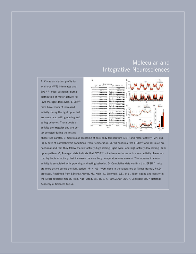
Molecular and Integrative Neurosciences A, Circadian rhythm profile for wild-type (WT) littermates and EP3R–/– mice. Although diurnal distribution of motor activity follows the light-dark cycle, EP3R–/– mice have bouts of increased activity during the light cycle that are associated with grooming and eating behavior. Those bouts of activity are irregular and are better detected during the resting phase (see carets). B, Continuous recording of core body temperature (CBT) and motor activity (MA) during 5 days at normothermic conditions (room temperature, 30°C) confirms that EP3R–/– and WT mice are nocturnal and that they follow the low activity–high resting (light cycle) and high activity–low resting (dark cycle) pattern. C, Averaged data indicate that EP3R–/– mice have an increase in motor activity characterized by bouts of activity that increases the core body temperature (see arrows). The increase in motor activity is associated with grooming and eating behavior. D, Cumulative data confirm that EP3R–/– mice are more active during the light period. *P = .03. Work done in the laboratory of Tamas Bartfai, Ph.D., professor. Reprinted from Sánchez-Alavez, M., Klein, I., Brownell, S.E., et al. Night eating and obesity in the EP3R-deficient mouse. Proc. Natl. Acad. Sci. U. S. A. 104:3009, 2007. Copyright 2007 National Academy of Sciences U.S.A. Cindy Ehlers, Ph.D., Professor, Gina Stouffer, Research Assistant, and José Criado, Jr., Ph.D., Staff Scientist MOLECUL AR AND INTEGRATIVE NEUROSCIENCES MOLECULAR AND I N T E G R AT I V E NEUROSCIENCES DEPAR TMENT S TA F F Tamas Bartfai, Ph.D. Chairman and Professor Director, Harold L. Dorris Neurological Research Institute Serge Ahmed, Ph.D. Adjunct Assistant Professor Etienne Baulieu, Ph.D. Adjunct Professor Floyd Bloom, M.D. Professor Emeritus Executive Director, Science Communication 2008 THE SCRIPPS RESEARCH INSTITUTE Steven J. Henriksen, Ph.D. Adjunct Professor Amanda Roberts, Ph.D. Associate Professor Paul L. Herrling, Ph.D. Adjunct Professor Michael G. Rosenfeld, M.D. Adjunct Professor Tomas Hokfelt, M.D., Ph.D. Adjunct Professor Pietro P. Sanna, M.D. Associate Professor Danny Hoyer, Ph.D. Adjunct Professor George R. Siggins, Ph.D. Professor Koki Inoue, Ph.D. Adjunct Associate Professor Iustin Tabarean, Ph.D. Assistant Professor Harvey Karten, M.D. Adjunct Professor Antoine Tabarin, Ph.D. Adjunct Associate Professor Henri Korn, M.D., Ph.D. Adjunct Professor Lars Terenius, Ph.D. Adjunct Professor Thomas Krucker, Ph.D. Adjunct Assistant Professor Claes Wahlestedt, M.D., Ph.D. Adjunct Professor Stefan Kunz, Ph.D. Adjunct Professor Mehrdad Alirezaei, Ph.D. Michal Bajo, M.D., Ph.D. Hilda Bajova, D.V.M. Fulvia Berton, Ph.D. Vez Repunte Canonigo, Ph.D. Kazuki Hagihara, Ph.D. Izabella Klein, Ph.D. Kayo Mitsukawa, Ph.D. Olivia Osborn, Ph.D. Covadonga Paneda, Ph.D. Gurudutt Pendyala, Ph.D. Jerry Pinghwa Pian, Ph.D. Tammy Wall, Ph.D. Adjunct Associate Professor Jilla Sabeti, Ph.D. Friedbert Weiss, Ph.D. Professor V I S I T I N G I N V E S T I G AT O R S Cary Lai, Ph.D. Associate Professor Karen T. Britton, M.D., Ph.D. Adjunct Associate Professor Ulo Langel, Ph.D. Adjunct Professor Michael Buchmeier, Ph.D. Adjunct Professor Xiaoying Lu, Ph.D. Assistant Professor Iain L. Campbell, Ph.D. Adjunct Professor Jan O. Lundstrom, Ph.D. Adjunct Professor Zhen Chai, Ph.D. Adjunct Assistant Professor Athina Markou, Ph.D. Adjunct Professor Jerold Chun, M.D., Ph.D. Adjunct Professor Madis Metsis, Ph.D. Adjunct Associate Professor Bruno Conti, Ph.D. Associate Professor Benjamin Neuman, Ph.D. Adjunct Assistant Professor Cindy L. Ehlers, Ph.D. Professor Shirley M. Otis, M.D. Adjunct Professor Ralph Feuer, Ph.D. Adjunct Assistant Professor Tommy Phillips, Ph.D. Adjunct Assistant Professor Howard S. Fox, M.D., Ph.D. Associate Professor John Polich, Ph.D. Associate Professor Brendan Walker, Ph.D. Hermann H. Gram, Ph.D. Adjunct Associate Professor Luigi Pulvirenti, M.D. Adjunct Associate Professor S C I E N C E A S S O C I AT E S S TA F F S C I E N T I S T S Roberto Ciccocioppo, Ph.D. José Criado, Ph.D. Walter Francesconi, Ph.D. David Gilder, M.D. Salvador Huitrón-Reséndiz, Ph.D. M. Cecilia Marcondes, Ph.D. Teresa Reyes, Ph.D. Adjunct Assistant Professor R E S E A R C H A S S O C I AT E S Zhifeng Chen, Ph.D. Jason Botten, Ph.D. Assistant Professor Donna L. Gruol, Ph.D. Associate Professor 327 Remi Martin-Fardon, Ph.D. Tom Nelson, Ph.D. Manuel Sánchez-Alavaz, M.D., Ph.D. Mitra Rebek, Ph.D. Caroline Lanigan, Ph.D. Sam Madamba Hedieh Badie, Ph.D. Genomics Institute of the Novartis Research Foundation San Diego, California Persephone Borrow, Ph.D. Edward Jenner Institute for Vaccine Research Compton, England Urs Christen, Ph.D. La Jolla Institute for Allergy and Immunology La Jolla, California Jean E. Gairin, Ph.D. CNRS Toulouse, France Karine Guillem, Ph.D. University of Pennsylvania Philadelphia, Pennsylvania Katsuro Hagiwara, Ph.D. Rakuno Gakuen University Ebetsu, Japan Dirk Homann, M.D., Ph.D. University of Colorado Health Sciences Center Denver, Colorado 328 MOLECUL AR AND INTEGRATIVE NEUROSCIENCES Shinchi Iwasaki, M.D., Ph.D. Osaka City University Medical School Osaka, Japan Rolf Kiessling, Ph.D. Karolinska Institutet Stockholm, Sweden Denise Naniche, Ph.D., M.P.H. Universitat de Barcelona Barcelona, Spain Noemi Sevilla, Ph.D. Universidad Autonoma de Madrid Madrid, Spain Christina Spiropoulou, Ph.D. Centers for Disease Control and Prevention Atlanta, Georgia Elina Zuniga, Ph.D. University of California San Diego, California 2008 THE SCRIPPS RESEARCH INSTITUTE MOLECUL AR AND INTEGRATIVE NEUROSCIENCES Chairman’s Overview I n the past year, we experienced scientific successes as well as organizational and policy changes in the Molecular and Integrative Neurosciences Department. The scientific work of several faculty members resulted in high-significance, high-visibility publications and important new research grants and renewals of earlier grants from the National Institutes of Health. Tamas Bartfai, Ph.D. Particularly noteworthy because of their immediate clinical usefulness are the findings of George Siggins and his collaborators in the Committee on the Neurobiology of Addictive Disorders that the widely used antiepileptic compound gabapentin may be useful in treating alcohol addiction. Pietro Sanna published important findings on the molecular mechanisms of alcohol-induced adaptation of nerve cells. Friedbert Weiss expanded our knowledge of the pharmacologic potential of the subtype-selective antagonists that can block the endogenous anxiogenic stress signal corticotropin-releasing factor. Research by Cindy Ehlers in pharmacogenomics led to new conclusions about the genetic basis of vulnerability of Native Americans to alcohol addiction, and Donna Gruol added new data on the effects of the proinflammatory cytokine IL-6 in the brain. John Polich expanded his noninvasive studies on the human brain by using attentional tasks. Bruno Conti made important findings about the role of the cytokine IL-18 in the regulation of feeding behavior and energy efficiency and through these mechanisms, the control of body weight. He also collaborated with Manuel Sánchez-Alavez and Iustin Tabarean, who uncovered a previously undetected night-eating phenotype in the commonly studied strain of mice that lack the gene for prostanoid receptor 3. These mice may be good models of night bingeing. Xiaoying Lu, Amanda Roberts, and I have 2008 THE SCRIPPS RESEARCH INSTITUTE 329 added to the studies on galanin and galanin receptors in anxiety and in depressive behaviors. The scientists of the department have engaged in many intradepartmental and interdepartmental collaborations. Numerous high-impact invited lectures and seminars were presented by the faculty nationally and internationally. For example, I was the keynote speaker at the largest drug development meeting (12,000 attendees) in Shanghai in June 2007. Despite a difficult economic climate, scientific progress in the department was good, and our educational goals for our graduate students and postdoctoral fellows were all successfully met. Several faculty and students received prestigous stipends. 330 MOLECUL AR AND INTEGRATIVE NEUROSCIENCES Investigators’ Reports Inflammation and Obesity O. Osborn, S.E. Brownell, M. Sánchez-Alavez, D. Salomon, H. Gram, T. Bartfai he proinflammatory cytokine IL-1β is elevated in obese humans and rodents and is implicated in impaired insulin secretion, decreased cell proliferation, and apoptosis of pancreatic beta cells. We have investigated the therapeutic effects of an antibody to IL-1β in hyperglycemic mice with diet-induced obesity. After 13 weeks of treatment, compared with a group given a control antibody, the group given the antibody to IL-1β had a significant improvement in glycemic control and in beta cell function, suggesting this novel therapeutic approach may slow or prevent progression to type 2 diabetes. IL-1β is also a key mediator of impaired function and destruction of pancreatic beta cells during the development of type 1 diabetes. Our findings suggest that an antibody to IL-1β has therapeutic potential in the treatment of type 2 diabetes and may have beneficial effects in other forms of diabetes in which tight glucose control is essential to prevent induction of IL-1β and thus limit beta cell destruction. T Galanin and Stress: Involvement of Galanin Receptor Subtypes K. Mitsukawa, X. Lu, T. Bartfai tress-related disorders are some of the most serious and prevalent medical conditions. Because current clinical treatments for stress have limited efficacy and cause unwanted effects in many patients, much effort has been put into understanding the molecular basis of these devastating disorders. Recent studies indicate that the neuropeptide galanin plays a role in mood disorders through its central G protein–coupled receptors: galanin receptor subtypes 1–3 (GalR–GalR3). We evaluated the effects of galanin on restraint stress in mice, a model of psychogenic stress. Core body temperature and locomotor activity were monitored by using radio telemetry devices. Intracerebroventricular injection of galanin had a biphasic effect on stress-induced hyperthermia and the associated increase in the levels of the stress hormones cortico- S 2008 THE SCRIPPS RESEARCH INSTITUTE sterone and corticotropin; low doses of galanin increased the stress response, whereas high doses had the opposite effects. High doses of galanin activated neurons in specific brain regions implicated in stress-related behaviors. To further clarify which receptor subtype is involved in the effects on stress-induced hyperthermia and associated changes in hormone levels, we used mice that lack the gene for GalR1 or the genes for GalR1 and GalR2. Compared with a control group, these mice had no change in stress-induced hyperthermia or hormone levels after treatment with high doses of galanin. These results indicate that GalR1 plays a role in the galanin effects observed. Transcriptional Profiling of Single Warm-Sensitive Hypothalamic Neurons I. Klein, I. Tabarean, O. Osborn, M. Sánchez-Alavez, E. Gregorsson, B. Ross, B. Conti, T. Bartfai aintenance of core body temperature is one of the greatest sources of energy expenditure in mammals. A small set of warm-sensitive GABAergic neurons in the preoptic area of the anterior part of the hypothalamus are known to play a key role in thermoregulation. To facilitate detailed molecular characterization of warm sensitivity at the level of single cells, we used electrophysiologic methods to detect individual warm-sensitive neurons in mouse embryonic hypothalamic cell cultures and in hypothalamic slices from adult mice. The transcriptomes of warm-sensitive and warm-insensitive cells were amplified by linear amplification and subsequently hybridized to microarrays. We found that warm-sensitive neurons in slices from the anterior part of the hypothalamus have functional receptors for several signal substances involved in the regulation of metabolism, feeding, and inflammation as well as pyrogenic substances. We are validating the microarray data by using quantitative polymerase chain reaction and in situ hybridization. The functional relevance of some of the expressed transcripts is being further investigated by using available ligands. This approach accelerates pharmacologic characterization of these neurons because receptors expressed in these cells can now be investigated as putative drug targets for regulation of metabolic rates. The differential expression of transcripts in warm-sen- M MOLECUL AR AND INTEGRATIVE NEUROSCIENCES 2008 THE SCRIPPS RESEARCH INSTITUTE sitive and warm-insensitive cells may also provide the long-awaited tool for distinguishing and modifying temperature-sensitive cells in vivo. Detailed molecular characterization of these neurons at a single-cell level will provide new insights into the regulation of metabolic rate, body temperature, and, indirectly, aging. Development of Therapeutic Agents and Vaccines for Diseases Caused by Arenaviruses and Hantaviruses Effects of Core Body Temperature in Energy Homeostasis J. Botten, D. Do, C.T. Cornillez-Ty, J. Ting, J. Klaus, M. Sánchez-Alavez, I. Tabarean, B. Conti, T. Bartfai ore body temperature (CBT) in homeotherms is maintained at a constant level and is largely independent of the temperature of the surroundings. Small but persistent changes in CBT are associated with significant changes in energy demand and thus in metabolism. Some temperature-sensitive GABAergic neurons in the preoptic area are involved in sensing CBT and brain temperature and in response regulate metabolic rate to maintain CBT. Microarray analysis followed by quantitative polymerase chain reaction indicated that bombesin and prolactin receptors are expressed in warm-sensitive neurons in the preoptic area. We are using available ligands to determine if CBT can be affected by applying agonists of these receptors to the preoptic area. Injection of bombesin into the preoptic area induced profound hypothermia accompanied by a marked decrease in the respiratory exchange ratio and heat production for 4 hours. Motor activity was increased during the same period, but it could not prevent hypothermia. Injection of bombesin into the pallidus raphe, which is one target of preoptic area projecting neurons, induced slight hyperthermia for the next 3 hours, which coincided with a slight increase in respiratory exchange ratio and heat production. Injection of prolactin into the preoptic area or pallidus raphe induced a slight increase in CBT, respiratory exchange ratio, and heat production, but the increases did not differ significantly from those in controls. Our findings suggest that peptides can modulate activity in the preoptic area; we are determining the mechanisms responsible for these effects in the brain. C PUBLICATIONS Mitsukawa, K., Lu, X., Bartfai, T. Galanin, galanin receptors and drug targets. Cell. Mol. Life Sci. 65:1796, 2008. 331 A.J. Hessell, D.R. Burton, P. Barrowman, J.L. Whitton, A. Sette,* M.J. Buchmeier** * La Jolla Institute for Allergy and Immunology, San Diego, California ** University of California, Irvine, California renaviruses and hantaviruses are rodent-borne pathogens that cause marked morbidity and mortality in humans. Arenaviruses cause illnesses ranging from aseptic meningitis after infection with lymphocytic choriomeningitis virus to hemorrhagic fever syndromes after infection with Lassa, Junin, Machupo, and Guanarito viruses. Hantaviruses cause hantavirus cardiopulmonary syndrome, a disease with a mortality rate of 30%–50% in the Americas. Viruses in both genera have RNA genomes that encode 4 open reading frames in either negative-sense (hantavirus) or ambisense (arenavirus) fashion. Currently, no licensed vaccines are available for the prevention of disease caused by arenaviruses or hantaviruses. Our research interests in these viruses include understanding how the host immune response contributes to pathogenesis and/or protective immunity, identifying novel host-pathogen interactions, and developing novel therapeutic agents and vaccines. A recent goal in our laboratory has been to develop the capability to express the complete proteome of each of the 7 pathogenic arenaviruses and the 2 primary etiologic agents of hantavirus cardiopulmonary syndrome in the Americas. We have successfully cloned each of the open reading frames encoded by these viruses and shuttled the clones into several unique expression vectors (based on plasmids and recombinant viruses). To date, we have expressed 30 of the cloned 34 open reading frames. One highlight from these studies has been the development of a plasmid vector that can be used to successfully express extremely difficult proteins such as the New World hantavirus envelope glycoproteins. Using the new library of expression vectors, we recently began new research projects, including probing viral proteins for novel host cellular binding partners and panning phage display antibody libraries derived A 332 MOLECUL AR AND INTEGRATIVE NEUROSCIENCES from samples from patients with hantavirus cardiopulmonary syndrome for monoclonal antibodies specific for the envelope glycoproteins of the North and South American hantaviruses Sin Nombre and Andes, respectively. In the past year, we identified several monoclonal antibodies specific for Sin Nombre and Andes viral antigens, and we are testing the antibodies for the ability to neutralize the New World hantaviruses. Our hope is that neutralizing antibodies can be identified and used as therapeutic agents to successfully treat hantavirus cardiopulmonary syndrome. A final highlight from our recent studies has been the finding that HLA-restricted, cross-reactive epitopes do exist among diverse arenaviruses and that individual epitopes can be used as effective vaccine determinants for multiple pathogenic arenaviruses. PUBLICATIONS Botten, J., Kotturi, M.F. Adaptive immunity to lymphocytic choriomeningitis virus: new insights into antigenic determinants. Future Virol. 2:495, 2007. Mothe, B.R., Stewart, B., Oseroff, C., Bui, H., Stogiera, S., Garcia, Z., Dow, C., Rodriguez-Carreno, M., Kotturi, M., Pasquetto, V., Botten, J., Crotty, S., Janssen, E., Buchmeier, M.J., Sette, A. Chronic lymphocytic choriomeningitis virus infection actively down-regulates CD4+ T cell responses directed against a broad range of epitopes. J. Immunol. 179:1058, 2007. Temperature Homeostasis and Aging, Sites and Effects of IL-18 in the CNS B. Conti, H. Bajova, M. Sánchez-Alavez, T. Bartfai, B. Ross, S. Alboni, D. Cervia, E. Zorrilla, V. Zhukov C H A R A C T E R I Z AT I O N O F T R A N S G E N I C M I C E W I T H R E D U C E D C O R E B O D Y T E M P E R AT U R E A N D PROLONGED LONGEVITY e previously found that transgenic mice with a modest but prolonged reduction of core body temperature (CBT) have increased median life expectancy. The mechanisms contributing to the prolonged life span in these animals are being characterized. Specifically, we are investigating whether lowered CBT reduces damage caused by free radicals over time. In collaborative studies, we are evaluating the effects of minor temperature changes on mitochondrial function, with S. Ali, University of California, San Diego, and the contribution of antioxidant pathways, with P. Maher, Salk Institute for Biological Studies, La Jolla, California. The possibility that reduced temperature may prolong W 2008 THE SCRIPPS RESEARCH INSTITUTE life span by influencing specific pathways is also being evaluated by using transcriptomics and a mass spectrometry–based metabolomic approach that is being carried out in collaboration with W. Wikoff, W. Webb, and G. Siuzdak, Center for Mass Spectrometry. INFLUENCE OF GONADAL HORMONES ON CBT SET POINT To gain insight on the nature of the “central thermostat” that regulates CBT, we are investigating what determines the sex-specific differences in CBT in mice. Specifically, we assessed the roles of gonads and sex hormones in determining the temperature set point. Castration of males resulted in an elevation in CBT of 1°C only during the light part of the day, whereas ovariectomy of females eliminated the estrus-associated variations in CBT. Gonadectomized animals had identical temperature profiles, indicating that the set point is identical in both sexes but can be modulated by the gonads. SITES OF IL-18 ACTION IN THE CNS In collaboration with E. Zorrilla, Committee on the Neurobiology of Addictive Disorders, we previously showed that the pleiotropic inflammatory cytokine IL-18 is a central anorexigenic agent and a regulator of energy efficiency. To understand the sites and the mechanisms of action of IL-18 in the CNS, we characterized members of the IL-18 family in the brains of mice. IL-18 and the 2 IL-18 receptors (IL-18Rs), the heterodimer IL-18RI and IL-18 RacP, and the soluble inhibitor IL-18 binding protein (IL-18BP) were investigated in the CNS of normal C57B/6 mice. In situ hybridization showed extensive neuronal localization of IL-18Rs in the cortex, hippocampus, and hypothalamus. In addition to the canonical IL-18RI, a shorter variant of this receptor subunit was identified. The biological activity and the possible significance of the short IL-18RI form with respect to IL-18 in the CNS are being investigated in vitro by using hypothalamic and macrophage and T-cell lines and lentiviral short interfering RNAs. We are comparing several transduction pathways, including the nuclear factor κB and the signal transducer and activator of transcription 3 pathways known to mediate the action of IL-18. I L - 1 8 , S T R E S S , A N D AT H E R O S C L E R O S I S IL-18 has proatherogenic effects, and its circulating level is elevated in a tissue-specific manner via differential use of promoters after neurogenic stimulation, restraint-induced stress, or treatment with corticotropin. Clinical studies and research in animal models indicate that chronic stress is associated with aggrava- MOLECUL AR AND INTEGRATIVE NEUROSCIENCES tion of atherosclerosis, coronary heart disease, and stroke, although no mediators of atherosclerosis during stress have been identified. We hypothesized that IL-18 can contribute to atherosclerosis during chronic mental stress. To test this hypothesis, we crossed mice that lacked the gene for IL-18 with atherosclerosis-prone mice that lacked the gene for apolipoprotein E. The effects of stress on atherosclerosis in these crossbred animals will be evaluated after chronic exposure to 4 different psychologic stressors. The extent and the quality of atherosclerotic lesions will be evaluated. PUBLICATIONS Sugama, S., Conti, B. Interleukin 18 and stress. Brain Res. Rev. 58:85, 2008. Zorrilla, E.P., Sánchez-Alavez, M., Sugama, S., Brennan, M., Fernandez, R., Bartfai, T., Conti, B. Interleukin-18 controls energy homeostasis by suppressing appetite and feed efficiency. Proc. Natl. Acad. Sci. U. S. A. 104:11097, 2007. Laboratory of Translational Neurophysiology and the San Diego Substance Abuse and Minorities Project C.L. Ehlers, B.M. Walker, J.R. Criado, S. Sanchez, G. Berg, D. Wills, J. Walker, G. Stouffer, C. Agneta, L. Corey, J.P. Pian, D.A. Gilder, J.W. Havstad, P. Lau, R. Duro, S.L. Lopez, E. Phillips he current and main focus of the laboratory is to determine the CNS etiology of substance abuse. Our strategy in studying these disorders in patients has been to capitalize on the use of new accurate structured diagnostics, physiologic measures, and the latest genetic techniques to identify risk and protective factors for alcohol and drug dependence. Because the prevalence of substance dependence varies among certain racial/ethnic groups, we have also focused on studying a wide range of ethnic groups to evaluate genetic and cultural differences that may lead to new clues for the causes of the disorders. This work encompasses parallel studies in animal models, currently called “translational research.” This type of research allows investigators to simultaneously evaluate disorders in patients and model the condition in animals so that progress toward understanding the causes for these disabilities can be more rapidly pursued. T 2008 THE SCRIPPS RESEARCH INSTITUTE 333 The differences in prevalence rates of alcohol and drug use and abuse between ethnic groups and in alcohol-drinking preferences between different strains of rats provide an opportunity to investigate how genetic variation may influence these disorders. To find genes associated with alcohol and other drug dependencies, we did a genome scan for behaviors related to alcoholism and substance use in American Indian families with a high prevalence of substance dependence. We found that several sites in the genome were linked not only to multiple drugs of abuse but also to externalizing disorders and body mass, suggesting that the same selective pressure may have enriched for genetic variants that increase the risk for consumption of both food and drugs of abuse. We investigated 2 genes in 2 different populations that appear to affect substance dependence. One of the genes, OPRM1, codes for the receptor that binds opiate drugs. This gene influences a person’s tolerance to alcohol. The other gene, CNR1, codes for the receptor that binds marijuana and influences impulsivity, a personality trait associated with drug dependence. A crucial variable in the development of alcohol dependence is the age at which a person begins to consume alcohol. We found that alcohol dependence was 5–6 times more likely in youth who started drinking before age 13 years than in persons who did not start drinking until after age 16 years. Studying biobehavioral risk factors for and the consequences of underage alcohol use in young adults and in animal models is critical for several reasons. First, rapid development of alcohol dependence is more likely in underage drinkers than in older drinkers, suggesting that alcohol may be “more addicting” to underage drinkers, a finding also partly confirmed by our controlled studies in animal models of the disorder. Second, exposure to alcohol and other substances during adolescence may have long-lasting consequences. Rapid changes in neural organization occur during this period, and these changes in CNS organization may make the brain uniquely vulnerable to injury by drug use or abuse. Thus, during adolescence, drugs may be both more addicting and more neurotoxic, a combination that makes drug abuse particularly malignant for adolescents. To combat this problem, we are not only conducting more research into the etiology of underage drinking but also developing new strategies, called “environmental preventions,” to reduce underage drinking in high-risk populations. 334 MOLECUL AR AND INTEGRATIVE NEUROSCIENCES PUBLICATIONS Criado, J., Ehlers, C.L. Electrophysiological responses to affective stimuli in Mexican Americans: relationship to alcohol dependence and personality traits. Pharmacol. Biochem. Behav. 88:148, 2007. Criado, J., Ehlers, C.L. Electrophysiological responses to affective stimuli in Southwest California Indians: relationship to alcohol dependence. J. Stud. Alcohol Drugs 68:813, 2007. Ehlers, C.L., Gilder, D.A., Phillips, E. P3 components of the event-related potential and marijuana dependence in Southwest California Indians. Addict. Biol. 13:130, 2008. Ehlers, C.L., Gilder, D.A., Slutske, W.S., Lind, P., Wilhelmsen, K.C. Externalizing disorders in American Indians: comorbidity and a genome wide linkage analysis. Am. J. Med. Genet. B Neuropsychiatr. Genet. 147B:690, 2008. Ehlers, C.L., Lind, P.A., Wilhelmsen, K.C. Association between single nucleotide polymorphisms in the µ opioid receptor gene (OPRM1) and self-reported responses to alcohol in American Indians. BMC Med. Genet. 9:35, 2008. Ehlers, C.L., Slutske, W.S., Lind, P.A., Wilhelmsen, K.C. Association between single nucleotide polymorphisms in the cannabinoid receptor gene (CNR1) and impulsivity in Southwest California Indians. Twin Res. Hum. Genet. 10:805, 2007. Gilder, D.A., Lau, P., Corey, L., Ehlers, C.L. Factors associated with remission from alcohol dependence in an American Indian community group. Am. J. Psychiatry 165:1172, 2008. Gilder, D.A., Lau, P., Corey, L., Ehlers, C.L. Factors associated with remission from cannabis dependence in Southwest California Indians. J. Addict. Dis. 26:23, 2007. Gilder, D.A., Lau, P., Gross, A., Ehlers, C.L. A comorbidity of alcohol dependence with other psychiatric disorders in young adult Mexican Americans. J. Addict. Dis. 26:31, 2007. Hodgkinson, C.A., Yuan, Q., Xu, K., Shen, P.H., Heinz, E., Lobos, E.A., Binder, E.B., Cubells, J., Ehlers, C.L., Gelernter, J., Mann, J., Riley, B., Roy, A., Tabakoff, B., Todd, R.D., Zhou, Z., Goldman, D. Addictions biology: haplotype-based analysis for 130 candidate genes on a single array. Alcohol Alcohol. 43:505, 2008. Montane-Jaime, K.L., Shafe, S., Moore, S., Gilder, D.A., Crooks, H., Joseph, R., Ehlers, C.L. The clinical course of alcoholism in Trinidad and Tobago. J. Stud. Alcohol Drugs, in press. Pian, J.P., Criado, J.R., Walker, B.M., Ehlers, C.L. Differential effects of acute alcohol on EEG and sedative responses in adolescent and adult Wistar rats. Brain Res. 1194:28, 2008. Scott, D.M., Williams, C.D., Cain, G.E., Kwagyan, J., Kalu, N., Hesselbrock, V., Ehlers, C.L., Taylor, R.E. Clinical course of alcohol dependence in African Americans. J. Addict. Dis., in press. Valladares, E.M., Eljammal, S.M., Motivala, S., Ehlers, C.L., Irwin, M.R. Sex differences in cardiac sympathovagal balance and vagal tone during nocturnal sleep. Sleep Med. 9:310, 2008. Walker, B.M., Walker, J.L., Ehlers, C.L. Dissociable effects of ethanol consumption during the light and dark phase in adolescent and adult Wistar rats. Alcohol 42:83, 2008. Neuroinflammation and Neurodegeneration: Mechanism and Biomarkers H.S. Fox, M. Alirezaei, A. Coutinho, C. Flynn, C. Marcondes, G. Pendyala, D. Watry, M. Zandonatti T he brain is a unique organ, not only functionally but also in terms of the host response to events such as damage and infection. We study processes 2008 THE SCRIPPS RESEARCH INSTITUTE in which the host response leads to neurodegeneration and brain dysfunction. Because the course of damage and disease is chronic, long-term studies are required. Several of these studies reached fruition this year. Much of our recent work has focused on biomarkers, objectively measured markers that correlate with disease states. These markers are useful for predicting risk of disease, development of disease, response to therapy, and other medical indications. In addition, they can give important clues to pathogenic mechanisms, leading to ways to prevent and treat disease. Neurodegenerative diseases have a distinct lack of such biomarkers, representing a marked gap in biomedical knowledge. We model the development of neurodegeneration leading to dementia by using a brain disease that occurs after a known stimulus, infection with HIV. Using infection of rhesus monkeys with simian immunodeficiency virus (SIV) as a model of neuroAIDS, we are studying the virology, immunology, pathology, neurobiology, and molecular basis of the resulting CNS disease. Two examples of this research are given here. First, using “-omic” technologies, we have discovered novel biomarkers for CNS disease, leading to mechanistic insights into neurodegenerative processes. For example, we found that osteopontin is a marker for HIVinduced dementia and that the increased expression of one of its receptors on monocytes, CD44v6, correlates with SIV encephalitis. Mechanistically, we found that osteopontin increases macrophage accumulation within tissues, paralleling the increases in macrophages in the brain that are responsible for HIV dementia. Second, in collaboration with G. Siuzdak’s group, Department of Molecular Biology, we are assessing whether metabolites in the cerebrospinal fluid would be useful as candidate biomarkers. Indeed the metabolites are, and using a combination of metabolomic and transcriptomic profiling, we have identified multiple new biomarkers in the cerebrospinal fluid, a finding that led to the discovery of a distinct mechanism of SIV-induced CNS disease. This research is now being expanded to encompass other CNS disorders and most likely will have important basic and applied effects. PUBLICATIONS Alirezaei, M., Watry, D.D., Flynn, C.F., Kiosses, W.B., Masliah, E., Williams, B.R.G., Kaul, M., Lipton, S.A., Fox, H.S. Human immunodeficiency virus-1/surface glycoprotein 120 induces apoptosis through RNA-activated protein kinase signaling in neurons. J. Neurosci. 27:11047, 2007. Burdo, T.H., Ellis, R.J., Fox, H.S. Osteopontin is increased in HIV-associated dementia. J. Infect. Dis. 198:715, 2008. MOLECUL AR AND INTEGRATIVE NEUROSCIENCES Gaskill, P.J., Zandonatti, M., Gilmartin, T., Head, S.R., Fox, H.S. Macrophagederived simian immunodeficiency virus exhibits enhanced infectivity by comparison with T-cell-derived virus. J. Virol. 82:1615, 2008. Huitron-Resendiz, S., Marcondes, M.C., Flynn, C., Lanigan, C.M., Fox, H.S. Effects of simian immunodeficiency virus on the circadian rhythms of body temperature and gross locomotor activity. Proc. Natl. Acad. Sci. U. S. A. 104:15138, 2007. Katner, S.N., Von Huben, S.N., Davis, S.A., Lay, C.C., Crean, R.D., Roberts, A.J., Fox, H.S., Taffe M.A. Robust and stable drinking behavior following long-term oral alcohol intake in rhesus monkeys. Drug Alcohol Depend. 91:236, 2007. Marcondes, M.C., Lanigan, C.M,. Burdo, T.H., Watry, D.D., Fox, H.S. Increased expression of monocyte CD44v6 correlates with the development of encephalitis in rhesus macaques infected with simian immunodeficiency virus. J. Infect. Dis. 197:1567, 2008. Marcondes, M.C., Sopper, S., Sauermann, S., Burdo, T.H., Watry, D.D., Zandonatti, M.A., Fox, H.S. CD4 deficits and disease course acceleration can be driven by a collapse of the CD8 response in rhesus macaques infected with simian immunodeficiency virus. AIDS 22:1441, 2008. Marcondes, M.C., Watry, D., Zandonatti, M., Flynn, C., Taffe, M.A.. Fox, H.S. Chronic alcohol consumption generates a vulnerable immune environment during early SIV infection in rhesus macaques. Alcohol. Clin. Exp. Res., in press. Wikoff, W.R., Pendyala, G., Siuzdak, G., Fox. H.S. Metabolomic analysis of the cerebrospinal fluid reveals changes in phospholipase expression in the CNS of SIVinfected macaques. J. Clin. Invest. 118:2661, 2008. Yadav, M.C., Burudi, E.M.E., Alirezaei, M., Flynn, C.C., Watry, D.D., Lanigan, C.M., Fox, H.S. IFN-γ induced IDO and WRS expression in microglia is differentially regulated by IL-4. Glia 55:1385, 2007. CNS Actions of Inflammatory Factors D.L. Gruol, T.E. Nelson, J. Sabeti, J. Cho,* M. Kuijpers,** H. Bajova, S. Chow, N. Bednorz, S. Michael, E. Vereyken,** P.N.E. de Graan,** J. Netzeband*** 2008 THE SCRIPPS RESEARCH INSTITUTE 335 ever, the functional consequence of glial-to-neuronal signaling via inflammatory factors is a relatively unexplored area. Information about the consequences is crucial to an understanding of the impact of neuroinflammation on the CNS. To address this issue, we are investigating the CNS actions of several inflammatory factors known to play an important role in CNS disorders. During the past year, we focused on IL-6, a cytokine produced at elevated levels in the CNS during a variety of abnormal conditions, including neurodegenerative disorders (e.g., Alzheimer’s disease, Parkinson’s disease), seizures, hypoxia-ischemia, trauma, and bacterial or viral infection. Results of Western blot analysis of cultured rodent hippocampal neurons chronically exposed to IL-6 to simulate neuroinflammatory conditions indicated that several proteins critical for normal neuronal function were downstream targets of the action of IL-6. These targets included proteins involved in synaptic transmission, neuron excitability, and intracellular calcium homeostasis. Western blot analysis of protein levels in hippocampi from transgenic mice that express elevated levels of IL-6 in the CNS and from nontransgenic littermate control mice showed changes in the hippocampal protein levels of the transgenic mice similar to those observed in the hippocampal cultures chronically exposed to IL-6. To determine if the changes in protein levels resulted in altered hippocampal function, we used electrophysiologic recordings (Fig. 1) to study synaptic transmission * Dongguk University, Gyeong Buk, Korea ** University Medical Center Utrecht, Utrecht, the Netherlands *** Molecular Devices, Union City, California onsiderable evidence indicates that neuroinflammation and the innate production of inflammatory factors are characteristic of a variety of CNS disorders, including disease and injury as well as neurodegenerative conditions and aging. Several inflammatory factors are produced in the CNS during neuroinflammation, and accumulating evidence implicates these factors as important contributors to, if not as a substrate for, the altered cognitive function that occurs in CNS disorders with a neuroinflammatory component. Glial cells are a primary source of inflammatory factors during CNS neuroinflammation, but CNS neurons also produce inflammatory factors under certain conditions. CNS neurons express receptors for inflammatory factors, consistent with neurons as an important target for inflammatory factors produced by glial cells. How- C F i g . 1 . Electrophysiologic recording from hippocampal slices. Dia- gram shows a hippocampal slice and the positioning of stimulating and recording electrodes. Representative field potential recordings of somatic (population spike) and dendritic (fEPSP) synaptic responses generated in area CA1 by stimulation of Schaffer collaterals are also shown. Downward arrows indicate the moment of electrical stimulation of Schaffer collateral afferents. The presynaptic volley (PSV), representing the summed activation of Schaffer collateral axons, is evident in the dendritic recording. 336 MOLECUL AR AND INTEGRATIVE NEUROSCIENCES at the synapse from Schaffer collaterals to the CA1 pyramidal neurons in isolated tissue slices of hippocampus obtained from IL-6 transgenic mice and nontransgenic mice. The synaptic pathway of the Schaffer collaterals to the CA1 pyramidal neurons is of interest because it plays a central role in hippocampal memory and learning. Electrical stimulation of the Schaffer collaterals elicited 3 electrical responses in field potential recordings from the hippocampal slices: a presynaptic volley, which reflects a summation of the action potentials occurring in the stimulated Schaffer collateral afferents; a field excitatory postsynaptic potential, which reflects a summation of excitatory postsynaptic responses occurring in the dendrites of CA1 pyramidal neurons activated by synaptic transmission; and a population spike, which represents a summation of the action potentials evoked by the synaptic response and occurring in the somata of the pyramidal neurons. The amplitudes of the 3 electrical responses were all significantly larger in the hippocampal slices from IL-6 transgenic mice than in the slices from the nontransgenic mice. These results show that chronic exposure to IL-6 can significantly alter the function of CNS neurons and indicates that mechanisms that mediate synaptic transmission and neuronal excitability are important targets of IL-6 action. These effects of IL-6 may play an important role in the cognitive dysfunction associated with neuroinflammation. For example, the IL-6–induced enhancement of synaptic transmission could saturate mechanisms mediating memory and learning and thereby impair cognitive function. Moreover, the enhanced synaptic transmission could lead to hyperexcitability, which is an important contributor to seizure activity. PUBLICATIONS Bajova, H., Nelson, T.E., Gruol, D.L. Chronic CXCL10 alters the level of activated ERK1/2 and transcriptional factors CREB and NF-κB in hippocampal neuronal cell culture. J. Neuroimmunol. 195:36, 2008. Netzeband, J.G., Gruol, D.L. mGluR1 agonists elicit a Ca2+ signal and membrane hyperpolarization mediated by apamin-sensitive potassium channels in immature rat Purkinje neurons. J. Neurosci. Res. 86:293, 2008. Sabeti, J., Gruol, D.L. Emergence of NMDA-receptor-independent long-term potentiation at hippocampal CA1 synapses following early-adolescent exposure to chronic intermittent ethanol: role for σ-receptor function. Hippocampus 18:148, 2007. 2008 THE SCRIPPS RESEARCH INSTITUTE Role of the Neuregulins in the Nervous System C. Lai, J.L. Weber, D. Aumann, C. Challis, D. Hom, K. Schaukowitch he focus of our research is understanding the signaling mechanisms that underlie the establishment and maintenance of mature neuronal and glial cell phenotypes. We are studying the roles played by a subfamily of receptor protein-tyrosine kinases, the ErbBs (EGFR, ErbB2, ErbB3, and ErbB4), and their ligands, the neuregulins (NRG-1–NRG-4). In the developing peripheral nervous system, NRG-1 was first recognized as the Schwann cell mitogen glial growth factor. NRG-1 was also termed ARIA (for acetylcholine receptor inducing activity), which was thought to regulate the expression of acetylcholine receptors at developing neuromuscular junctions. These distinct functions are now thought to be mediated by discrete types of NRG-1 (I, II, and III) that arise by alternative splicing. A primary goal of our research is to understand the specific roles of each of these types of NRG-1 in the nervous system. NRG-1 supports survival of Schwann cells and regulates the number of premyelinating Schwann cells. The results of genetic studies suggested that the type III isoform serves in this capacity, and we have helped determine that this isoform also plays a key role in regulating the thickness of the myelin sheath. The emerging picture is that different NRG-1 isoforms serve as signaling molecules to carry out distinct biological activities. We are also investigating the roles of these NRG-1 isoforms in the brain, which became an area of considerable interest after NRG-1 was identified as a susceptibility gene for schizophrenia. We have 4 areas of primary interest. The first is the roles of the 3 types of NRG-1 in the developing and mature nervous system. We developed transgenic mice that permit the tetracycline-regulated expression of specific NRG-1 isoforms. With these mice, we can assess the distinct biological functions served by each isoform. The second area is neurogenesis and migration. We found that the NRG receptor ErbB4 is expressed by multiple tangentially migrating populations of neurons in the developing and mature nervous systems. ErbB4 is expressed at high levels in the mature subventricular zone and rostral migratory stream, one of the few regions in the brain in rats where neurogenesis occurs in the adults. We are searching for the endoge- T MOLECUL AR AND INTEGRATIVE NEUROSCIENCES nous ligands and testing the effects of the NRGs on cells derived from the subventricular zone. Our data suggest that ErbB4 influences both the proliferation of neural progenitor cells and migration of neuroblasts in the rostral migratory stream The third area of interest is the effects of the loss of ErbB4 function in the mature brain. We are analyzing the phenotype of mice that lack the gene for ErbB4 in the nervous system. These animals have a reduction in anxiety-like behavior, and we are testing the hypothesis that the loss of ErbB4 in the amygdala underlies this defect. Last, we are developing novel, bacterial artificial chromosone–based transgenic tools that permit regulated gene expression in specific subsets of neurons. We have successfully developed lines of mice that permit regulated gene expression in cholinergic neurons, and we are evaluating similar lines that permit regulated expression in the medium spiny neurons of the striatum. These animal models may be useful for studies of neural development and neurodegenerative disorders such as Alzheimer’s, Parkinson’s, and Huntington’s diseases. PUBLICATIONS Prieto, A.L., O’Dell, S., Varnum, B., Lai, C. Localization and signaling of the receptor protein tyrosine kinase Tyro3 in cortical and hippocampal neurons. Neuroscience 150:319, 2007. Woo, R.S., Li, X.M., Tao, Y., Carpenter-Hyland, E., Huang, Y.Z., Weber, J., Neiswender, H., Dong, X.P., Wu, J., Gassmann, M., Lai, C., Xiong, W.C., Gao, T.M., Mei, L. Neuregulin-1 enhances depolarization-induced GABA release. Neuron 54:599, 2007. Galanin Receptors in Antidepressant and Anxiolytic-Like Actions X. Lu, F. Xia, B. Ross, K. Mitsukawa, J. Rebek, E. Roberts, T. Bartfai W e focus on receptors (GalRs) for galanin, a neuropeptide, in depression and anxiety and on developing small-molecule synthetic ligands for GalRs. We are using molecular, cellular, and behavioral techniques to study the role of GalR2 and GalR3 signaling in antidepressant and anxiolytic-like actions. GalR2 and GalR3 are G protein–coupled receptors expressed at high levels in brain areas implicated in mood regulation. Previously, we showed that clinically effective antidepressant treatments induced the expression of 2008 THE SCRIPPS RESEARCH INSTITUTE 337 galanin and upregulated GalR2 signaling in several areas of the brain in rats, including the hippocampus and dorsal raphe nucleus. We hypothesized that GalR2 signaling mediates antidepressant-like effects. In behavioral studies, compared with wild-type mice, mice lacking the gene for GalR2 had a more persistent depressive-like phenotype in the learned helplessness model and increased immobility in the tail suspension test. These data are consistent with an antidepressant-like effect of GalR2. We are also interested in the role of GalR2 signaling in hippocampal neurogenesis in adults, a process that mediates antidepressant-like effects. We found that GalR2 is expressed at high levels in progenitor/stem cells isolated from the hippocampus of adult mice; signaling through GalR2 is associated with an increase in the proliferative activity of these cells. Effects of GalR2 signaling on differentiation and survival of the progenitor cells are under investigation. We also plan to deliver lentiviral short hairpin RNAs against GalR2 to the hippocampus in rats to study the effect of GalR2 signaling in neurogenesis in adults in vivo. We are also interested in the physiologic function of GalR3, in particular, its potential involvement in antidepressant and anxiolytic actions. GalR3 is the least studied receptor subtype of all 3 galanin receptors. Despite a recent report that a class of putative GalR3 antagonists has antidepressant and anxiolyticlike effects in rodent models, our understanding of the physiologic function of GalR3, including its signaling mechanism and precise distribution pattern, is limited. We are developing mice that lack the gene for GalR3 that express a green fluorescent protein–LacZ fusion reporter at the GalR3 locus. Phenotypic assessment of these mice should provide valuable insight into the function of GalR3. This molecular approach is also important for establishing GalR3 as a novel drug target for treatment of depression and anxiety. In collaboration with E. Roberts, Department of Chemistry, and J. Rebek, Skaggs Institute, we are developing small-molecule synthetic ligands for GalR2 and GalR3. PUBLICATIONS Lu, X., Ross, B., Sanchez-Alavez, M., Zorrilla, E.P., Bartfai, T. Phenotypic analysis of GalR2 knockout mice in anxiety- and depression-related behavioral tests. Neuropeptides 42:387, 2008. Mitsukawa, K., Lu, X., Bartfai, T. Galanin, galanin receptors and drug targets. Cell. Mol. Life Sci. 65:1796, 2008. 338 MOLECUL AR AND INTEGRATIVE NEUROSCIENCES Cognitive Aging, Physical Fitness, and Attention Brain Waves 2008 THE SCRIPPS RESEARCH INSTITUTE scalp; the target stimulus elicits the P3b potential over the parietal areas. This difference in scalp topography is hypothesized to reflect distinct neural generating systems related to attention and memory processing of the stimuli. AT T E N T I O N B R A I N WAV E S J. Polich, C.H. Hillman* * University of Illinois, Champaign-Urbana, Illinois Figure 1 illustrates scalp topography amplitude from the application of a visual 3-stimulus task to each of COGNITIVE AGING AND PHYSICAL FITNESS major question in cognitive aging is how healthoriented behaviors can mitigate age-related mental decline. Recent evidence indicates that participation in aerobic exercise may ameliorate or protect against declines in the brain and cognitive function associated with aging. Event-related brain potential (ERP) methods were used to obtain initial information on the neural mechanisms that underlie exercise-induced changes in cognition. Exercise-related changes in cognition may be due in part to changes in the attention system that contributes to the P300 component. When coupled with recent neuroanatomic evidence indicating age-related atrophy of the neural network involved in attentional control, ERPs can provide insight into the effects of exercise on cognition. In order to assess this hypothesis, neuroelectric measures were used to assess younger and older (20.2 vs 66.8 years) individuals who were classified as low fit and high fit on the basis of a cardiovascular fitness evaluation in which volume of oxygen intake during maximal physical stress was used as a measure of fitness. A P3a AND P3b BRAIN POTENTIALS ERPs enable the direct evaluation of CNS activity during processing of stimulus information. The P3a and P3b subcomponents of the P300 ERP reflect the operation of frontal attentional and temporoparietal memory operations that appear to be associated with dopaminergic and variation in locus coeruleus norepinephrine pathways, respectively. A 3-stimulus visual model was used to elicit both of these components by presenting stimuli once every 2 seconds in a series. The infrequently occurring target stimuli were detected and responded to in the context of more frequently occurring standard stimuli; the stimuli were constructed to be difficult to discriminate (e.g., circles 5.5 and 5.0 cm in diameter). When an infrequently presented “distracter” stimulus randomly occurs (e.g., a large checkerboard pattern), the attentional focus induced by the discrimination task is disrupted and produces a P3a potential that has its maximum amplitude over frontal-central areas of the F i g . 1 . Topographic scalp amplitude mappings from the distracter and target stimuli from the younger and older lower- and higher-fit groups. The nose is upward and the scales differ for the P3a (top row, 20 µV) and P3b (bottom row, 10 µV) components. P3a from the distracter stimuli was larger in younger participants compared with older participants but did not differ between fitness levels in either group. P3b from the target stimuli was larger for the higherfit younger participants but did not differ for the older participants. See text for explanation. the 4 groups of participants. Fitness did not alter the P3a component, suggesting that attentional orienting processes are unaffected by fitness. Larger P3b amplitudes were found only in higher-fit younger adults, suggesting that aerobic fitness does not protect against age-related cognitive deficits. Cardiorespiratory fitness may be associated with an increased ability to suppress extraneous neural activity to facilitate attentional processing, because fitness is related to the increased health of neural tissue and increased ability to allocate attentional resources for higher-fit relative to lower-fit participants. Higher levels of cardiorespiratory fitness were associated with increased P3b amplitude only for the higher-fit younger adults relative to all other groups. Hence, higher-fit older adults may be unable to overcome deficits associated with cognitive aging, for example, loss of gray and white matter that is especially predominant in the prefrontal cortex. Fitness-related changes in cognitive aging therefore do not originate from a general change but appear specific to attentional processing. Although the mechanisms underlying differences in fitness-related aging in attentional process- MOLECUL AR AND INTEGRATIVE NEUROSCIENCES ing remain unclear, results from animal studies have suggested a link between aerobic exercise and neuronal proliferation as well as increases in monoamines such as norepinephrine and dopamine. Chronic exercise training may mediate and even reverse age-related decreases in neuronal tissue loss. Thus, cognitive decline from the depletion of available neural structures could affect attentional resource availability and result in a reduced ability to inhibit extraneous neural activity when task demands increase because of perceptual difficulty. PUBLICATIONS Allison, B.Z., Polich, J. Workload assessment of computer gaming using a singlestimulus event-related potential paradigm. Biol. Psychol. 77:277, 2008. Azizian, A., Polich, J. Evidence for attentional gradient in the serial position memory curve from event-related potentials. J. Cogn. Neurosci. 19:2071, 2007. Cahn, B.R., Polich, J. Meditation (Vipassana) and the P3a event-related brain potential. Int. J. Psychophysiol., in press. Cano, M.E., Class, Q.A, Polich, J. Affective valence, stimulus attributes, and P300: color vs black/white and normal vs scrambled images. Int. J. Psychophysiol., in press. Courtney, K.E., Polich, J. Binge drinking in young adults: data, definitions, and determinants. Psychol. Bull., in press. Duncan, C.C., Barry, R.J., Connolly, J.F., Fischer, C., Michie, P.T., Näätänen, R., Polich, J., Reinvang, I., Van Petten, C. Event-related potentials in clinical research: guidelines for eliciting, recording, and quantifying mismatch negativity, P300, and N400. Clin. Neuropsychol., in press. Olofsson, J.K., Nordin, S., Sequeira, H., Polich, J. Affective picture processing: an integrative review of ERP findings. Biol. Psychol. 77:247, 2008. Polich, J. Neuropsychology of P300. In: Handbook of Event-Related Potential Components. Luck, S.J., Kappenman, E.S. (Eds.). Oxford University Press, New York, in press. Pontifex, M.B., Hillman, C.H., Polich, J. Age, physical fitness, and attention: P3a and P3b. Psychophysiology, in press. Rossini, P.M., Rossi, S., Babiloni, C., Polich, J. Clinical neurophysiology of aging brain: from normal aging to neurodegeneration. Prog. Neurobiol. 83:375, 2007. Rozenkrants, B., Olofsson, J.K., Polich, J. Affective visual event-related potentials: arousal, valence, and repetition effects for normal and distorted pictures. Int. J. Psychophysiol. 67:114, 2008. Rozenkrants, B., Polich, J. Affective ERP processing in a visual oddball task: arousal, valence, and gender effects. Clin. Neurophysiol. 119:2260, 2008. Mouse Behavioral Assessment Core Facility and Alcohol and Drug Self-Administration A.J. Roberts, C.L. Levy, S. Huitron-Resendiz, K. Rios, D. Cates, L. Pockros W e continue to run the Mouse Behavioral Assessment Core facility, which is partially supported by a grant from the Blueprint for 2008 THE SCRIPPS RESEARCH INSTITUTE 339 Neurosciences Research, National Institutes of Health. This grant brings together more 100 laboratories from 4 San Diego institutions through the expansion of core service availability. The goal is to promote the study of how the nervous system works and the development of treatments for nervous system diseases. The Mouse Behavioral Assessment Core provides quality test batteries relevant to anxiety disorders, depressive disorders, disorders of learning and memory, disorders of motor functioning, drug and alcohol abuse and dependence, eating disorders, and other compulsive and impulsive disorders. This past year we provided services for investigators in the Molecular and Integrative Neurosciences Department (Drs. Lu, Sanna, Bartfai, and Conti), the Committee on the Neurobiology of Addictive Disorders (Drs. Koob and Zorrilla), and the Departments of Molecular and Experimental Medicine (Dr. Buxbaum), Cell Biology (Drs. Mueller, Miles, Stowers, and Baldwin), Genetics (Dr. Beutler), Molecular Biology (Dr. Hedlund), and Chemistry (Drs. Roberts, Schultz, and Janda). Several investigators from outside institutions also have used our services. We are continuing studies of drug and alcohol abuse and dependence. We found that neuropeptide S, a recently discovered neuroactive peptide with intriguing behavioral properties and brain localization, increases cocaine-seeking behavior. This effect appears to depend on stimulation of corticotropin-releasing factor receptor 1. Mice lacking the receptor do not respond to neuropeptide S, and pharmacologic antagonism of the receptor blocks this effect of neuropeptide S. Our results suggest that receptors for neuropeptide S may be an important target for drug abuse research and treatment. In addition, we developed a high-throughput model of increased alcohol drinking after alcohol dependence in mice, and we are expanding this model in several ways. For example, we are exploring sex differences in dependence and the interrelationships between dieting behavior and alcohol drinking and circadian rhythms and alcohol dependence. For instance, alternating periods of fasting and free feeding appear to increase later alcoholdrinking behavior, suggesting that dieting may increase alcohol acceptance and thus the potential for abuse. In other studies in which we use the Comprehensive Laboratory Animal Monitoring System with telemetry, we found that mice have lasting changes in circadian function after multiple bouts of alcohol dependence. We are currently exploring the relationship between circadian changes and proclivity to consume ethanol in dependent animals. 340 MOLECUL AR AND INTEGRATIVE NEUROSCIENCES PUBLICATIONS Buxbaum, J.N., Ye, Z., Reixach, N., Friske, L., Levy, C., Das, P., Golde, T., Masliah, E., Roberts, A.J., Bartfai, T. Transthyretin protects Alzheimer’s mice from the behavioral and biochemical effects of Aβ toxicity. Proc. Natl. Acad. Sci. U. S. A. 105:2681, 2008. Katner, S.N., Von Huben, S.N., Davis, S.A., Lay, C.C., Crean, R.D., Roberts, A.J., Fox, H.S., Taffe, M.A. Robust and stable drinking behavior following long-term oral alcohol intake in rhesus macaques. Drug Alcohol Depend. 91:236, 2007. Papaleo, F., Ghozland, S., Roberts, A.J., Koob, G.F., Contarino, A. Disruption of the CRF2 receptor pathway decreases the somatic expression of opiate withdrawal. Neuropsychopharmacology, in press. Adaptive and Maladaptive Neuronal Plasticity P.P. Sanna, F. Berton, K. Hagihara, V. Repunte-Canonigo, L. van der Stap, W. Francesconi euronal plasticity is the ability of neuronal functions to be persistently modified by normal experience or as a consequence of injury or drugs. This plasticity may involve changing both synaptic efficacy and/or the intrinsic excitability of neurons. Mechanisms behind changes in synaptic efficacy have received greater attention, whereas forms of plasticity of the intrinsic excitability of neurons have only recently begun to be explored. As part of our effort to better understand the neurobiological mechanisms of compulsive drug taking, we discovered a new form of plasticity of the intrinsic excitability of neurons in the bed nucleus of the stria terminalis. The bed nucleus is a brain region implicated in the reinforcing actions of drugs of abuse and stress. As its name indicates, it is embedded in the stria terminalis, an axonal pathway that helps connect various brain regions involved in the processing and behavioral output of emotions, such as the amygdala; the hypothalamus, which is an interface between stress and autonomic systems; and the prefrontal cortex, which is key in the brain’s executive functions, that is, the cognitive control of behavior. This new form of plasticity is characterized by a long-lasting change in the intrinsic excitability of neurons due to a shift toward hyperpolarization of the threshold for the generation of action potentials or firing, resulting in a greater likelihood of firing in response to stimulation. Increased likelihood of firing of neurons of the bed nucleus of the stria terminalis was accompanied by a significant reduction in the “jitter” of the spike, a measure of the precision in which stimulation of neurons results in their activation. Together, these obser- N 2008 THE SCRIPPS RESEARCH INSTITUTE vations indicate that changes in the intrinsic properties of neurons in the bed nucleus mediate the extent and the precision of the neurons’ responses to stimulation. We also observed that this form of plasticity is impaired in rats with a history of dependent self-administration of drugs of abuse, including alcohol, cocaine, and heroin. The subdivision of the bed nucleus where this form of plasticity of intrinsic excitability was observed—referred to as juxtacapsular because of its proximity to the internal capsula, a main white matter structure in the brain—receives dense glutamatergic projections from the basolateral nucleus of the amygdala and, in turn, projects back to the central nucleus of the amygdala and to other brain regions. The basolateral nucleus of the amygdala is activated in humans and in experimental animals by exposure to drug-related environmental cues that elicit drug-seeking behavior. Thus, reduced ability of neurons of the bed nucleus to synchronously increase their activity during protracted withdrawal in abstinent individuals with histories of abuse could result in inefficient regulation of the central amygdala and increased emotional arousal, contributing to greater vulnerability to relapse to drug taking. In other studies, we are continuing our investigation of the neurobiology of drug addiction with highthroughput gene expression analyses in animal models of drug abuse. In particular, we are monitoring the effects of drugs of abuse on the regulation of microRNAs, short RNA molecules that do not code for proteins themselves but regulate protein synthesis by regulating the stability of the messenger RNAs that code for specific proteins and DNA methylation, a form of gene regulation called epigenetic because it causes altered gene expression without changes in the structures of the genes themselves. We found that distinct patterns of DNA methylation and microRNA expression are induced by chronic exposure to either alcohol or cocaine, revealing potential mechanisms of gene regulation by these drugs of abuse. PUBLICATIONS Williams, J.V., Chen, Z., Cseke, G., Wright, D.W., Keefer, C.J,. Tollefson, S.J., Hessell, A., Podsiad, A., Shepherd, B.E., Sanna, P.P., Burton, D.R., Crowe, J.E., Jr., Williamson, R.A. A recombinant human monoclonal antibody to human metapneumovirus fusion protein that neutralizes virus in vitro and is effective therapeutically in vivo. J. Virol. 81:8315, 2007. MOLECUL AR AND INTEGRATIVE NEUROSCIENCES Neurotransmission, Neuropeptides, and Drugs of Abuse G.R. Siggins, P. Schweitzer, M. Roberto,* M. Bajo, S. Madamba, T. Bartfai, Z. Nie, L. Sharkey, L. Parsons,* N. Gilpin,* A. Roberts, S. Moore,** R. Messing*** * Committee on Neurobiology of Addictive Disorders, Scripps Research ** Duke University, Raleigh-Durham, North Carolina *** University of California, San Francisco, California e investigate the role of neuropeptides and the actions of abused drugs on electrophysiologic and molecular mechanisms of neuronal and synaptic function. We use extracellular, intracellular, and patch recording of brain neurons in vitro to measure membrane and synaptic effects of transmitters, peptides, drugs, and neurotoxins applied via micropipettes and superfusion or released by stimulation of synaptic pathways. We also use molecular methods to assess druginduced alterations of receptors. We primarily study 2 brain regions, the dorsal raphe nucleus and the central amygdala, because they are involved in stress, anxiety, and drug abuse. Our previous studies revealed that opioid peptides and nociceptin had inhibitory effects on synaptic transmission in the hippocampus, nucleus accumbens, and central amygdala. Our patch-clamp studies have shown that these peptides act primarily by decreasing vesicular release of transmitters, particularly the inhibitory transmitter γ-aminobutyric acid (GABA), via activation of δ and µ opioid and nociceptin receptors, with little postsynaptic effect. Our new studies suggest that acute morphine treatment, like µ opioid receptor agonists, also presynaptically reduces vesicular GABA release in neurons of the central amygdala. However, chronic morphine treatment appears to result in neuroadaptations of µ opioid receptors, and even a reversal of the effects of µ opioid agonists in half of the neurons of the central amygdala, to an augmentation of GABAergic transmission via increased GABA release. Our past studies also showed that brief ethanol treatment increased the amplitude of GABAergic inhibitory postsynaptic potentials (IPSPs) and diminished glutamatergic excitatory postsynaptic potentials (EPSPs), reciprocally altering GABAergic and glutamatergic systems. In collaborative studies with M. Roberto and L. Parsons, Committee on Neurobiology of Addictive W 2008 THE SCRIPPS RESEARCH INSTITUTE 341 Disorders, quantal analysis of miniature IPSPs and microdialysis studies indicated that ethanol’s augmentation of IPSPs is predominantly presynaptic, enhancing vesicular GABA release, whereas the EPSP effect largely involves postsynaptic receptors for N-methyl-D-aspartate. Corticotropin-releasing factor (CRF), likely involved in stress-induced alcohol drinking, also presynaptically augmented IPSPs in mouse and rat central amygdala. CRF1 receptor antagonists and a mutation that eliminated the gene for CRF1 abolished the effects of both CRF and ethanol, indicating that CRF1 activation mediates ethanol’s effect. CRF augmentation of IPSPs was greater after chronic ethanol treatment, suggesting a novel synaptic neuroadaptation underlying ethanol dependence. CRF also augmented EPSPs in both rat and mouse central amygdala, although probably via a postsynaptic site involving CRF2 receptors. In a new collaboration with R.M. Cruz and Dr. Roberto, Committee on Neurobiology of Addictive Disorders, and R. Messing, University of California, San Francisco, we investigated the signaling mechanism underlying the augmenting effect of ethanol and CRF on IPSPs in the central amygdala. We found that a specific antagonist of protein kinase C ε or a mutation that eliminated the gene for the kinase totally blocked the effect of ethanol and CRF on IPSPs, suggesting that this kinase pathway is activated by CRF1 receptors and required for the ethanol stimulation of GABA release. In another collaborative study with Dr. Roberto, N. Gilpin, and G. Koob, Committee on Neurobiology of Addictive Disorders, the drug gabapentin, previously approved for other nervous disorders, was assessed for interactions with ethanol on the GABAergic system in the central amygdala. Gabapentin augmented evoked IPSPs in neurons in the central amygdala of nondependent rats but diminished IPSPs in the central amygdala of ethanol-dependent rats. A GABAB receptor antagonist blocked gabapentin effects in control central amygdala, but less so after chronic ethanol, suggesting that ethanol-induced neuroadaptations of GABAB receptors may account for the altered effects of gabapentin following ethanol dependence. Gabapentin injected systemically and into the central amygdala reduced ethanol intake in dependent rats, but not nondependent rats, and reversed the anxiogenic-like effects of ethanol abstinence in an acute dependence model. Thus, gabapentin reverses both cellular and behavioral measures of ethanol dependence and may provide a medication for treatment of alcoholism. 342 MOLECUL AR AND INTEGRATIVE NEUROSCIENCES Past collaborative studies with S. Moore, Duke University, indicated that ethanol increases vesicular GABA release in central amygdala neurons from mice with δ opioid receptor null mutations significantly more than in central amygdala neurons from control mice. Further, ethanol augmented vesicular GABA release more after pharmacologic block of δ opioid receptors, and δ opioid agonists diminished IPSPs, indicating that endogenous opioids act opposite to CRF, presynaptically opposing ethanol effects on GABAergic synapses. More recently, we found that a null mutation for µ opioid receptors enhanced baseline GABA release in central amygdala neurons. However, ethanol significantly increased GABA release to a comparable extent in the central amygdala of both wild-type mice and mice genetically deficient in µ opioid receptors. The genetic deficiency in µ opioid receptors had little effect on EPSPs or on their response to ethanol. The differences between the effect of activation of δ opioid receptors and µ opioid receptors on IPSPs in the central amygdala may underlie the behavioral differences between mice that lack the gene for δ opioid receptors and mice that lack the gene for µ opioid receptors with regard to anxiety and ethanol self-administration. In collaboration with T. Bartfai, Molecular and Integrative Neurosciences Department, we have continued studies on the effects of the neuropeptide galanin on neurons of the dorsal raphe nucleus and central amygdala, both known to contain galanin and several of its receptors. We found that galanin decreases the size of evoked IPSPs in dorsal raphe, probably via a presynaptic decrease in GABA release, but augments evoked IPSPs in central amygdala neurons. Notably, galanin receptor 3 antagonists, reported to have antidepressant effects, partially antagonize the effect of galanin in central amygdala. Also, galanin and ethanol have additive effects in augmenting IPSPs in central amygdala neurons. The role of these galanin actions in anxiety and depression is being investigated in the behavioral studies done by Dr. Bartfai and A. Roberts, Molecular and Integrative Neurosciences Department. These combined studies have suggested new hypotheses on the cellular and synaptic mechanisms underlying anxiogenesis and stress-related alcoholism, based on disfacilitation of a disynaptic GABAergic pathway projecting from central amygdala to downstream target areas in the brain. PUBLICATIONS Bajo, M., Cruz, M.T., Siggins, G.R., Messing, R., Roberto, M. Protein kinase C epsilon mediation of CRF- and ethanol-induced GABA release in central amygdala. Proc. Natl. Acad. Sci. U. S. A. 105:8410, 2008. 2008 THE SCRIPPS RESEARCH INSTITUTE Kang-Park, M.-H., Kieffer, B.L., Roberts, A.J., Roberto, M., Madamba, S., Siggins, G.R., Moore, S.D. Mu opioid receptors selectively regulate basal inhibitory transmission in the central amygdala: lack of ethanol interactions. J. Pharmacol. Exp. Ther., in press. Roberto, M., Gilpin, N.W., O’Dell, L.E., Cruz M.T., Morse, A.C., Siggins, G.R., Koob, G.F. Cellular and behavioral interactions of gabapentin with alcohol dependence. J. Neurosci. 28:5762, 2008. Sharkey, L.M., Madamba, S.G., Siggins, G.R., Bartfai, T. Galanin alters GABAergic neurotransmission in the dorsal raphe nucleus. Neurochem. Res., 33:285, 2008. Histamine and Thermoregulation I.V. Tabarean, M. Sánchez-Alavez, B. Ross, H. Korn* * Institut Pasteur, Paris, France he histaminergic neurons of the tuberomammillary nucleus are the major source of histamine in the brain. They project their axons throughout the brain, and they control arousal, energy expenditure, feeding, and thermoregulation. In the hypothalamus, the histaminergic fibers are particularly dense in the anterior region. Early studies in mammals indicated a role of hypothalamic histamine in the control of body temperature. Studies on behavioral temperature selection suggest that hypothalamic histamine signaling affects both the thermoregulatory set point and heat loss mechanisms. The cellular and molecular mechanisms of these actions are not understood. The preoptic area/anterior part of the hypothalamus, a region that contains temperature-sensitive neurons and regulates the thermoregulatory set point, appears to be the main locus at which histamine affects body temperature. Warm-sensitive neurons in this region are involved in thermoregulation by sensing local temperature, integrating peripheral thermal information, and responding to endogenous pyrogens and cryogens. We have shown that the endogenous pyrogens prostaglandin E2 and IL-1β can modulate the activity and thermosensitivity of neurons in the preoptic area. We found that histamine infused into the median preoptic nucleus induced a strong (2°C–3°C), long-lasting hyperthermia. This response was larger than that observed when histamine was infused intracerebroventricularly. In hypothalamic slices containing the median preoptic nucleus, histamine induced bidirectional effects on distinct populations of neurons. The neurotransmitter decreased the firing rates in some neurons. A different population of preoptic neurons was depolarized, and their firing rate was enhanced by histamine. This depolarization persisted in the presence of tetrodotoxin or blockade T MOLECUL AR AND INTEGRATIVE NEUROSCIENCES of fast synaptic potentials, suggesting that it is a postsynaptic effect. However, increased frequency and amplitude of spontaneous excitatory potentials often occurred in the neurons depolarized by histamine, suggesting that reciprocal connections exist between these neurons. These results indicate that histamine modulates the core body temperature by acting at 2 distinct populations of preoptic neurons. The receptor subtypes involved and the pathways activated are being investigated. 2008 THE SCRIPPS RESEARCH INSTITUTE 343 substantial conservation across species. We have also shown that many small noncoding RNAs and antisense transcripts have differential expression under various conditions and can affect conventional gene expression. These novel RNA transcripts also likely are affected by a range of disease processes in humans. A fruitful study during the past year has been the investigation of RNA transcripts in the FMR1 locus, which is related to fragile X syndrome and to autism spectrum disorders. RNA INTERFERENCE AND DEVELOPMENT OF PUBLICATIONS Calabrese, B., Shaked, G.M., Tabarean, I.V., Braga, J., Koo, E.H., Halpain, S. Rapid, concurrent alterations in pre- and postsynaptic structure induced by naturally-secreted amyloid-β protein. Mol. Cell. Neurosci. 35:183, 2007. Sánchez-Alavez, M., Klein, I., Brownell, S.E., Tabarean, I.V., Davis, C.N., Conti, B., Bartfai, T. Night eating and obesity in the EP3R-deficient mouse. Proc. Natl. Acad. Sci. U. S. A. 104:3009, 2007. Neuroscience Discovery and Pharmacogenomics C. Wahlestedt, M.A. Faghihi, J. Huang, J. Kocerha, A.M. Khalil, S. Brothers, F. Modarresi ur research involves aspects of Alzheimer’s disease, schizophrenia, alcohol addiction, fragile X syndrome, autism, and aging. In addition to drug discovery efforts, we focus on basic aspects of mammalian genomics, genetics, and transcriptomics (RNA research). O HIGH-THROUGHPUT GENOMICS TECHNOLOGY RNA interference has become one of the most important gene manipulation technologies. Short interfering RNA (siRNA), the inducer of RNA interference in mammals, can be used to elucidate gene functions by rapidly silencing expression of a target gene. Today, siRNAs are widely used and have potential for becoming therapeutic agents. We have built up a powerful and versatile portfolio of siRNA technology. Moreover, we have introduced the use of locked nucleic acids as components of siRNAs (and antisense oligonucleotides) and have shown a range of beneficial properties of these modified agents. G PROTEIN–COUPLED RECEPTORS AS DRUG TA R G E T S F O R C N S D I S O R D E R S I D E N T I F I C AT I O N A N D F U N C T I O N A L A N A LY S I S O F More than half of known drugs bind to G protein– coupled receptors. We have continued our long-standing work on these receptors. Currently, we are focusing on neuropeptide Y and nociceptin receptors. This research involves ultra-high-throughput screening. R E G U L AT O R Y R N A T R A N S C R I P T S HUMAN GENETICS AND PHARMACOGENOMICS We are among the few neuroscientists who have been and continue to be involved in high-throughput sequencing of transcriptomes (i.e., all the RNA transcripts in a cell) of humans and mice. Such efforts have provided strong evidence that in contrast to earlier understanding, in mammalian cells, a majority of the genome is transcribed. Analysis of such data sets has indicated that most mammalian RNA transcripts are noncoding. Thus, conventional protein-coding genes appear to account for only a minority of human RNA transcripts. A substantial component of the full-length mouse and human cDNA sets that we and others have analyzed does not contain an annotated protein- coding sequence and likely corresponds to noncoding RNA. In addition to small RNAs, many of the noncoding RNAs constitute natural antisense RNA transcripts. We have shown that many noncoding RNAs identified to date have We are also involved in several genotyping and genome-wide association studies related to human CNS disorders, including schizophrenia. We wish to understand what makes certain individuals susceptible to disease and how their responses to drug treatment may differ (pharmacogenomics). One of our goals is to identify biomarkers associated with human disorders, including Alzheimer’s disease. PUBLICATIONS Dahlgren, C., Zhang, H.Y., Du, Q., Grahn, M., Norstedt, G., Wahlestedt, C., Liang, Z. Analysis of siRNA specificity on targets with double-nucleotide mismatches. Nucleic Acids Res. 36:e53, 2008. Faghihi, M.A., Modarresi, F., Khalil, A.M., Wood, D.E., Sahagan, B.E., Morgan, T.E., Finch, C.E., St-Laurent, G. III, Kenny, P.J., Wahlestedt, C. Expression of a noncoding RNA is elevated in Alzheimer’s disease and drives rapid feed-forward regulation of β-secretase. Nat. Med. 14:723, 2008. Hong, J., Wei, N., Chalk, A., Wang, J., Song, Y., Yi, F., Qiao, R.P., Sonnhammer, E.L., Wahlestedt, C., Liang, Z., Du, Q. Focusing on RISC assembly in mammalian cells. Biochem. Biophys. Res. Commun. 368:703, 2008. 344 MOLECUL AR AND INTEGRATIVE NEUROSCIENCES Huang, J., Young, B., Pletcher, M.T., Heilig, M., Wahlestedt, C. Association between the nociceptin receptor gene (OPRL1) single nucleotide polymorphisms and alcohol dependence. Addict. Biol. 13:88, 2008. Kemmer, D., Podowski, R.M., Yusuf, D., Brumm, J., Cheung, W., Wahlestedt, C., Lenhard, B., Wasserman, W.W. Gene characterization index: assessing the depth of gene annotation. PLoS ONE 3:e1440, 2008. Khalil, A.M., Faghihi, M.A., Modarresi, F., Brothers, S.P., Wahlestedt, C. A novel RNA transcript with antiapoptotic function is silenced in fragile X syndrome. PLoS ONE 3:e1486, 2008. Khalil, A.M., Wahlestedt, C. Epigenetic mechanisms of gene regulation during mammalian spermatogenesis. Epigenetics 3:21, 2008. Scheele, C., Nielsen, A.R., Walden, T.B., Sewell, D.A., Fischer, C.P., Brogan, R.J., Petrovic, N., Larsson, O., Tesch, P.A., Wennmalm, K., Hutchinson, D.S., Cannon, B., Wahlestedt, C., Pedersen, B.K., Timmons, J.A. Altered regulation of the PINK1 locus: a link between type 2 diabetes and neurodegeneration? FASEB J. 21:3653, 2007. St-Laurent, G. III, Wahlestedt, C. Noncoding RNAs: couplers of analog and digital information in nervous system function? Trends Neurosci. 30:612, 2007. Neurobiology of Addiction F. Weiss, R. Ciccocioppo,* M.F. Miles,** G. González-Cuevas,*** M. Navarro,*** R. Martin-Fardon, E.P. Zorrilla, C.V. Dayas, H. Aujla, N. Sidhpura, Y. Zhao, Y. Hao, T.M. McGranahan, T.M. Kerr, S.M. Henley, J.R. Lewis, S.K. Letchworth * University of Camerino, Camerino, Italy ** Virginia Commonwealth University, Richmond, Virginia *** Universidad Complutense de Madrid, Madrid, Spain e focus on neural systems that mediate the addictive actions of drugs of abuse. Our emphasis is on understanding the neurobiological basis of vulnerability to relapse and identifying potential treatment targets to prevent relapse. In this past year, our main interests were the role of group I and II metabotropic glutamate receptors (mGluRs) in drug addiction and their potential as targets in treatment of addiction. These receptors are abundant in brain regions that mediate incentive motivation and drive and in brain circuits that regulate anxiety and behavioral responses to stress. Because of their location in specific glutamatergic cellular and synaptic compartments within these circuits, these receptors have been implicated in the fine-tuning of the synaptic efficacy of glutamatergic transmission. Abnormalities in the functioning of these receptors therefore may play important roles in psychiatric disorders, including drug addiction, and, conversely, these receptors may be effective treatment targets for such disorders. Indeed, we found earlier that pharmacologic reduction of mGluR-mediated neural excitability at either the W 2008 THE SCRIPPS RESEARCH INSTITUTE presynaptic level (by an mGluR2/3 agonist) or the postsynaptic level (by an mGluR5 antagonist) effectively blocked drug seeking in animal models of relapse. Moreover, these “antirelapse” effects persisted for 3 different classes of drugs of abuse (cocaine, ethanol, and, as shown by others, opiates) and for 2 risk factors for relapse (stress and “craving” induced by exposure to drug-associated environmental cues). Additionally, we found that chronic cocaine or ethanol intoxication neuroadaptively alters the function of group II mGluRs, with greatly enhanced sensitive to agonists for these receptors. To advance understanding of mGluRs in drug addiction, we have evaluated the potential of mGluR2/3 and mGluR5 to serve as targets not only for relapse prevention but also for the treatment of ongoing substance abuse. For these studies, we used a selective mGluR5 antagonist, 3-((2-methyl-1,3-thiazol-4-yl)ethynyl)pyridine (MTEP), and a selective mGluR2/3 agonist, LY379268, as pharmacologic probes and compared the effects of these agents on drug seeking induced by stress. We also explored the molecular basis of the antistress and the anxiolytic actions of the agents, and we examined whether they preferentially modify drug-directed responses or exert general suppressant effects on motivated behavior. We found that similar to our earlier observations with LY379268, MTEP decreases self-administration of cocaine and ethanol. Interestingly, in ethanol-dependent rats, the dose-response profiles of LY379268 and MTEP were shifted in opposite directions, an increase in potency for LY379268 and a decrease in potency for MTEP. This finding suggests that a history of ethanol dependence differentially alters the sensitivity and/or expression of mGluR2/3 and mGluR5, leading to greater efficacy of the presynaptically acting mGluR2/3 agonist (LY379268) in attenuating ethanol intake compared with the postsynaptically acting mGluR5 antagonist (MTEP). Second, consistent with previous reports that MTEP and LY379268 attenuate behavioral manifestations of stress and have anxiolytic actions, we have now established that these effects extend to attenuation of cocaine seeking in an animal model of stress-induced cocaine and ethanol relapse. Third, we found that MTEP attenuated cocaine and ethanol seeking induced by drug-related contextual stimuli and behavior directed at obtaining a palatable nondrug reward to an equal extent. These effects differ from those of LY379268, which we previously found selectively attenuated drugdirected behavior. MOLECUL AR AND INTEGRATIVE NEUROSCIENCES Last, in earlier studies we established that LY379268 effectively reduces increased susceptibility to anxiogenic stimuli for up to 12 weeks in rats after withdrawal from a chronic high-dose cocaine self-administration regimen. To investigate the molecular mechanisms underlying this long-lasting consequence of cocaine withdrawal, we have conducted microarray studies in collaboration with M. Miles at Virginia Commonwealth University. To date, analysis of amygdala tissue samples has indicated significant differences between chronic cocaine-exposed rats and control rats at both 14 and 42 days after cocaine withdrawal and has revealed a number of genes known to function in neuronal plasticity, such as brain-derived neurotrophic factor and adenylate cyclase activating peptide 1. Currently, we are comparing the gene networks regulated by chronic cocaine self-administration and extending these studies to other regions of the brain. In summary, we have established a role for group I and II mGluRs in drug seeking linked to 2 critical risk factors for relapse by showing that pharmacologic manipulation of these receptors reduces drug-seeking responses in rats exposed to stress or drug-associated contextual stimuli. A third factor implicated in risk for relapse is neuroadaptation induced by ethanol intoxication resulting in mood dysregulation, anxiety, and heightened stress sensitivity. Our data show that ethanol intoxication neuroadaptively alters the function of group I and II mGluRs in opposite directions. These changes likely are important for understanding the neurobiological basis of alcohol dependence and susceptibility to relapse. Additionally, we have shown that manipulation of these receptors attenuates cocaine and ethanol intake. Overall, the results confirm that agents interacting with group I or II mGluRs have therapeutic potential both for preventing relapse and for reducing ongoing drug use. Questions exist, however, about the selectivity of LY379268 and MTEP for reducing drug seeking as opposed to affecting appetitively motivated behavior in general. Studies addressing this question are under way. PUBLICATIONS Aujla, H., Martin-Fardon, R., Weiss, F. Rats with extended access to cocaine exhibit increased stress reactivity and sensitivity to the anxiolytic-like effects of the mGluR 2/3 agonist LY379268 during abstinence. Neuropsychopharmacology 33:1818, 2008. Economidou, D., Hansson, A.C., Weiss, F., Terasmaa, A., Sommer, W.H., Cippitelli, A., Fedeli, A., Martin-Fardon, R., Massi, M., Ciccocioppo, R., Heilig, M. Dysregulation of nociceptin/orphanin FQ activity in the amygdala is linked to excessive alcohol drinking in the rat. Biol. Psychiatry 64:211, 2008. González-Cuevas, G., Aujla, H., Martin-Fardon, F., López-Merono, J.A., Navarro, M., Weiss, F. Subchronic cannabinoid agonist (WIN 55,212-2) treatment during cocaine abstinence alters subsequent cocaine seeking behavior. Neuropsychopharmacology 32:2260, 2007. 2008 THE SCRIPPS RESEARCH INSTITUTE 345 Martin-Fardon, R., Ciccocioppo, R., Aujla, H., Weiss, F. The dorsal subiculum mediates the acquisition of conditioned reinstatement of cocaine-seeking. Neuropsychopharmacology 33:1827, 2008. Zhao, Y., Valdez, G.R., Fekete, E.M., Rivier, J.E., Vale, W.W., Rice, K.C., Weiss, F., Zorrilla, E.P. Subtype-selective corticotropin-releasing factor receptor agonists exert contrasting, but not opposite, effects on anxiety-related behavior in rats. J. Pharmacol. Exp. Ther. 323:846, 2007. Zhao, Y., Weiss, F., Zorrilla, P. Remission and resurgence of anxiety-like behavior across protracted withdrawal stages in ethanol-dependent rats. Alcohol. Clin. Exp. Res. 31:1505, 2007.

