Simple methods for isolating homoeologous loci from allopolyploid genomes
advertisement
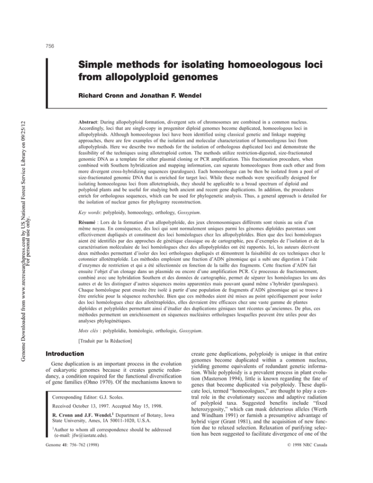
756 Simple methods for isolating homoeologous loci from allopolyploid genomes Richard Cronn and Jonathan F. Wendel Genome Downloaded from www.nrcresearchpress.com by US National Forest Service Library on 09/25/12 For personal use only. Abstract: During allopolyploid formation, divergent sets of chromosomes are combined in a common nucleus. Accordingly, loci that are single-copy in progenitor diploid genomes become duplicated, homoeologous loci in allopolyploids. Although homoeologous loci have been identified using classical genetic and linkage mapping approaches, there are few examples of the isolation and molecular characterization of homoeologous loci from allopolyploids. Here we describe two methods for the isolation of orthologous duplicated loci and demonstrate the feasibility of the techniques using allotetraploid cotton. The methods utilize restriction-digested, size-fractionated genomic DNA as a template for either plasmid cloning or PCR amplification. This fractionation procedure, when combined with Southern hybridization and mapping information, can separate homoeologues from each other and from more divergent cross-hybridizing sequences (paralogues). Each homoeologue can be then be isolated from a pool of size-fractionated genomic DNA that is enriched for target loci. While these methods were specifically designed for isolating homoeologous loci from allotetraploids, they should be applicable to a broad spectrum of diploid and polyploid plants and be useful for studying both ancient and recent gene duplications. In addition, the procedures enrich for orthologous sequences, which can be used for phylogenetic analysis. Thus, a general approach is detailed for the isolation of nuclear genes for phylogeny reconstruction. Key words: polyploidy, homoeology, orthology, Gossypium. Résumé : Lors de la formation d’un allopolyploïde, des jeux chromosomiques différents sont réunis au sein d’un même noyau. En conséquence, des loci qui sont normalement uniques parmi les génomes diploïdes parentaux sont effectivement dupliqués et constituent des loci homéologues chez les allopolyploïdes. Bien que des loci homéologues aient été identifiés par des approches de génétique classique ou de cartographie, peu d’exemples de l’isolation et de la caractérisation moléculaire de loci homéologues chez des allopolyploïdes ont été rapportés. Ici, les auteurs décrivent deux méthodes permettant d’isoler des loci orthologues dupliqués et démontrent la faisabilité de ces techniques chez le cotonnier allotétraploïde. Les méthodes emploient une fraction d’ADN génomique qui a subi une digestion à l’aide d’enzymes de restriction et qui a été sélectionnée en fonction de la taille des fragments. Cette fraction d’ADN fait ensuite l’objet d’un clonage dans un plasmide ou encore d’une amplification PCR. Ce processus de fractionnement, combiné avec une hybridation Southern et des données de cartographie, permet de séparer les homéologues les uns des autres et de les distinguer d’autres séquences moins apparentées mais pouvant quand même s’hybrider (paralogues). Chaque homéologue peut ensuite être isolé à partir d’une population de fragments d’ADN génomique qui se trouve à être enrichie pour la séquence recherchée. Bien que ces méthodes aient été mises au point spécifiquement pour isoler des loci homéologues chez des allotétraploïdes, elles devraient être efficaces chez une vaste gamme de plantes diploïdes et polyploïdes permettant ainsi d’étudier des duplications géniques tant récentes qu’anciennes. De plus, ces méthodes permettent un enrichissement en séquences nucléaires orthologues lesquelles peuvent être utiles pour des analyses phylogénétiques. Mots clés : polyploïdie, homéologie, orthologie, Gossypium. [Traduit par la Rédaction] Cronn and Wendel Gene duplication is an important process in the evolution of eukaryotic genomes because it creates genetic redundancy, a condition required for the functional diversification of gene families (Ohno 1970). Of the mechanisms known to Corresponding Editor: G.J. Scoles. Received October 13, 1997. Accepted May 15, 1998. R. Cronn and J.F. Wendel.1 Department of Botany, Iowa State University, Ames, IA 50011-1020, U.S.A. 1 Author to whom all correspondence should be addressed (e-mail: jfw@iastate.edu). Genome 41: 756–762 (1998) 762 create gene duplications, polyploidy is unique in that entire genomes become duplicated within a common nucleus, yielding genome equivalents of redundant genetic information. While polyploidy is a prevalent process in plant evolution (Masterson 1994), little is known regarding the fate of genes that become duplicated via polyploidy. These duplicate loci, termed “homoeologues,” are thought to play a central role in the evolutionary success and adaptive radiation of polyploid taxa. Suggested benefits include “fixed heterozygosity,” which can mask deleterious alleles (Werth and Windham 1991) or furnish a presumptive advantage of hybrid vigor (Grant 1981), and the acquisition of new function due to relaxed selection. Relaxation of purifying selection has been suggested to facilitate divergence of one of the © 1998 NRC Canada Genome Downloaded from www.nrcresearchpress.com by US National Forest Service Library on 09/25/12 For personal use only. Cronn and Wendel 757 Fig. 1. Organismal and genome relationships of diploid and allopolyploid taxa in the genus Gossypium. (Left) Diploid progenitors of the extant allotetraploids diverged approximately 6–11 million years before present (mybp), giving rise to two lineages represented by the A-genome diploids in Africa and D-genome diploids in the New World. Allopolyploid cotton formed 1–2 million years ago in the New World, when A- and D-genomes were combined in a common nucleus (Endrizzi et al. 1985; Wendel 1989). (Right) Types of homology among related sequences within and between diploids and their derived allopolyploids. For simplicity, only a single chromosome having three “similar” sequences (clear, grey, and black fill, respectively) is shown for the diploids; both homoeologues are illustrated in the allopolyploid. At the diploid level, loci with the same fill pattern are orthologues, reflecting the speciation event that created the opportunity for genic divergence. Genic relationships reflecting past duplication events rather than speciation events are paralogous, as exemplified by all comparisons among loci having different fill colors, be they within or between genomes. When orthologues become united in a common nucleus as a consequence of allopolyploid formation, homoeologous relationships are established, as illustrated for three sets of orthologues (“clear,” “grey,” and “black”) in this example. two homoeologues, possibly leading to novel gene function (Ohno 1970; Li 1983). In addition to the possibility of functional divergence, duplicated loci in polyploids may maintain their original function (Hughes 1994), or one copy may be silenced or degenerate into a non-functional pseudogene (Walsh 1995). While the importance of gene duplication in polyploidy has been recognized for almost half a century (Stephens 1951), molecular genetic analysis of homoeologous loci has been hampered by the problem of distinguishing loci whose close relationship reflects a recent polyploidization event from loci related by other duplication processes or more ancient cycles of whole-genome duplication. In this respect, it is noteworthy that diploid plant genomes can contain considerable genetic redundancy (Cavalier-Smith 1985; Tanksley and Pichersky 1988; Pickett and Meeks-Wagner 1995), as is commonly evidenced in Southern hybridization analyses and RFLP-mapping studies (e.g., Bonierbale et al. 1988; Jena et al. 1994). Accordingly, many if not most genes in plant genomes occur as members (i.e., paralogous copies, or paralogues) of small multi-gene families whose relationships reflect a complex evolutionary history of duplication and divergence. This history poses a stumbling block for those wishing to isolate a specific member of a gene family, or the “same” member of a gene family (i.e., strictly orthologous copies) from two or more taxa. This technical difficulty is compounded in polyploid plants, since for each gene family the entire set of paralogues becomes duplicated during polyploidization (Fig. 1). To examine the fate of specific loci that became duplicated by virtue of allopolyploidization (homoeologues), it becomes necessary to isolate each locus not just from its corresponding copy in the other subgenome, but also from divergent paralogues that exist in both subgenomes. It is this task, confidently deciphering whether a pair of sequences isolated from an allopolyploid are related by homoeology (duplication via polyploidization) versus paralogy (duplication via other mechanisms or more ancient cycles of polyploidy), that remains the crux of homoeologue analysis. With the goal of assessing evolutionary fate of duplicated loci in allopolyploid cotton (Gossypium hirsutum L.), we devised two approaches for isolating homoeologous genes and genomic regions. Using mapped loci revealed by anonymous PstI genomic DNA fragments, we adapted standard cloning and sequence-tagged-site (STS) PCR methods to create pools of DNA enriched for homoeologous target loci while minimizing contamination from paralogues present in the genome. Both methods utilize restriction enzyme-digested genomic DNA which has been size-fractionated by agarose gel electrophoresis. This simple fractionation step can separate two homoeologous loci into different DNA size pools, thereby affording a means for isolating and characterizing of © 1998 NRC Canada Genome Downloaded from www.nrcresearchpress.com by US National Forest Service Library on 09/25/12 For personal use only. 758 each duplicated copy. Two approaches are described here, one entailing the construction of plasmid libraries and the other using STS-PCR. In conjunction with Southern hybridization and RFLP-mapping data, the methods allow the homoeology and subgenomic origin of each particular homoeologue to be confidently assessed. To demonstrate the feasibility of the methods, we show how they were applied to the isolation of a pair of homoeologous P-glycoprotein genes from allotetraploid cotton. In addition, we isolated the orthologous copies of these genes from the two diploid species that represent the ancestral genome donors to the allopolyploids. While these methods were specifically designed for isolating homoeologous loci from allotetraploid cotton, the methods appear to be applicable for studying ancient and recent gene duplications in a broad spectrum of diploid and polyploid plants. Genome Vol. 41, 1998 D-genome, and AD-genome populations confirms that the loci are orthologous in diploids and homoeologous in the allotetraploids (Fig. 2B). Specifically, marker loci revealed by G1262 fall onto a single homoeologous assemblage of linkage groups corresponding to diploid linkage groups A3 and D10 and tetraploid chromosomes 6A and 25D. Size-fractionation of genomic DNA Approximately 10 µg of genomic DNA from allotetraploid and diploid cottons was digested to completion with HindIII, and the products were separated on a 1.0% agarose gel (SeaPlaque GTG; FMC BioProducts) in low-EDTA TAE buffer (0.1 mM EDTA, 40 mM tris-acetate). After staining with ethidium bromide, DNAs were visualized under long-wavelength UV light. Using a 1-kb DNA ladder for reference, each lane of the gel was carefully divided at 1-kb intervals (e.g., 1–2 kb, 2–3 kb, etc., up to 10 kb), and the DNA was isolated from each agarose slice using either Gene-Clean™ (Bio-101; for DNA larger than 4 kb) or DEAE-cellulose membrane (Schleicher & Schuell; for DNA smaller than 4 kb). Organismal context and plant materials Evidence suggests that allotetraploid species of cotton (AD-genome, 2n = 4x = 52) formed 0.5–2 million years ago in the New World following hybridization between two divergent diploid parents, one similar to the African species G. herbaceum L. (A-genome; 2n = 2x = 26) and the other to the Peruvian species G. raimondii Ulbrich (D-genome; 2n = 2x = 26) (Wendel and Albert 1992; Endrizzi et al. 1985). After polyploidization, the AD-genome group diverged into three distinct lineages (Brubaker and Wendel 1994; Small et al. 1998) presently represented by five species, including the economically important cottons G. hirsutum L., and G. barbadense L. The interrelationships between diploid and allopolyploid cottons and their respective genomes are depicted in Fig. 1. Genetic linkage maps for allotetraploid cotton (Reinisch et al. 1994) and the diploid A- and D-genome groups (Brubaker, unpublished data) have been constructed. Comparisons among the maps demonstrate that a high degree of synteny is conserved between the two diploid genomes and within the allopolyploids. Thus, homoeologous linkage groups have been demonstrated for allopolyploid Gossypium, with clear counterparts in diploid cottons. For the present study, we chose one representative from each of the populations used for constructing the RFLP maps described in Reinisch et al. (1994). Gossypium hirsutum race palmeri was used to represent allopolyploid, AD-genome cottons, while G. herbaceum and G. raimondii were selected for A-genome and D-genome diploids, respectively. DNAs were extracted using methods detailed in Paterson et al. (1993). Locus selection Homoeologous linkage groups and their constituent loci were revealed using clones from several libraries including anonymous cDNA and genomic clones (Reinisch et al. 1994). We focus on the loci revealed by the anonymous PstI probe G1262 because this gene appears to exist in a small gene family of 1–2 copies per diploid genome and 2–4 copies in the allopolyploids. This 1018 bp clone, originally isolated from PstI-fractionated G. raimondii DNA (Brubaker and Wendel 1994), shows high sequence similarity to the transcribed P-glycoprotein gene pgp1 from A. thaliana (Genbank number 99741; Dudler and Hertig 1992). When hybridized with probe G1262 under conditions of high stringency, HindIII-digested genomic DNAs from the A-genome and D-genome diploids show single fragments of approximately 1.9 kb and 4.5 kb, respectively (Fig. 2A). These fragments are additive in allotetraploid cottons, providing indirect evidence of homoeology. Comparative linkage mapping of this locus in A-genome, Isolation of homoeologues: Assumptions of the approach Two methods were designed to isolate homoeologues, one employing selective cloning and the other STS-PCR. For either approach to succeed, two criteria must be satisfied: (1) Restriction enzyme and PCR primer sites must be conserved in the allopolyploid subgenomes. Both the PstI-selective cloning and STS-PCR approaches require sequence conservation of homoeologous loci in regions corresponding to the 5′ and 3′ ends of the probe of interest. In addition, due to the methylation sensitivity of PstI, the homoeologous loci of interest must be unmethylated. Restriction site conservation and methylation status can be verified experimentally by checking the susceptibility of subgenomic sequences to a restriction enzyme digestion prior to cloning. In the case of PstI-probe G1262, digestion of allotetraploid G. hirsutum genomic DNA with PstI yields a homogeneous digestion product of ~1.0 kb (Fig. 2A). This band is identical in size to the original PstI probe, confirming that the PstI sites that flank locus G1262 are conserved with respect to nucleotide sequence and methylation status in both the A- and D-subgenomes of the allotetraploid. It is more difficult to evaluate conservation of PCR priming sites a priori, although in some cases other evidence may be used to infer the degree of similarity between homoeologues. In the present example, the high level of nucleotide conservation between P-glycoprotein-like genes of plants and animals (Dudler and Hertig 1992) suggested that the primer sites had a similarly high likelihood of conservation. (2) Mapping probes must be fully contained within the RFLP bands they reveal. Since the PstI-selective cloning and STS-PCR approaches require conservation of the regions corresponding to the termini of the cloned probe, both PstI sites and PCR primer sites must be located internal to the enzyme sites used to generate RFLPs. The probability of an RFLP probe being entirely contained within an RFLP band depends both upon the size of the probe (X) and the RFLP band (Y). This relationship can be expressed mathematically as P ≅ (Y – X)/(X + Y), where the numerator describes the number of possible “alignment states” for containment of the probe within the RFLP band, and the denominator describes the total number of alignment states possible for the probe and the RFLP. For the 1018 bp probe G1262, the probability that both PstI sites lie within the HindIII digestion products are 0.30 for the 1.9-kb G. herbaceum band and 0.63 for the 4.5-kb G. raimondii band. While these probabilities appear low, se© 1998 NRC Canada Cronn and Wendel 759 Genome Downloaded from www.nrcresearchpress.com by US National Forest Service Library on 09/25/12 For personal use only. Fig. 2. Evidence for homoeology and orthology relationships of loci revealed by probe G1262. (A) Southern hybridization of HindIIIand PstI-digested genomic DNA from diploid and allotetraploid cottons using probe G1262. Hybridization reveals a single band in the A- and D-genome diploids while two bands are evident in the AD-allotetraploid, one from each progenitor genome. Digestion of AD-allotetraploid DNA with PstI generates a single band of 1 kb, equal in size to the original mapping probe G1262. Abbreviations of taxa: G. raimondii = D5, G. hirsutum = AD1, and G. herbaceum = A1. Size marker (M) is lambda DNA digested with AccI and HindIII. (B) Comparative linkage mapping supports orthology of locus G1262 in the two diploid genomes (linkage groups D10 and A3 in the D- and A-genome, respectively) and homoeology of the corresponding loci on two syntenic chromosomes (25D and 6A) in the allotetraploid genomes. quencing of probe G1262 revealed no internal HindIII sites, providing evidence that the probe is fully contained within the RFLP band from its source genome (G. raimondii) and suggesting that it may be similarly internal in the other relevant genomic HindIII fragments. PstI-selective cloning of homoeologues To isolate homoeologues of locus G1262 by selective cloning, we digested ~500 ng of the 1–2 kb and 4–5 kb HindIII size-fractionated DNA pools from G. hirsutum with the restriction enzyme PstI. Size-fractionated DNAs from G. herbaceum (from the 1–2 kb HindIII pool) and G. raimondii (4–5 kb HindIII pool) were treated similarly for isolation of orthologous diploid loci. Following digestion, products were cleaned with Gene-Clean and ligated into the PstI site of the plasmid vector pZero (InVitrogen) as per manufacturer’s instructions. After ligation, mixtures were heated to 65oC, diluted 1/10 in water and aliquots (1 µL) were electroporated into TOP10F′ E. coli cells. Cells were incubated overnight at 37oC on LB-agar plates containing Zeocin (0.1 mM) and IPTG (1 mM). Once colonies had grown to ~ 0.5–1.0 mm in diameter, a replica was made onto a second LB-agar – Zeocin – IPTG plate and these petri dishes were incubated for 8 h at 37oC. After incubation, cells from the original transformation plate were used to make a permanent mini-library by elution in a minimal volume (1–2 mL) of LB medium – Zeocin (0.1 mM / 35% glycerol) and the cells were stored at –70oC. The second replica plate was used to make a colony lift onto nylon membrane (Nytran, Schleicher & Schuell) using the methods described in Ausubel (1994). PCR-amplification of homoeologues To amplify the homoeologues and diploid orthologues of locus G1262, clone G1262 was sequenced using universal sequencing primers of the vector and primers were designed. The priming sites for “G1262F” (5′-GGCGGCAGGCTAAGCACTTCY-3′) and “G1262R” (5′-CGGAGGTCATACTTCCAGCTTY-3′) were selected to minimize intermolecular primer dimers and to maintain an annealing temperature >54oC. Amplification cycling parameters were similar to those described in Don et al. (1991); a 2 min hotstart © 1998 NRC Canada Genome Downloaded from www.nrcresearchpress.com by US National Forest Service Library on 09/25/12 For personal use only. 760 Fig. 3. Agarose gel analysis of G1262 PCR-amplification products using HindIII size-fractionated DNA as a template for amplification. (A) Amplification products from the 1–8 kb HindIII size-fractions (lanes 1 through 7) of the A-genome diploid G. herbaceum; product is only amplified from the 2–3 kb pool (lane 2). (B) Amplification products from the 1–8 kb HindIII size-fractions (lanes 1 through 7) of the D-genome diploid G. raimondii; amplification is apparent only in the 4–5 kb pool (lane 4). (C) Amplification products from the 1–8 kb HindIII size-fractions (lanes 1 through 7) of allotetraploid G. hirsutum; products are apparent in lanes corresponding to 1–2, 2–3, 4–5, 5–6, and 6–7 kb pools. (D) Homogeneous DNA sequences obtained from G1262 amplification products isolated from the G. hirsutum 1–2 kb (left) and 4–5 kb (right) HindIII size-fractionated DNA pools (from panel C). The arrow indicates a region of locus G1262 where the sequences from the A- and D-subgenomes have diverged, showing the homogeneity of amplfication products that result from size-fractionation. (80oC) was followed by 10 cycles of 1 min at 94oC for denaturation, 1 min at 60oC (with a decrease of 0.6oC per cycle) for primer annealing, and a 1 min extension at 72oC. An additional 25 cycles were run once the annealing temperature reached 54oC. Each reaction mixture (25 µL) contained: 10 pM (picomoles) of each primer, 2.5 mM MgCl2, and approximately 10 ng HindIII size-fractionated DNA. DNA sequencing Isolated clones and PCR products were sequenced manually using the Amersham cycle sequencing kit with [33P]ddNTP terminators. Sequencing reactions typically used 0.25 pM of template and 2 pM of either the G1262F or G1262R primer. Cycle sequencing parameters were as follows: 45 cycles of 95oC for 30 s, 54oC for 30 s, and 72oC for 45 s. Sequencing products were resolved on 5% acrylamide gels (Long Ranger, FMC) and visualized by autoradiography. Sequences from this study are deposited in Genbank under accession numbers AF061085–AF061089. Genome Vol. 41, 1998 Selective cloning As shown in Fig. 2, high stringency southern hybridization of HindIII-digested DNA with probe G1262 yields a pattern where the A-genome diploid G. herbaceum shows one band of 1.9 kb, the D-genome diploid G. raimondii shows one band of 4.5 kb, and the allotetraploid G. hirsutum shows an additive pattern. In addition, hybridization of probe G1262 against PstI-digested G. hirsutum DNA yields a single band of identical size to the original probe. These data indicate that the loci corresponding to the PstI clone G1262 are conserved in all four genomes (A-, D-, and both subgenomes of the allopolyploid) with respect to PstI sites, suggesting that it would be possible to clone all four loci using a selection strategy employing PstI and size-fractionated genomic DNA. Using the PstI selective cloning approach described above, transformations yielded between 75 to several thousand primary recombinants for each of the size-fractionated DNA libraries, with 1:75 to ~1:2000 colonies containing inserts similar to probe G1262. For example, the 4–5 kb HindIII pool from G. hirsutum contained ~ 800 primary recombinants, six of which hybridized to probe G1262. To verify the subgenomic origin of these clones, plasmids were isolated and inserts were digested with SpeI, an enzyme that cleaves D-subgenome sequences at nucleotide 427. As expected, all three clones were susceptible to digestion with SpeI, confirming they had originated from the D-subgenome (data not shown). These results indicate that HindIII size-fractionation had effectively separated the A- and D-subgenome G1262 homoeologues into different size pools, allowing us to isolate each locus in the absence of its homoeologue. STS-PCR approach Using genomic DNA as the template for PCR reactions with primers G1262F and G1262R, amplification products of uniform size (~1000 bp) were readily obtained from the diploids G. herbaceum, G. raimondii, and from the allotetraploid G. hirsutum. Direct DNA sequencing showed the PCR products from the diploids to be homogeneous; on the other hand, sequences from the allotetraploid were consistently polymorphic, showing “additivity” of the A- and D-genome diploid sequences. These results indicated that both the A- and D-subgenomic loci had been amplified, and that isolation of specific homoeologues could be facilitated by gel fractionation prior to PCR. When HindIII sizefractionated DNA was used as a template in PCR reactions, amplification products were again obtained from the diploids G. herbaceum (from the 2–3 kb DNA pool) and G. raimondii (from the 4–5 kb DNA pool), and from the allotetraploid G. hirsutum (from these two pools and from adjacent size fractions; Fig. 3). Sequences of PCR products from the diploids were identical to those obtained when genomic DNA was used in amplification. However, in contrast to the polymorphic sequence obtained when using genomic G. hirsutum DNA as a PCR template, amplification products from the 1–2 kb (A-subgenome) and 4–5 kb (D-subgenome) HindIII-digested size fractions of G. hirsu© 1998 NRC Canada Cronn and Wendel Genome Downloaded from www.nrcresearchpress.com by US National Forest Service Library on 09/25/12 For personal use only. tum showed no polymorphism and were nearly identical to the sequences from their respective diploids (Fig. 3D). These results indicate that HindIII size- fractionation effectively separated the A- and D-subgenome G1262 homoeologues into different size pools, making it possible to amplify homoeologues separately and to generate uncontaminated sequences. We have described two methods for the isolation of homoeologous loci from an allotetraploid and for their orthologous counterparts from progenitor diploids. The basic techniques used to isolate homoeologues (selective cloning and STS-PCR amplification) are easily executed, yet the application of these approaches to size-fractionated genomic DNA facilitates the isolation of specific loci. Rather than attempting to amplify or clone homoeologues from a genomic DNA pool which contains one homoeologue per subgenome (and numerous unwanted paralogues), the size fractions are considerably less complex and are highly enriched for the locus of interest. When combined with Southern hybridization analysis and mapping information, these procedures offer a powerful tool for the analysis of orthologous and homoeologous loci. Their use increases confidence that a specific target orthologue has been isolated from other related sequences in a genome, and that its direct duplicate in an allopolyploid or counterparts in other diploids have been similarly isolated. The example provided here, involving the loci corresponding to probe G1262, was selected because Southern hybridization and RFLP mapping data indicated that this was a simple system with few interfering paralogous copies (1–2 at low stringency hybridization conditions). We have had success, however, applying these same techniques to other gene families, indicating that the methods are useful in more complex situations. A number of authors have used sequence-tagged primers to amplify mapped loci from allopolyploid plant genomes (Zhu et al. 1994; Talbert et al. 1995; Liu et al. 1996). Using similar approaches, we find that the majority of PCR amplification products from sequence-tagged sites (including locus G1262) are polymorphic when allotetraploid cotton genomic DNA is used as a template. This indicates that many STS primers will not be subgenome-specific in Gossypium, perhaps as a consequence of the limited sequence divergence between subgenomes of allotetraploid cotton (1.8% for locus G1262). This outcome does not appear to be unique to polyploid cotton since a similar absence of genome-specificity was observed in STS-PCR amplification from allotetraploid Stylosanthes (Liu et al. 1996). In addition, nearly all other STS primer pairs examined in both diploid and allopolyploid cottons lead to heterogeneous amplification pools. Considering the inherent redundancy of allopolyploid and most diploid genomes, lack of genome and locus specificity may prove to be the rule rather than the exception. In these cases, size-fractionation of restriction-digested template DNA combined with Southern hybridization offers a convenient route to enhanced locus selectivity. 761 While the use of size-fractionated DNA in cloning and STS-PCR has many advantages over the use of total genomic DNA, there are disadvantages to the approaches described. For the STS-PCR method, abundant unsheared genomic DNA is paramount for success. Sheared genomic DNA and (or) partially digested genomic DNA can yield unwanted paralogues in the desired size pool, or anomalous migration of fragments that include the desired orthologue or homoeologue. Due to the inherently high sensitivity of PCR, these anomalously migrating targets can appear in unexpected size pools, even from DNA preparations that appear to be reasonably free from shearing (e.g., Fig. 3C). A second factor that influences the success of amplifying a target from a discrete size fraction involves the accuracy with which genomic digests are fractionated. Bands that migrate near a zone of fractionation (e.g., the A-genome/subgenome RFLPs of ~1.9 kb) can be divided into either one of two adjacent pools (e.g., the 2–3 kb pool of A1; Fig. 3A) or both adjacent pools (Fig. 3C). For this reason, gel-fractionation yields the best results when bands of interest are separated by a large distance on the gel (i.e., two or more size fractions). It should be noted that the selective cloning approach appears less sensitive to the influence of sheared genomic DNA, as “contamination” in incorrect size-fractionated libraries has not been observed. This is due, at least in part, to the low cloning efficiency of inserts from the heterogeneous size-fractionated DNA mixtures. This low efficiency provides a selective filter for sequences present in high abundance, making low frequency homoeologue “contaminants” rare. A practical consideration dictated by the low efficiency is that the cloning vector chosen should, ideally, provide presence/absence selection through the use of a cell death gene (such as ccdB in pZero). The loci corresponding to G1262 account for less than a millionth of the DNA present in allotetraploid cotton (~2 kb out of 2.9 × 10⁶ kb total; Michaelson et al. 1991; Gomez et al. 1993). Isolation of both homoeologues from the allotetraploid and the corresponding orthologues from the parental diploids, via a two-enzyme, size-fractionation method, represents a significant enrichment relative to random library screens. For example, to have a 90% probability of obtaining both G1262 homoeologues in YAC or cosmid libraries (assuming insert sizes of 150 kb and 35 kb, respectively), approximately 58 000 independent YAC or 250 000 cosmid clones would need to be surveyed (Ausubel 1994). Despite the disadvantages of the selective cloning method, it remains a simple and flexible approach for cloning one or both homoeologues for mapped PstI loci. The methods described in this paper were designed to isolate homoeologous loci from allopolyploid genomes, although they clearly are applicable to the isolation of target orthologues from diploids as well. Thus, the methods provide a general tool for isolating a specific nuclear sequence from a suite of diploid and polyploid taxa, as well as a means for accessing divergent members of a given gene family from a single taxon. In the arena of genome mapping, an important assumption of STS-PCR analyses is that PCR amplification products are orthologous to the locus originally mapped (Olson et al. 1989). While this assumption may be warranted for many loci (particularly diploids with © 1998 NRC Canada Genome Downloaded from www.nrcresearchpress.com by US National Forest Service Library on 09/25/12 For personal use only. 762 small genome sizes; Inoue et al. 1994), it appears to be less reliable in diploids with larger genomes. In a survey of 99 barley-specific STS-primers, for example, Blake et al. (1996) found that amplification products from 36 primer pairs did not correspond to the loci originally identified in the RFLP map. This illustrates how frequently un-mapped paralogues of sequence-tagged sites may be amplified if total genomic DNA is used as the PCR template. As demonstrated here, the use of size-fractionated DNA can obviate this problem, thereby improving the accuracy of STS-amplifications. Finally, we note that the ability to target specific nuclear sequences may find utility in evolutionary and phylogenetic analyses. The use of nuclear sequences for phylogenetic purposes has frequently been hampered by intractable problems associated with paralogy/orthology conflation. Isolation of strict orthologues is an essential prerequisite to the use of nuclear sequences in phylogeny reconstruction; the methods detailed here should prove useful in this regard. The authors thank C.L. Brubaker for sharing comparative mapping results prior to publication, A. Paterson and X. Zhao for assistance in probe selection, and J. Ryburn for technical assistance. This research was supported by the National Science Foundation. Ausubel, F.M. (ed). 1994. Current Protocols in Molecular Biology. John Wiley and Sons, N.Y. Blake, T.K., Kadyrzhanova, D., Shepherd, K.W., Islam, A.K.M.R., Langridge, P.L., McDonald, C.L., Erpelding, J., Larson, S., Blake, N.K., and Talbert, L.E. 1996. STS-PCR markers appropriate for wheat-barley introgression. Theor. Appl. Genetics, 93: 826–832. Bonierbale, M.W., Plaisted, R.L., and Tanksley, S.D. 1988. RFLP maps based on a common set of clones reveal modes of chromosomal evolution in potato and tomato. Genetics, 120: 1095–1103. Brubaker, C.L., and Wendel, J.F. 1994. Reevaluating the origin of domesticated cotton (Gossypium hirsutum: Malvaceae) using nuclear restriction fragment length polymorphisms (RFLPs). Amer. J. Bot. 81: 1309–1326. Cavalier-Smith, T. 1985. The Evolution of Genome Size. John Wiley and Sons, N.Y. Don, R.H., Cox, P.T., Wainwright, B.J., Baker, K., and Mattick, J.S. 1991. “Touchdown” PCR to circumvent spurious priming during gene amplification. Nucleic Acids Res. 19: 4008. Dudler, R., and Hertig, C. 1992. Structure of an mdr-like gene from Arabidopsis thaliana. Evolutionary implications. J. Biol. Chem. 267: 5882–5888. Endrizzi, J.E., Turcotte, E.L., and Kohel, R.J. 1985. Genetics, cytology, and evolution of Gossypium. Adv. Genet. 23: 271–375. Gomez, M., Johnston, J.S., Ellison, J.R, and Price, H.J. 1993. Nuclear 2C DNA content of Gossypium hirsutum L. accessions determined by flow cytometry. Biol. Zentralbl. 112: 351–357. Grant, V. 1981. Plant speciation. Columbia, N.Y. Genome Vol. 41, 1998 Hughes, A.L. 1994. The evolution of functionally novel proteins after gene duplication. Proc. Royal Soc. Lond. B, 256: 119–124. Inoue, T., Zhong, H.S., Miyao, A., Ashikawa, I., Monna, L., Fukuoka, S., Miyadera, N., Nagamura, Y., Kurata, K., Sasaki, T., and Minobe, Y. 1994. Sequence-tagged sites (STSs) as standard landmarkers in the rice genome. Theor. Appl. Genet. 89: 728–734. Jena, K., Khush, G.S., and Kochert, G. 1994. Comparative RFLP mapping of a wild rice, Oryza officinalis, and cultivated rice, O. sativa. Genome, 37: 382–389. Li, W.-H. 1983. Evolution of duplicate genes and pseudogenes. In Evolution of genes and proteins. Edited by M. Nei., and R.K. Koehn. Sinauer, Sunderland, Mass. Liu, C.J., Musial, J. M., and Smith, F.W. 1996. Evidence for a low level of genomic specificity of sequence-tagged-sites in Stylosanthes. Theor. Appl. Genet. 93: 864–868. Masterson, J. 1994. Stomatal size in fossil plants: Evidence for polyploidy in majority of angiosperms. Science, 264: 421–424. Michaelson, M.J., Price, H.J., Ellison, J.R., and Johnston, J.S. 1991. Comparison of plant DNA contents determined by Feulgen microspectrophotometry and laser flow cytometry. Amer. J. Bot. 78: 183–188. Ohno, S. 1970. Evolution by gene duplication. Springer, N.Y. Olson, M., Hood, L., Cantor, C., and Botstein, D. 1989. A common language for physical mapping of the human genome. Science, 245: 1434–1435. Paterson, A.H., Brubaker, C.L., and Wendel, J.F. 1993. A rapid method for extraction of cotton (Gossypium spp.) genomic DNA suitable for RFLP or PCR analysis. Plant Mol. Biol. Reptr. 11: 122–127. Pickett, F.B., and Meeks-Wagner, D.R. 1995. Seeing double: appreciating genetic redundancy. Plant Cell, 7: 1347–1356. Reinisch, A.J., Dong, J-M., Brubaker, C.L., Stelly, D.M., Wendel, J.F., and Paterson, A.H. 1994. A detailed RFLP map of cotton, Gossypium hirsutum × Gossypium barbadense: chromosome organization and evolution in a disomic polyploid genome. Genetics, 138: 1–19. Small, R.L., Ryburn, J.A., Cronn, R.C., Seelanan, T., and Wendel, J.F. 1998. The tortoise and the hare: choosing between noncoding plastome and nuclear Adh sequences for phylogeny reconstruction in a recently diverged plant group. Amer. J. Bot. 85: 1301–1315. Stephens, S.G. 1951. Possible significance of duplication in evolution. Adv. Genetics, 4: 247–265. Talbert, L.E., Blake, N.K., Storlie, E.W., and Lavin, M. 1995. Variability in wheat based on low-copy DNA sequence comparisons. Genome, 38: 951–957. Tanksley, S.D., and Pichersky, E. 1988. Organization and evolution of sequences in the plant nuclear genome. In Plant evolutionary biology. Edited by L.D. Gottlieb, and S.K. Jain. Chapman and Hall, N.Y. Walsh, J.B. 1995. How often do duplicated genes evolve new functions? Genetics, 139: 421–428. Wendel, J.F., and Albert, V.A. 1992. Phylogenetics of the cotton genus (Gossypium L.): Character-state weighted parsimony analysis of chloroplast DNA restriction site data and its systematic and biogeographic implications. Syst. Bot. 17: 115–143. Werth, C.R., and Windham, M.D. 1991. A model for divergent, allopatric speciation of polyploid pteridophytes resulting from silencing of duplicate-gene expression. Amer. Nat. 137: 515–526. Zhu, T., Shi, L., Doyle, J.J., and Keim, P. 1995. A single nuclear locus phylogeny of soybean based on DNA sequence. Theor. Appl. Genet. 90: 991–999. © 1998 NRC Canada Genome Downloaded from www.nrcresearchpress.com by US National Forest Service Library on 09/25/12 For personal use only. This article has been cited by: 1. Sukhjiwan Kaur, Michael G. Francki, John W. Forster. 2011. Identification, characterization and interpretation of singlenucleotide sequence variation in allopolyploid crop species. Plant Biotechnology Journal no-no. [CrossRef] 2. M. Rousseau-Gueutin, A. Gaston, A. Aïnouche, M.L. Aïnouche, K. Olbricht, G. Staudt, L. Richard, B. Denoyes-Rothan. 2009. Tracking the evolutionary history of polyploidy in Fragaria L. (strawberry): New insights from phylogenetic analyses of low-copy nuclear genes. Molecular Phylogenetics and Evolution 51:3, 515-530. [CrossRef] 3. R. L. Small, J. F. Wendel. 2002. Differential Evolutionary Dynamics of Duplicated Paralogous Adh Loci in Allotetraploid Cotton (Gossypium). Molecular Biology and Evolution 19:5, 597-607. [CrossRef] 4. R. C. Cronn, R. L. Small, J. F. Wendel. 1999. Duplicated genes evolve independently after polyploid formation in cotton. Proceedings of the National Academy of Sciences 96:25, 14406-14411. [CrossRef]

