Anti-Filamin A antibody ab11074 Product datasheet 5 Abreviews 3 Images
advertisement
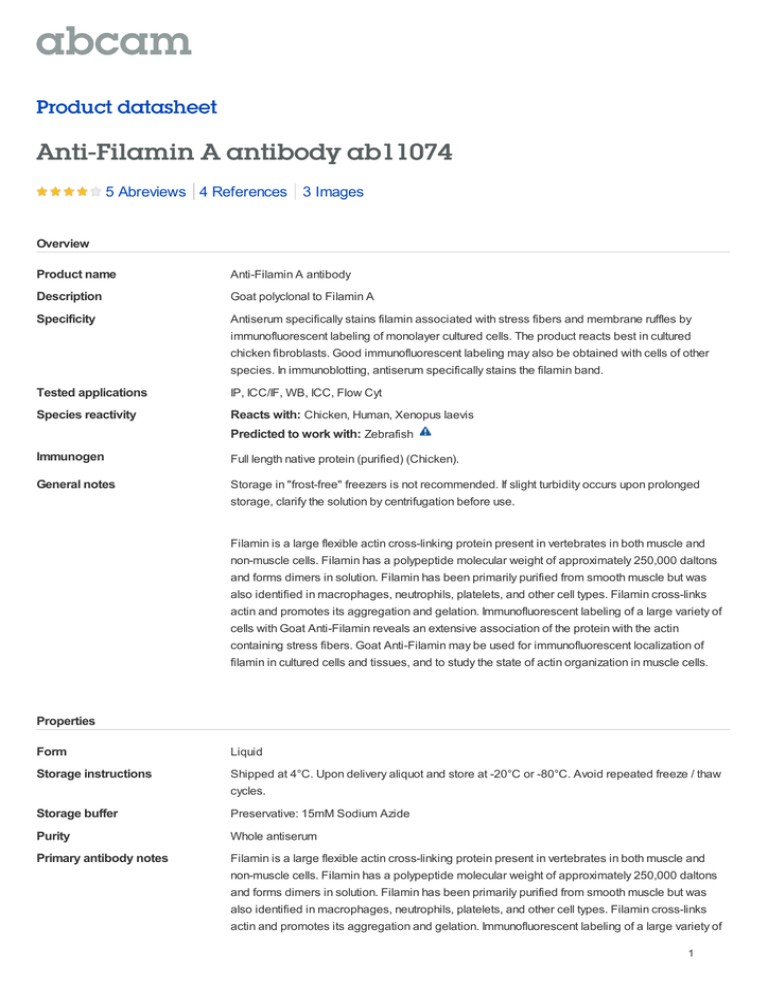
Product datasheet Anti-Filamin A antibody ab11074 5 Abreviews 4 References 3 Images Overview Product name Anti-Filamin A antibody Description Goat polyclonal to Filamin A Specificity Antiserum specifically stains filamin associated with stress fibers and membrane ruffles by immunofluorescent labeling of monolayer cultured cells. The product reacts best in cultured chicken fibroblasts. Good immunofluorescent labeling may also be obtained with cells of other species. In immunoblotting, antiserum specifically stains the filamin band. Tested applications IP, ICC/IF, WB, ICC, Flow Cyt Species reactivity Reacts with: Chicken, Human, Xenopus laevis Predicted to work with: Zebrafish Immunogen Full length native protein (purified) (Chicken). General notes Storage in "frost-free" freezers is not recommended. If slight turbidity occurs upon prolonged storage, clarify the solution by centrifugation before use. Filamin is a large flexible actin cross-linking protein present in vertebrates in both muscle and non-muscle cells. Filamin has a polypeptide molecular weight of approximately 250,000 daltons and forms dimers in solution. Filamin has been primarily purified from smooth muscle but was also identified in macrophages, neutrophils, platelets, and other cell types. Filamin cross-links actin and promotes its aggregation and gelation. Immunofluorescent labeling of a large variety of cells with Goat Anti-Filamin reveals an extensive association of the protein with the actin containing stress fibers. Goat Anti-Filamin may be used for immunofluorescent localization of filamin in cultured cells and tissues, and to study the state of actin organization in muscle cells. Properties Form Liquid Storage instructions Shipped at 4°C. Upon delivery aliquot and store at -20°C or -80°C. Avoid repeated freeze / thaw cycles. Storage buffer Preservative: 15mM Sodium Azide Purity Whole antiserum Primary antibody notes Filamin is a large flexible actin cross-linking protein present in vertebrates in both muscle and non-muscle cells. Filamin has a polypeptide molecular weight of approximately 250,000 daltons and forms dimers in solution. Filamin has been primarily purified from smooth muscle but was also identified in macrophages, neutrophils, platelets, and other cell types. Filamin cross-links actin and promotes its aggregation and gelation. Immunofluorescent labeling of a large variety of 1 cells with Goat Anti-Filamin reveals an extensive association of the protein with the actin containing stress fibers. Goat Anti-Filamin may be used for immunofluorescent localization of filamin in cultured cells and tissues, and to study the state of actin organization in muscle cells. Clonality Polyclonal Isotype IgG Applications Our Abpromise guarantee covers the use of ab11074 in the following tested applications. The application notes include recommended starting dilutions; optimal dilutions/concentrations should be determined by the end user. Application Abreviews Notes IP Use at an assay dependent concentration. (PMID 18495106). ICC/IF Use at an assay dependent concentration. WB 1/1000. Predicted molecular weight: 250 kDa. ICC Use at an assay dependent concentration. Flow Cyt 1/250. (see Abreview). ab37373-Goat polyclonal IgG, is suitable for use as an isotype control with this antibody. Target Function Promotes orthogonal branching of actin filaments and links actin filaments to membrane glycoproteins. Anchors various transmembrane proteins to the actin cytoskeleton and serves as a scaffold for a wide range of cytoplasmic signaling proteins. Interaction with FLNA may allow neuroblast migration from the ventricular zone into the cortical plate. Tethers cell surfacelocalized furin, modulates its rate of internalization and directs its intracellular trafficking. Tissue specificity Ubiquitous. Involvement in disease Defects in FLNA are the cause of periventricular nodular heterotopia type 1 (PVNH1) [MIM:300049]; also called nodular heterotopia, bilateral periventricular (NHBP or BPNH). PVNH is a developmental disorder characterized by the presence of periventricular nodules of cerebral gray matter, resulting from a failure of neurons to migrate normally from the lateral ventricular proliferative zone, where they are formed, to the cerebral cortex. PVNH1 is an X-linked dominant form. Heterozygous females have normal intelligence but suffer from seizures and various manifestations outside the central nervous system, especially related to the vascular system. Hemizygous affected males die in the prenatal or perinatal period. Defects in FLNA are the cause of periventricular nodular heterotopia type 4 (PVNH4) [MIM:300537]; also known as periventricular heterotopia Ehlers-Danlos variant. PVNH4 is characterized by nodular brain heterotopia, joint hypermobility and development of aortic dilation in early adulthood. Defects in FLNA are the cause of otopalatodigital syndrome type 1 (OPD1) [MIM:311300]. OPD1 is an X-linked dominant multiple congenital anomalies disease mainly characterized by a generalized skeletal dysplasia, mild mental retardation, hearing loss, cleft palate, and typical facial anomalies. OPD1 belongs to a group of X-linked skeletal dysplasias known as oto-palato2 digital syndrome spectrum disorders that also include OPD2, Melnick-Needles syndrome (MNS), and frontometaphyseal dysplasia (FMD). Remodeling of the cytoskeleton is central to the modulation of cell shape and migration. FLNA is a widely expressed protein that regulates reorganization of the actin cytoskeleton by interacting with integrins, transmembrane receptor complexes and second messengers. Males with OPD1 have cleft palate, malformations of the ossicles causing deafness and milder bone and limb defects than those associated with OPD2. Obligate female carriers of mutations causing both OPD1 and OPD2 have variable (often milder) expression of a similar phenotypic spectrum. Defects in FLNA are the cause of otopalatodigital syndrome type 2 (OPD2) [MIM:304120]; also known as cranioorodigital syndrome. OPD2 is a congenital bone disorder that is characterized by abnormally modeled, bowed bones, small or absent first digits and, more variably, cleft palate, posterior fossa brain anomalies, omphalocele and cardiac defects. Defects in FLNA are the cause of frontometaphyseal dysplasia (FMD) [MIM:305620]. FMD is a congenital bone disease characterized by supraorbital hyperostosis, deafness and digital anomalies. Defects in FLNA are the cause of Melnick-Needles syndrome (MNS) [MIM:309350]. MNS is a severe congenital bone disorder characterized by typical facies (exophthalmos, full cheeks, micrognathia and malalignment of teeth), flaring of the metaphyses of long bones, s-like curvature of bones of legs, irregular constrictions in the ribs, and sclerosis of base of skull. Defects in FLNA are the cause of X-linked congenital idiopathic intestinal pseudoobstruction (CIIPX) [MIM:300048]. CIIPX is characterized by a severe abnormality of gastrointestinal motility due to primary qualitative defects of enteric ganglia and nerve fibers. Affected individuals manifest recurrent signs of intestinal obstruction in the absence of any mechanical lesion. Defects in FLNA are the cause of FG syndrome type 2 (FGS2) [MIM:300321]. FG syndrome (FGS) is an X-linked disorder characterized by mental retardation, relative macrocephaly, hypotonia and constipation. Defects in FLNA are the cause of terminal osseous dysplasia (TOD) [MIM:300244]. A rare Xlinked dominant male-lethal disease characterized by skeletal dysplasia of the limbs, pigmentary defects of the skin and recurrent digital fibroma during infancy. A significant phenotypic variability is observed in affected females. Defects in FLNA are the cause of cardiac valvular dysplasia X-linked (CVDX) [MIM:314400]. A rare X-linked heart disease characterized by mitral and/or aortic valve regurgitation. The histologic features include fragmentation of collagenous bundles within the valve fibrosa and accumulation of proteoglycans, which produces excessive valve tissue leading to billowing of the valve leaflets. Sequence similarities Belongs to the filamin family. Contains 1 actin-binding domain. Contains 2 CH (calponin-homology) domains. Contains 24 filamin repeats. Domain Comprised of a NH2-terminal actin-binding domain, 24 internally homologous repeats and two hinge regions. Repeat 24 and the second hinge domain are important for dimer formation. Post-translational modifications Phosphorylated upon DNA damage, probably by ATM or ATR (By similarity). Phosphorylation extent changes in response to cell activation. The N-terminus is blocked. Cellular localization Cytoplasm > cell cortex. Cytoplasm > cytoskeleton. Anti-Filamin A antibody images 3 ab11074 staining Filamin A in Human platelet cells by Flow cytometry. Cells were fixed in paraformaldehyde and permeabilized using 0.1% Triton-X-100 in 2% BSA for 15 minutes. Primary antibody used at a 1/250 dilution and incubated for 18 hours at 4°C. The secondary antibody used was an Alexa Fluor®488 conjugated donkey anti-goat IgG (H+L) at a 1/500 dilution. US: Unstained, Red Peak; Flow Cytometry - Filamin A antibody (ab11074) Image courtesy of Dr Mahesh Shivananjappa by Abreview. IgG Ms: IgG Mouse (Isotype Control), Blue Peak; Filamin: Filamin A, Green peak. ab11074 at a 1/2000 dilution staining Filamin A in XTC embryonic polyclonal fibroblasts by Immunocytochemistry/ Immunofluorescence. Cells were fixed with formaldehyde, permeabilized using Triton X-100 and blocked with 1% BSA. The secondary used was a Texas Red conjugated anti-goat, 1/200 dilution. Immunocytochemistry/ Immunofluorescence Filamin A antibody (ab11074) Image courtesy of Helene Cousin by Abreview. 4 Anti-Filamin A antibody (ab11074) at 1/6000 dilution + Human platelet lysate at 20 µg Secondary Rabbit anti-Goat HRP at 1/10000 dilution developed using the ECL technique Performed under reducing conditions. Predicted band size : 250 kDa Observed band size : >250 kDa Western blot - Anti-Filamin A antibody (ab11074) Exposure time : 30 seconds This image is courtesy of an Abreview submitted by Mahesh Shivananjappa, United States Please note: All products are "FOR RESEARCH USE ONLY AND ARE NOT INTENDED FOR DIAGNOSTIC OR THERAPEUTIC USE" Our Abpromise to you: Quality guaranteed and expert technical support Replacement or refund for products not performing as stated on the datasheet Valid for 12 months from date of delivery Response to your inquiry within 24 hours We provide support in Chinese, English, French, German, Japanese and Spanish Extensive multi-media technical resources to help you We investigate all quality concerns to ensure our products perform to the highest standards If the product does not perform as described on this datasheet, we will offer a refund or replacement. For full details of the Abpromise, please visit http://www.abcam.com/abpromise or contact our technical team. Terms and conditions Guarantee only valid for products bought direct from Abcam or one of our authorized distributors 5
![Anti-Filamin A (phospho S1458) antibody [EP2309Y] ab68424](http://s2.studylib.net/store/data/012742938_1-8b93a434b309b510e061b30b53123285-300x300.png)
![Anti-muscle Actin antibody [HHF35], prediluted ab838](http://s2.studylib.net/store/data/012732226_1-b0960fb9f879f68010a9c7299d6f4518-300x300.png)
