The Breakdown of Olivine to Perovskite and Magnesiowu¨stite Yanbin Wang,* Isabelle Martinez,
advertisement
REPORTS
The Breakdown of Olivine to Perovskite
and Magnesiowüstite
Yanbin Wang,* Isabelle Martinez,† François Guyot,‡
Robert C. Liebermann
San Carlos olivine crystals under laboratory conditions of 26 gigapascals and 973 to 1473
kelvin (conditions typical of subducted slabs at a depth of 720 kilometers) for periods of
a few minutes to 19 hours transformed to the phase assemblage of perovskite and
magnesiowüstite in two stages: (i) the oxygen sublattice transformed into a cubic closepacked lattice, forming a metastable spinelloid, and (ii) at higher temperatures or longer
run durations, this spinelloid broke down to perovskite and magnesiowüstite by redistributing silicon and magnesium while maintaining the general oxygen framework. The
breakdown was characterized by a blocking temperature of 1000 kelvin, below which
olivine remained metastable, and by rapid kinetics once the reaction was activated.
Lithospheric materials are brought into the
deep mantle through subduction processes at
a rate of tens of thousands of cubic kilometers per year. As the slabs descend, phase
transformations occur; the kinetics and
mechanisms of these transformations play a
central role in controlling the mechanical
properties of the slabs and have been suggested to be responsible for deep-focus earthquakes (1). Among the various minerals
present in the subducting lithosphere, olivine is believed to be the most abundant. It
is also the dominant phase in the upper
mantle, and its phase transformation into
the b phase is considered to be the major
contributor to the seismic discontinuity at
410 km (2). The seismic discontinuity at 660
km is a manifestation of the disproportionation reaction of (Mg,Fe)2SiO4-spinel into
perovskite and magnesiowüstite (3). Studies
of the mechanism and kinetics of this reaction are critical to understanding the dynamics of the mantle and the fate of the subducted slabs.
We compressed San Carlos olivine crystals to 26 GPa (as found at depths of about
720 km) and heated them to temperatures
between 973 and 1473 K (corresponding to
the range of estimated cold and hot slab
temperatures) for run durations from 0 to 19
hours (4). The recovered samples were examined with the use of transmission electron microscopy (TEM) (5).
At 973 K, olivine crystals remained metaCenter for High Pressure Research and Mineral Physics
Institute, State University of New York, Stony Brook, NY
11794 –2100, USA.
*Present address: Consortium for Advanced Radiation
Sources, The University of Chicago, Chicago, IL 60637,
USA.
†Present address: Laboratoire de Geochemie des Isotopes Stables and Institut de Physique du Globe de Paris,
4 place Jussieu, 75252 Paris Cedex 05, France.
‡Present address: Laboratoire de Mineralogie-Cristallographie and Institut de Physique du Globe de Paris, 4
place Jussieu, 75252 Paris Cedex 05, France.
510
stable even after 19 hours, suggesting that
this temperature is not sufficient to activate
the transformation process, even under tremendous overpressure (about 14 and 10 GPa
above the olivine-b and b-spinel boundaries,
respectively). At 1473 K, however, a single
crystal of olivine transformed in less than
30 s into a complex phase assemblage containing perovskite, magnesiowüstite, and
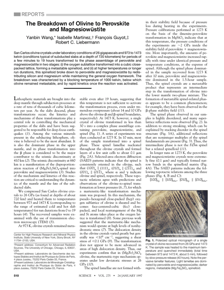
spinel (Fig. 1). A series of experiments was
then conducted at 1273 K. In 10 min, most
of the sample transformed into a spinel
phase. These spinel lamellae nucleated
throughout the olivine crystals and formed
layers of a few unit cells to about 0.1 mm
(Fig. 2A). Selected-area electron diffraction
(SAED) patterns indicate that the spinel is
consistently oriented in the olivine, such
that (100)a \ (111)g, (010)a \ (211)g, and
(001)a \ (011)g, where a and g indicate
olivine and spinel, respectively. These topotactic relations agree with predictions and
observations of the olivine-to-spinel transformation at lower pressures (6, 7), for which
a martensitic-like transformation mechanism was proposed. In this mechanism, the
pseudo– hexagonal close-packed (hcp) oxygen sublattice of olivine is sheared and becomes face-centered-cubic (fcc) closepacked, and local rearrangement of the Mg
and Si atoms takes place as the oxygen lattice is transformed (8). Some previous work
suggested that the martensitic-like mechanism would operate at high levels of nonhydrostatic stress (7). The dislocation density
in the olivine crystals varied greatly but generally was ,108 cm22, suggesting a shear
stress of ,0.1 GPa (9). The transformation
does not appear to be more advanced in
areas of high dislocation density. Thus, our
experiments confirm that in (Mg,Fe)2SiO4
olivine, the martensitic-type mechanism operates under low deviatoric stresses at 26
GPa (10).
The spinel lamellae are not formed withSCIENCE
z
VOL. 275
z
24 JANUARY 1997
in their stability field because of pressure
loss during heating in the experiments.
Pressure calibrations performed at 1273 K
on the basis of the ilmenite-perovskite
transformation in MgSiO3 indicate that at
this temperature, the pressure conditions of
the experiments are ;2 GPa inside the
stability field of perovskite 1 magnesiowüstite. More importantly, the amounts of perovskite and magnesiowüstite increased rapidly with time under identical pressure and
temperature conditions, at the expense of
spinel. Although the spinel phase dominated in the sample recovered from 1273 K
after 10 min, perovskite and magnesiowüstite dominated in the 1.5-hour sample.
Thus, the spinel crystals are a metastable
product that represents an intermediate
step in the transformation of olivine into
the lower mantle two-phase mixture. The
formation of metastable spinel-related phases appears to be a common phenomenon;
for example, they have been observed in the
b-phase stability field (11).
The spinel phase observed in our samples is highly disordered, and many superlattice reflections were observed (Fig. 2). In
addition to strong streaking, which can be
explained by stacking disorder in the spinel
structure (Fig. 3A), additional reflections
that are noninteger multiples of the spinel
fundamentals are present (Fig. 3). Thus, the
intermediate phase is not the Fd3m spinel
but a related spinelloid (11).
In longer runs at 1273 K, the perovskite
and magnesiowüstite crystals were extremely fine (0.1 mm) and typically formed narrow bands in the spinelloid matrix (Fig.
4A). The SAED patterns indicate the following topotactic relations among the three
phases (Fig. 4, B and C):
[100]Sp \ [100]Mw, [010]Sp \ [010]Mw,
0.4 mm
Fig. 1. Polarized optical micrograph of a single
crystal of olivine recovered from 26 GPa and 1473
K. The sample was heated to the maximum temperature and quenched immediately (total time
between 973 and 1473 K, about 5 min), followed
by slow pressure release (40 hours). Note the pervasive lamellar features. Light lamellae are dominated by perovskite and magnesiowüstite; darker
regions, metastable (Mg,Fe)2SiO4 spinelloid.
[001]Sp \ [001]Mw and [100]Mw \ [110]cPv,
[010]Mw \ [110]cPv, [001]Mw \ [001]cPv, where
Sp, Mw, and cPv indicate spinelloid, magnesiowüstite, and cubic perovskite, respectively. The orthorhombic perovskite structure can be derived from the cubic form by
continuous tilting of the SiO6 octahedra,
with the general topology remaining unchanged (12).
The oxygen sublattices for spinel, magnesiowüstite, and perovskite are similar
(Fig. 5). For spinel, the sequence of stacking
layers A, B, C, and D (about 2.05 Å between layers at zero pressure) completes one
unit cell. All of these layers contain only
Mg and O atoms, with a Mg population half
that of O; the tetrahedrally coordinated Si
atoms are halfway between these Mg-O layers. For MgO, two layers E and F form a fcc
oxygen sublattice identical to that of the
spinel; in these layers, the Mg and O sites
are fully occupied, and the interlayer distance is about 2.10 Å at ambient conditions. The cubic perovskite has a layer G
identical to the layer B in MgO (but with
shorter Mg-O distances) and a layer H with
much more closely packed oxygens and octahedrally coordinated Si; the interlayer
distance is about 1.7 Å at zero pressure.
Our observations of the breakdown reaction may be explained by a two-step mech-
anism. At relatively low temperatures (below
1473 K), the hcp oxygen sublattice of olivine
first transforms into a fcc arrangement with a
spinelloid structure by a martensitic-like
mechanism (6–8). This spinelloid then
breaks down to form perovskite and magnesiowüstite. An energetically economic way
for the breakdown reaction to proceed is to
maintain most of the common fcc oxygen
framework and to redistribute the Si and Mg
atoms within the spinel lattice. As a Si41
cation moves into a Mg-O layer by a small
displacement of 1⁄8^111&g (arrow in layer A),
the four surrounding O22 anions in the layer
are pulled closer together, thereby forming a
part of layer H (the Si-O layer) of the perovskite. This movement causes the Mg-O
bond length to increase within the layer,
thus decreasing the bond strength and making it easier for the Mg21 cations (M1 and
M2 in layer A) to join the adjacent Mg-O
layer (positions M1 and M2 in layer B) by a
displacement of 1⁄4^110&g. This shift doubles
the Mg occupancy in layer B, making it an
Mg-O layer like those in MgO and perovskite. Thus, the metastable transformation in
the oxygen sublattice provides a low–activation energy path for the breakdown to proceed because little diffusion of the oxygen
atoms is required.
If all the rearrangements occurred coher-
Fig. 2. Electron micrographs of a sample treated at 1273 K for 10 min.
(A) Spinel lamellae in the
olivine host, parallel to
the (100)ol planes. Some
extremely thin lamellae
are indicated by arrows.
Note the strain contours
across the lamella interface. (Inset) SAED pattern indicating topotactic
relations between olivine
(ol) and spinel (sp). (B)
SAED pattern taken
across an interface between olivine and the
spinel, showing well-defined topotactic relations
between the two phases. The foil was tilted
slightly from the orientation in (A). The spinel
phase is heavily twinned
[see (C)]; superlattice reflections and streaking
indicate significant structural disorder. (C) SAED
pattern taken within a
spinel lamellae, showing
pervasive {111} twinning. Spots from two
crystal orientations are
labeled by subscripts 1
and 2, respectively. Note also superlattice reflections in the spinel (arrows), indicating a threefold repeat
along the ^111& axes.
SCIENCE
z
VOL. 275
z
24 JANUARY 1997
ently while maintaining the entire topology
of the oxygen framework, the result would
be a structure of Mg2SiO4 known as the
K2NiF4 structure, which can be viewed as
being composed of alternating layers of
rocksalt and perovskite (13). We do not
have observational evidence for the existence of K2NiF4-structured Mg2SiO4, although it could be a transient and nonquenchable phase. Our model does not require formation of this phase; rather, small
nuclei of perovskite and magnesiowüstite
may be formed based on the fcc oxygen
sublattice of the spinelloid, and then
growth processes may take over. The finegrained microstructure (Fig. 4A) supports
this mechanism.
This model explains the topotactic relations among the three phases. The observed
diffused satellite diffraction spots in the
spinelloid may be an indication of structural
disorder resulting from incomplete cation
reordering after a shear mechanism affecting the oxygen sublattice. In turn, this disordering could be an additional driving
force for the breakdown reaction. Experimentally, the kinetics of this reaction are
Fig. 3. SAED patterns of spinelloid indicating
complex disordering and possible incommensurate behavior. (A) Pattern taken along the ^111&
zone axis showing streaking perpendicular to
{110} resulting from the large number of stacking
faults in these planes. Also notice satellite reflections (arrowheads). (B) The [001] zone axis. Note
streaking in the vertical direction and numerous
satellite reflections.
511
Fig. 4. (A) Electron micrograph showing microstructure following the
breakdown reaction after
1.5 hours at 1273 K; finegrained perovskite and
magnesiowüstite form a
band (upper left to lower
right) within the spinelloid. Fine, dark crystals in
the band are magnesiowüstite (Fe-enriched) in
the perovskite (light gray,
partly amorphous). (B)
SAED pattern showing
topotactic relations between spinelloid (sp) and
magnesiowüstite (mw).
Spinelloid spots are labeled by arrowheads
pointing to the central spot; magnesiowüstite spots are labeled with arrowheads pointing outward. Notice
the absence of (220) type reflections in the spinelloid and the appearance of the (4⁄3 8⁄3 4⁄3) type spots (thin
arrows). (C) SAED pattern showing topotactic relation between perovskite (pv) and magnesiowüstite (mw).
Fig. 5. Plane views of stacked layers A through
H of spinel, MgO, and cubic perovskite. Large
open circles represent oxygen; small open circles, Mg; and solid circles, Si atoms. All unit cell
dimensions in the layers are outlined by solid
lines. The third dimension is built by stacking
layers on top of one another. Note that the perovskite unit cell is rotated by 45° with respect to
spinel and MgO, in the same fcc oxygen framework.
512
extremely fast; this observation is also consistent with our model as very little diffusion is needed during nucleation of the
higher pressure phase assemblage.
In subduction zones, the disproportionation reaction will weaken the slab. The
breakdown reaction, originated by the martensitic-like mechanism forming the spinelloid, should reduce the shear moduli of the
olivine along the slip systems that transform
the hcp oxygen sublattice into a fcc arrangement (14), thereby weakening the slab at the
transformation front. In addition, the material is composed of fine grains immediately
after the transformation and may behave like
a superplastic material (15); thus, the slab
may thicken, and subduction would be inhibited. However, because the perovskite
and magnesiowüstite grains coarsen rapidly
at higher temperatures (16), the weakening
of the slab is localized. Once the transformation is complete, the slab will regain its
strength (now controlled only by the relatively lower temperature in the slab as compared to the surrounding mantle) and be
able to continue its descent into the lower
mantle.
The possible existence of a blocking
temperature was recognized early on in experiments on the olivine-spinel transformations (17, 18). Sung and Burns (17) analyzed various effects on the transformation
kinetics and concluded that within the
framework of the mechanisms under consideration, a blocking temperature would be
expected for the Mg-rich olivine system.
They suggested that the blocking temperature is essentially independent of pressure
and the speed of subduction, although this
conclusion has been challenged (18).
Our observations suggest that the concept of a blocking temperature is also appliSCIENCE
z
VOL. 275
z
24 JANUARY 1997
cable to the breakdown reaction to perovskite and magnesiowüstite. The fact that
olivine first transforms into a spinel-related
structure suggests that the temperature dependence in the breakdown kinetics is related to the blocking temperature for the
olivine-spinel transformation. An additional constraint on the thermal structure of
subducting slabs may thus be obtained.
Global seismicity ceases at a depth of 690
km; if the phase transformations of olivine
are responsible for the deep-focus earthquakes, then slab temperatures at these
depths must be above the blocking temperature.
REFERENCES AND NOTES
___________________________
1. S. H. Kirby, S. Stein, E. Okal, D. C. Rubie, Rev.
Geophys. 34, 261 (1996); S. H. Kirby, J. Geophys.
Res. 92, 13789 (1987); H. W. Green II and P. C.
Burnley, Nature 341, 733 (1989); H. W. Green II, T. E.
Young, D. Walker, C. H. Scholz, ibid. 348, 720
(1990); P. C. Burnley, H. W. Green, D. J. Prior, J.
Geophys. Res. 96, 425 (1991); H. W. Green II, T. E.
Young, D. Walker, C. H. Scholz, in High-Pressure
Research: Implications in Earth and Planetary Sciences, M. H. Manghnani and Y. Syono, Eds. (American Geophysical Union, Washington, DC, 1992), pp.
229 –235.
2. See, for example, T. Katsura and E. Ito, J. Geophys.
Res. 94, 15663 (1989).
3. See, for example, E. Ito and E. Takahashi, in HighPressure Research in Mineral Physics, M. H. Manghnani and Y. Syono, Eds. (American Geophysical
Union, Washington, DC, 1987), pp. 211–229; J.
Geophys. Res. 94, 10637 (1989).
4. The experiments were conducted in a 2000-ton
uniaxial split-sphere apparatus (USSA-2000) at the
Stony Brook High Pressure Laboratory. Sample assembly and experimental techniques were described previously (16). In the current experiments,
a mixture of coarse crystals (from 0.2 to 1.0 mm in
linear dimensions) and fine powder (about 5 mm) of
San Carlos olivine was used as the starting material. Microprobe analyses on the starting olivine gave
an average composition of (Mg0.9Fe0.1)2SiO4.
5. Samples were polished into standard 30-mm thin
sections and then thinned by Ar ion beam. Because
of the unstable nature of the silicate perovskite, a
low-tension Ar beam was used (2.5 keV and 0.25
mA), and about 100 to 150 hours were required to
prepare each thin foil for TEM observations. Details
of the preparation techniques are described in (16).
6. For predictions, see J. P. Poirier, Phys. Earth Planet.
Inter. 26, 179 (1981); B. G. Hyde, T. J. White, M.
O’Keeffe, A. W. S. Johnson, Z. Kristallogr. 160, 53
(1982). For observations, see, for example, A.
Lacam, M. Madon, J. P. Poirier, Nature 228, 155
(1980); J. N. Boland and L. G. Liu, ibid. 303, 233
(1983); M. Madon, F. Guyot, J. Peyronneau, J. P.
Poirier, Phys. Chem. Miner. 16, 320 (1989).
7. P. C. Burnley and H. W. Green, Nature 338, 753
(1989).
8. M. Madon and J. P. Poirier, Phys. Earth Planet. Inter.
33, 31 (1983).
9. See, for example, D. L. Kohlstedt, C. Goetze, W. B.
Durham, in Physics and Chemistry of Minerals and
Rocks, R. G. J. Strens, Ed. ( Wiley, New York, 1976),
pp. 35 – 49; R. C. Liebermann and Y. Wang, in HighPressure Research: Implications in Earth and Planetary Sciences, M. H. Manghnani and Y. Syono, Eds.
(American Geophysical Union, Washington, DC,
1992), pp. 19 –31.
10. Similar observations were made by L. Kerschhofer,
T. G. Sharp, and D. C. Rubie [Science 274, 79
(1996)] in (Mg,Fe)2SiO4 olivine up to 20 GPa and by
P. C. Burnley [Am. Mineral. 80, 1293 (1995)] in the
Mg2GeO4 system.
11. A. J. Brearley and D. C. Rubie, Phys. Earth Planet.
REPORTS
Inter. 86, 45 (1994); Y. Wang, R. C. Liebermann, D.
Cuny, L. Galoisy, Eos 75, 613 (1994).
12. See, for example, Y. Wang, F. Guyot, A. YeganehHaeri, R. C. Liebermann, Science 248, 468 (1990).
13. R. W. G. Wyckoff, Crystal Structures (Interscience,
New York, ed. 2, 1965), vol. 3. For earlier speculation
of K2NiF4-structured (Mg,Fe)2SiO4, see, for example, A. E. Ringwood and A. F. Reid, Earth Planet. Sci.
Lett. 5, 67 (1968).
14. J. P. Poirier, in Anelasticity in the Earth, F. D. Stacey,
M. S. Patterson, A. Nicholas, Eds. (Geodynamics
Series 4, American Geophysical Union, Washington,
DC, 1981), pp. 113–117.
15. D. C. Rubie, Nature 308, 505 (1984); E. Ito and H.
Sato, ibid. 351, 140 (1991).
16. Y. Wang, F. Guyot, R. C. Liebermann, J. Geophys.
Res. 97, 12327 (1992); I. Martinez, Y. Wang, F.
Guyot, R. C. Liebermann, J. C. Doukhan, ibid., in
press.
17. C.-M. Sung and R. G. Burns, Tectonophysics 31, 1
(1976). Coincidentally, their rate equations predicted
a blocking temperature of 973 K for the olivine-spinel
Transformation in Garnet from
Orthorhombic Perovskite to LiNbO3 Phase
on Release of Pressure
Nobumasa Funamori, Takehiko Yagi, Nobuyoshi Miyajima,
Kiyoshi Fujino
High-pressure in situ x-ray diffraction and transmission electron microscopy on
quenched samples show that natural garnet transforms to orthorhombic perovskite (and
minor coexisting phases) containing increasing amounts of aluminum with increasing
pressure. This suggests that the perovskite is the dominant host mineral for aluminum
in Earth’s lower mantle. Orthorhombic perovskite is quenched from ;35 gigapascals but,
because of the increased aluminum content, transforms to the LiNbO3 structure upon
quenching from ;60 gigapascals.
In 1974, Liu (1) reported the transformation of a natural garnet, the host mineral of
Al in Earth’s upper mantle, into the orthorhombic perovskite structure at 30 GPa.
Since then, the transformation of garnets of
various compositions has been investigated
to clarify the host mineral of Al in the
lower mantle. The results, however, are
complicated and the host remains uncertain, whereas the hosts of the other main
cations (Mg, Si, and Fe) in the mantle—
(Mg,Fe)SiO3 orthorhombic perovskite and
(Mg,Fe)O magnesiowüstite—are well identified. Weng et al. (2) carried out experiments on pyrope (Mg3Al2Si3O12)–grossular
(Ca3Al2Si3O12) garnet at 40 GPa and suggested that it transforms to an assemblage of
orthorhombic perovskite and unquenchable
Ca-rich perovskite. O’Neill and Jeanloz (3)
reported the coexistence of garnet with orthorhombic perovskite up to 50 GPa in a
pyrope-almandine (Fe3Al2Si3O12) system.
Irifune et al. (4) found the decomposition of
pyrope into a sub-aluminous (Al-deficient
relative to garnet) orthorhombic perovskite
and a corundum-ilmenite solid solution at
pressures greater than 26.5 GPa. They also
found an increase of Al in the perovskite
phase with increasing pressure, and predicted the formation of an aluminous perovskite with pyrope composition above 30 to
40 GPa. Recently, Kesson et al. (5) reported
N. Funamori and T. Yagi, Institute for Solid State Physics,
University of Tokyo, Minato-ku 106, Japan.
N. Miyajima and K. Fujino, Department of Earth and Planetary Sciences, Hokkaido University, Sapporo 060, Japan.
that rhombohedral perovskite, rather than
orthorhombic perovskite, is the stable phase
of pyrope-almandine garnet at 55 to 70
GPa. On the other hand, Ahmed-Zaı̈d and
Madon (6) suggested, from their experiments at 40 to 50 GPa, that the main host
mineral of Al varies depending on chemical
composition, pressure, and temperature.
The candidates they proposed are
(Ca,Mg,Fe)Al2Si2O8 with the hollandite
structure, Al2SiO5 with the V3O5 structure,
and (Ca,Mg)Al2SiO6 with an unknown
structure.
To clarify the host mineral of Al under
lower mantle conditions, we carried out
high-pressure in situ x-ray diffraction experiments on natural garnet with the use of a
diamond anvil cell and synchrotron radiation (7). Garnet from the Udachnaya kimberlite pipe in the Sakha Republic (8) with
the composition Py49Alm29Gro21Sp1 [Py,
pyrope; Alm, almandine; Gro, grossular; Sp,
spessartine (Mn3Al2Si3O12)] was ground to
a powder and used as the starting material.
The samples were heated by a yttrium-aluminum-garnet (YAG) laser at two different
pressures (9). The recovered samples were
examined by transmission electron microscopy (TEM) (10).
The sample was compressed to 67.5 GPa
and heated by the YAG laser. Pressure decreased to 52.8 GPa, measured after heating. The x-ray diffraction profile (Fig. 1A)
obtained after heating shows that most of
the intense lines can be indexed as orthorhombic perovskite, with intensities similar
SCIENCE
z
VOL. 275
z
24 JANUARY 1997
transformation.
18. D. C. Rubie and C. R. Ross II, Phys. Earth Planet.
Inter. 86, 223 (1994).
19. The Stony Brook High Pressure Laboratory is jointly
supported by the State University of New York at
Stony Brook and the NSF Science and Technology
Center for High Pressure Research (EAR 89-20239)
and by NSF grant EAR 93-04502. Mineral Physics
Institute contribution 183.
3 September 1996; accepted 21 November 1996
to those of MgSiO3 perovskite (11). Unit
cell parameters of this phase are a 5
4.539(4) Å, b 5 4.761(5) Å, c 5 6.622(5)
Å, and V 5 143.1(2) Å3. The other lines
can be assigned to Ca-rich perovskite,
stishovite, and garnet. A trace amount of
stishovite is often observed as a metastable
phase during transformation, but it is not
clear whether the stishovite observed in
this sample is the metastable phase (12).
The garnet may be a residual of the starting
material that has not been heated to a high
enough temperature (9, 13). The orthorhombic perovskite phase was observed on
decompression down to 9.6 GPa, although
splitting of the characteristic triplet
02011121200 became unclear with decreasing pressure (Fig. 1, B and C). Both a
decrease of the orthorhombic distortion
from cubic symmetry (14) and an increase
of the pressure gradient across the sample
during decompression can explain this phenomenon. Diffraction from the orthorhombic perovskite is not observed in the profile
obtained after complete decompression
(Fig. 1D). The main peaks can be indexed
on the basis of rhombohedral symmetry
Fig. 1. X-ray diffraction profiles of the ;60 GPa
sample obtained during decompression at room
temperature. The star indicates the characteristic
triplet 02011121200 of orthorhombic perovskite.
513
