– Ion Sources CH908, Problem set ?
CH908, Problem set ?
– Ion Sources
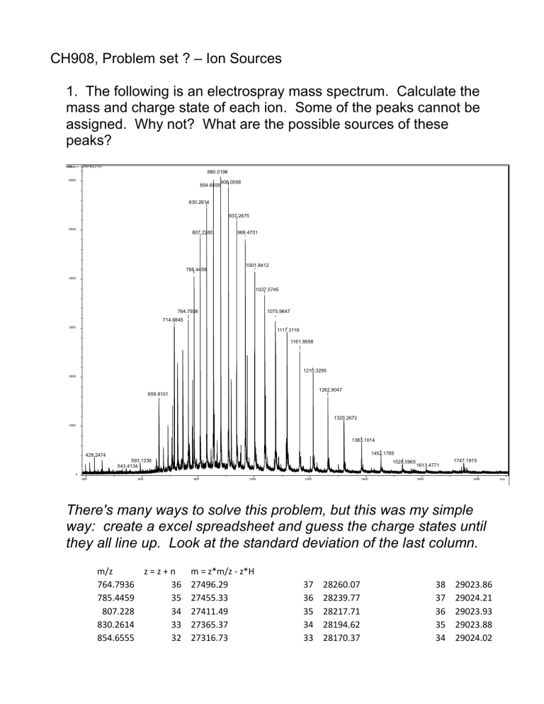
1. The following is an electrospray mass spectrum. Calculate the mass and charge state of each ion. Some of the peaks cannot be assigned. Why not? What are the possible sources of these peaks?
6000
880.5196
5000
830.2614
807.2280
937.2675
968.4701
4000
3000
785.4459
764.7936
714.6845
1001.8412
1037.5745
1075.9647
1117.3119
1161.9558
2000
1210.3295
1262.9047
659.8101
1320.2672
1000
1383.1014
429.2474
1400
1452.1785
1528.5965
1613.4771
1600
1747.1915
0
400
593.1336
543.4134
600 800 1000 1200
There's many ways to solve this problem, but this was my simple
1800 m/z way: create a excel spreadsheet and guess the charge states until they all line up. Look at the standard deviation of the last column. m/z
764.7936
785.4459
807.228
830.2614
854.6555 z = z + n m = z*m/z - z*H
36 27496.29
35 27455.33
34 27411.49
33 27365.37
32 27316.73
37 28260.07
36 28239.77
35 28217.71
34 28194.62
33 28170.37
38 29023.86
37 29024.21
36 29023.93
35 29023.88
34 29024.02
n
H
880.5196
908.0058
937.2675
968.4701
1001.841
1037.575
1075.965
1117.312
1161.956
1210.33
1262.905
1320.267
1383.101
1452.179
1528.597
1613.477
σ
31 27264.87
30 27209.94
29 27151.53
28 27088.94
27 27022.5
26 26950.73
25 26873.92
24 26791.3
23 26701.8
22 26605.08
21 26499.83
20 26385.19
19 26259.78
18 26121.07
17 25969.01
16 25799.51
512.6327
15
1.007825
σ
32 28144.38
31 28116.94
30 28087.79
29 28056.41
28 28023.33
27 27987.3
26 27948.88
25 27907.6
24 27862.75
23 27814.4
22 27761.73
21 27704.45
20 27641.87
19 27572.24
18 27496.6
17 27411.98
256.3191
16
σ
17
Some of the small, interspersed peaks, like 714 and 659, and such cannot be assigned because we don't have the resolution to determine their charge state, and we don't know if there are more
33 29023.89
32 29023.94
31 29024.05
30 29023.87
29 29024.17
28 29023.87
27 29023.84
26 29023.91
25 29023.7
24 29023.72
23 29023.63
22 29023.71
21 29023.97
20 29023.41
19 29024.18
18 29024.45
0.225443 than one charge state of them.
They are almost certainly nozzle-skimmer (in source) CAD fragments from the main protein. However, they could also be contaminants or from a previous sample (carry-over).
2. Calculate the repeat unit mass and end group masses of the polymer in the following MALDI spectrum.
1581.914
1405.812
8000 1361.785
1758.019
1802.049
1317.760
1846.074
6000
1273.735
1890.098
1229.709
1934.124
4000
1978.143
1185.686
2000
1141.659
1097.626
1053.586
2022.169
2066.187
2110.212
2198.245
0
1000 1200 1400 1600 1800 2000 2200 m/z
Again, there's many ways to solve this, but I used a simple excel spreadsheet:
MALDI TOF spectrum of a polymer guestimate oligomer number residual mass delta mass dm/median
1053.586
1097.626
1141.659
1185.686
1229.709
1273.735
1317.76
1361.785
44.04 1.000313734
44.033 1.000154737
44.027 1.000018455
44.023 0.9999276
44.026 0.999995741
44.025 0.999973027
44.025 0.999973027
44.027 1.000018455
1405.812 176.102 3.999937537
1581.914 176.105 4.000005678
1758.019
1802.049
1846.074
1890.098
44.03 1.000086596
44.025 0.999973027
44.024 0.999950314
44.026 0.999995741
31
35
39
40
41
42
27
28
29
30
23
24
25
26
40.9836875
40.9975
41.0043125
41.005125
41.0019375
41.00175
41.0005625
40.999375
41.0001875
40.9974375
40.9976875
41.0015
41.0003125
40.998125
1934.124
1978.143
2022.169
2066.187
2110.212
2198.045
44.019 0.999836745
44.026 0.999995741
44.018 0.999814031
44.025 0.999973027
87.833 1.99501717
43
44
45
46
47
49
40.9979375
40.99075
40.9905625
40.982375
40.9811875
40.7618125 median 44.0262 guess oligomer number N
= average
23.9308934
40.9847
This spreadsheet says that the oligomer mass is 44.0262
(polyethylene glycol) with a residual mass of 40.9847. This residual will be the sum of the two endgroups plus the charge carrier. The sample is singly charged, so there's only one of the latter.
This is a strange value, but it corresponds to water (18.011) plus a sodium ion (22.989) = 41.000. This is an error of 15 millidaltons, which is not bad for a long extrapolation. It would be better with higher resolution data.
3. Using polyacrylamide gel electrophoresis, a silver-stained gel spot yields a dark band for a protein of about 40 kDa. Could you get a usable signal from ESI - FTMS? ESI – qTOF? MALDI –
TOF?
A dark band on a silver stained gel will have something like 1-10 nanograms of material. At 40 kDa, that's 250 - 2500 femtomoles. If the sample is clean, and if you can extract most of that protein, that's plenty for detection on all three instruments. The FTMS will be the worst of the three, and will often struggle with 1-10 femtomoles of material.
