Rli1/ABCE1 Recycles Terminating Ribosomes and Controls Translation Reinitiation in 3 Article
advertisement
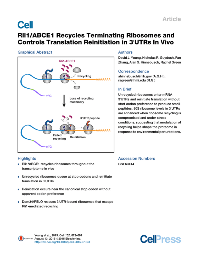
Article Rli1/ABCE1 Recycles Terminating Ribosomes and Controls Translation Reinitiation in 30UTRs In Vivo Graphical Abstract Authors David J. Young, Nicholas R. Guydosh, Fan Zhang, Alan G. Hinnebusch, Rachel Green Correspondence ahinnebusch@nih.gov (A.G.H.), ragreen@jhmi.edu (R.G.) In Brief Unrecycled ribosomes enter mRNA 30 UTRs and reinitiate translation without start codon preference to produce small peptides. 80S ribosome levels in 30 UTRs are enhanced when ribosome recycling is compromised and under stress conditions, suggesting that modulation of recycling helps shape the proteome in response to environmental perturbations. Highlights d Rli1/ABCE1 recycles ribosomes throughout the transcriptome in vivo d Unrecycled ribosomes queue at stop codons and reinitiate translation in 30 UTRs d Reinitiation occurs near the canonical stop codon without apparent codon preference d Dom34/PELO rescues 30 UTR-bound ribosomes that escape Rli1-mediated recycling Young et al., 2015, Cell 162, 872–884 August 13, 2015 ª2015 Elsevier Inc. http://dx.doi.org/10.1016/j.cell.2015.07.041 Accession Numbers GSE69414 Article Rli1/ABCE1 Recycles Terminating Ribosomes and Controls Translation Reinitiation in 30UTRs In Vivo David J. Young,1,3 Nicholas R. Guydosh,2,3 Fan Zhang,1 Alan G. Hinnebusch,1,* and Rachel Green2,* 1Laboratory of Gene Regulation and Development, Eunice Kennedy Shriver National Institute of Child Health and Human Development, National Institutes of Health, Bethesda, MD 20892, USA 2Howard Hughes Medical Institute, Johns Hopkins University School of Medicine, Baltimore, MD 21205, USA 3Co-first author *Correspondence: ahinnebusch@nih.gov (A.G.H.), ragreen@jhmi.edu (R.G.) http://dx.doi.org/10.1016/j.cell.2015.07.041 SUMMARY To study the function of Rli1/ABCE1 in vivo, we used ribosome profiling and biochemistry to characterize its contribution to ribosome recycling. When Rli1 levels were diminished, 80S ribosomes accumulated both at stop codons and in the adjoining 30 UTRs of most mRNAs. Frequently, these ribosomes reinitiated translation without the need for a canonical start codon, as small peptide products predicted by 30 UTR ribosome occupancy in all three reading frames were confirmed by western analysis and mass spectrometry. Eliminating the ribosome-rescue factor Dom34 dramatically increased 30 UTR ribosome occupancy in Rli1 depleted cells, indicating that Dom34 clears the bulk of unrecycled ribosomes. Thus, Rli1 is crucial for ribosome recycling in vivo and controls ribosome homeostasis. 30 UTR translation occurs in wild-type cells as well, and observations of elevated 30 UTR ribosomes during stress suggest that modulating recycling and reinitiation is involved in responding to environmental changes. INTRODUCTION Ribosome recycling is a vital cellular process that dissociates post-termination ribosomes into small and large subunits for participation in new rounds of translation initiation. Recycling of ribosomes is essential for maintaining homeostasis of the pool of free ribosomes, and it has been well documented that mutations in ribosome rescue/recycling factors such as Dom34 and Hbs1 exhibit synthetic phenotypes with other deficiencies in ribosome production (Bhattacharya et al., 2010; Carr-Schmid et al., 2002). In eukaryotes, recycling effectively begins with termination on recognition of a stop codon in the A site of the 80S ribosome by release factors eRF1 and GTP-bound eRF3. Following hydrolysis of GTP, eRF3 dissociates, leaving eRF1 in the A site poised to hydrolyze the peptidyl-tRNA in the P site. This peptide release step is temporally coupled through the action of Rli1 (yeast)/ABCE1 872 Cell 162, 872–884, August 13, 2015 ª2015 Elsevier Inc. (mammals) to the first stage of recycling, where the 80S ribosome is separated into a free 60S subunit and a 40S subunit bound to deacylated tRNA and mRNA. In the second stage of recycling, the deacylated tRNA is removed from the ribosome with attendant dissociation of the remaining 40S$mRNA complex (Jackson et al., 2012; Dever and Green, 2012). In vitro studies in reconstituted mammalian and yeast translation systems defined this common pathway for ribosome recycling. While ribosome dissociation is promoted simply by eRF1 (and by the ribosome rescue factor Dom34) (Shoemaker et al., 2010), the rate of the reaction is greatly stimulated by Rli1/ ABCE1 resulting in efficient dissociation of 60S subunits over a wide range of Mg+2 concentrations (Pisarev et al., 2010; Shoemaker and Green, 2011). The ribosome-splitting activity of Rli1/ABCE1 leaves mRNA and deacylated tRNA bound to the 40S subunit, and release of the tRNA in the second stage of recycling appears to be stabilized by eIF1, ligatin/eIF2D, or the interacting proteins MCT and DENR (Pisarev et al., 2007; Skabkin et al., 2010). Yeast Rli1 also stimulates translation termination (Khoshnevis et al., 2010; Shoemaker and Green, 2011), where its contribution is ATP-hydrolysis independent. The dual function of Rli1 in termination and recycling, gated by ATP hydrolysis, is consistent with its location in a cryo-EM structure of an 80S complex containing peptidyl-tRNA in the P site and eRF1 in the A site. In this structure, Rli1 interacts directly with eRF1 and with components of both the large and small ribosomal subunits in the intersubunit space (Preis et al., 2014). The impact of depleting Rli1/ABCE1 on ribosome recycling in vivo has not been addressed previously, and earlier publications suggest roles for the yeast factor in ribosome biogenesis (Yarunin et al., 2005; Strunk et al., 2012) and translation initiation (Dong et al., 2004). It is even plausible that in certain situations in the cell, destabilization of the subunit interface by eRF1 (Shoemaker et al., 2010) may be sufficient, with initiation factors acting to stabilize dissociated subunits (Pisarev et al., 2007), to provide recycling independently of Rli1. Other studies have probed biochemically the possible consequences of deficiencies in ribosome recycling. Early studies suggested that post-termination ribosomes, generated by puromycin treatment, remain associated with mRNA transcripts and could resume translation (Freedman et al., 1968). Using a mammalian reconstituted translation system, it was found that unrecycled 80S ribosomes, where peptide had been released, can migrate upstream or downstream from the stop codon and form stable complexes at nearby triplets that are complementary to the deacylated tRNA remaining in the P site (cognate to the penultimate codon of the open reading frame or ORF) (Skabkin et al., 2013). Other studies with yeast in vitro translation extracts argued that ribosomes terminating at a ‘‘premature stop codon’’ are inefficiently recycled and can migrate to nearby AUG codons (Amrani et al., 2004). Even in bacteria, impairment of ribosome recycling factor (RRF) evokes scanning and reinitiation by post-termination ribosomes (Janosi et al., 1998). These studies provide fodder for thinking about the fate of ribosomes in the absence of sufficient recycling activity in vivo. Rli1/ABCE1 can also function in ribosome splitting to rescue stalled elongating 80S ribosomes, acting in conjunction with the eRF1 and eRF3 paralogs Dom34(yeast)/PELO(mammals) and Hbs1(yeast)/HBS1L(mammals), respectively, to release stalled ribosomes in the no-go and non-stop mRNA decay pathways (Pisareva et al., 2011; Shoemaker et al., 2010; Tsuboi et al., 2012). Dom34/PELO$Hbs1 and Rli1/ABCE1 have also been implicated in subunit splitting of vacant 80S subunits (Pisareva et al., 2011). Such 80S couples cannot function in translation initiation and, under starvation, accumulate in yeast cells stabilized by the Stm1 protein (Ben-Shem et al., 2011). Recently, evidence was provided that Dom34 promotes translational recovery from starvation (van den Elzen et al., 2014). Thus, Rli1 appears to operate in the recycling of terminating, stalled elongating, or long-term-storage 80S ribosomes. Using ribosome profiling, we recently defined a role for Dom34$Hbs1 in recovering unrecycled 80S ribosomes in the 30 UTRs of 10% of all yeast genes in a dom34D mutant (Guydosh and Green, 2014). While the origin of these 30 UTR ribosomes was unclear, a defect in ribosome recycling seemed plausible because the phenomenon was enhanced by treating cells with diamide, an oxidizing agent known to inactivate Fe-S cluster proteins (Philpott et al., 1993) such as Rli1 (Yarunin et al., 2005). It appeared that some 30 UTR ribosomes present in dom34D cells scanned, rather than translated, the 30 UTR and eventually accumulated at the beginning of the poly(A) tail. However, translation by a fraction of the 30 UTR ribosomes, either by read-through of the main ORF stop codon or reinitiation, was not excluded (Guydosh and Green, 2014). In this study, we use ribosome profiling (Ingolia et al., 2012) and biochemistry to define the role of Rli1/ABCE1 in living cells. In an Rli1-depleted yeast strain (dubbed rli1-d), we find that 80S ribosomes accumulate at the stop codons and in the 30 UTRs of virtually all yeast genes, to a much greater extent than seen previously in dom34D cells. The distribution of 80S footprints strongly suggests that ribosomes in the 30 UTR of the rli1-d strain are frequently engaged in translation, displaying occupancy peaks that coincide with 30 UTR stop codons in all three reading frames. We detected the aberrant 30 UTR translation products for several genes and obtained strong evidence that they can originate from reinitiation in any frame rather than by read-through of the main ORF stop codon. We further find that reinitiation tends to occur in proximity to the main ORF stop codon, but that neither AUG codons nor triplets complementary to the penulti- mate P-site tRNA are preferred start sites. We show that overexpressing Rli1 diminishes the 30 UTR ribosomes detected previously in dom34D cells, demonstrating that they arise from incomplete recycling of terminating ribosomes at main ORF stop codons. Finally, the absence of Dom34 in rli1-d cells evokes an increase in ribosomes occupying main ORF stop codons and 30 UTRs, as well as a dramatic rise in the number arrested at the 30 UTR/poly(A) boundary; these observations indicate that Dom34 is critically required for rescuing ribosomes that escape normal termination/recycling. Our findings demonstrate that Rli1 is crucial for ribosome recycling in vivo and thus plays an essential role in controlling non-canonical 30 UTR translation. RESULTS Depletion of Rli1 Evokes Increased 80S Occupancies in 30 UTRs Transcriptome-Wide To determine the translational consequences of reduced Rli1 function in vivo, we conducted ribosome profiling of an rli1 degron strain that allows for conditional depletion of an unstable version of Rli1 tagged at the N terminus with ubiquitin followed by an arginine residue (Park et al., 1992). Transcription of this engineered allele PGAL-UBI-R-FH-RLI1 is under the GAL promoter and therefore is repressed with glucose as carbon source (Dong et al., 2004). Western analysis confirmed that the degron protein is expressed at lower levels than Rli1 expressed from the RLI1 promoter on galactose medium, and is undetectable only 2 hr following a shift to glucose medium (Figures S1A and S1B). By 8 hr on glucose medium, the degron mutant (rli1-d) ceases growth completely (Figure S1C) and displays a 40% reduction in polysomes compared to isogenic WT cells (Figure S1D). Accordingly, we chose these conditions to culture rli1-d and WT cells for ribosome profiling experiments. Cells were harvested by rapid filtration, lysed while frozen in liquid N2, and thawed in the presence of cycloheximide to arrest elongation (Guydosh and Green, 2014). Following digestion by RNase I, 80S ribosomes were isolated by sedimentation through a sucrose density gradient. Ribosome footprint levels (on ORFs) were highly reproducible between biological replicates (Spearman’s R2 = 0.98 for rli1-d cells) (Figure S2A). We also performed mRNA-Seq on the WT and rli1-d cells and found that the density of reads was broadly redistributed among coding sequences in the depletion strain and showed signs typical of stress, such as downregulation of ribosomal proteins (Figures S2B and S2C). As expected, these changes in gene expression at the transcript level were correlated with (and often amplified by) the observed redistribution in footprint density. Examination of the average level of ribosome footprint reads for all genes aligned at stop codons in WT cells revealed a 3-nt periodicity in the ORF, indicating the translated reading frame, with a peak occurring at the stop codon (Figure 1A), in accordance with previous results (Ingolia et al., 2009; Ingolia et al., 2011). The corresponding profile for rli1-d cells revealed two striking differences: the peak at stop codons was greatly magnified, and a second, smaller, peak appeared 30 nt upstream. Both phenomena are consistent with a delay in termination or recycling at the stop codon accompanied by queuing of the trailing elongating ribosomes behind those stalled at the stop codon. Cell 162, 872–884, August 13, 2015 ª2015 Elsevier Inc. 873 Figure 1. Ribosomes Accumulate at Stop Codons and in 30 UTRs of Most Genes When Rli1 Is Depleted A Stacked ribosomes (A) Normalized average ribosome footprint occupancy (each gene equally weighted) from all genes aligned at their stop codons for WT, SUP4-o (genes with UAA stop codons only) and rli1-d cells. (Inset) Demagnified view of (A) with schematic depicting ribosome stalling and queuing at the stop codon. (B) Ratio of footprint densities in 30 UTRs to the respective ORFs is plotted for rli1-d cells versus WT cells, for genes with > 5 rpkm in ORFs and > 0.5 rpkm in 30 UTRs. Each point represents the data for 1 gene. Normalized reads Stop 28 nt rli1-d SUP4-o WT -60 -40 0 -20 20 40 60 Distance of footprint 5' end from start of 3'UTR (nt) B rli1-d density ratio (3'UTR:ORF) ORF 10 10 10 10 3' UTR 2 0 -2 -4 10 -4 -2 0 10 10 10 WT density ratio (3'UTR:ORF) 2 Second, ribosome occupancies were elevated in 30 UTR regions, slightly rising for the first 50 nt of the 30 UTR and gently falling off thereafter (Figure 1A). Moreover, these 30 UTR ribosomes do not occupy a single reading frame like those in the coding sequence (no more than 35% of 28-nt reads mapped without mismatches to a single 30 UTR reading frame versus 94% in ORFs). A comparison of the ratio between ribosome density in 30 UTR versus the ORF for each transcript revealed a broad increase in 30 UTR density, by roughly an order-of-magnitude on average, when Rli1 was depleted (Figure 1B). We found that the level of ribosome density in 30 UTRs was correlated with that found in ORFs (Figure S2D) and, between biological replicates, averaged between 21%–34% of that found in ORFs (Figure S2E). The general increase in 30 UTR occupancies seen in rli1-d cells is consistent with a genome-wide failure in termination or recycling at stop codons that leads to either a continuation of translation without termination (‘‘read-through translation’’), reinitiation of new translation in the 30 UTR, or termination followed by 80S ‘‘scanning.’’ We define scanning as the movement of unrecycled ribosomes (for brevity dubbed 80S post-termination complexes or post874 Cell 162, 872–884, August 13, 2015 ª2015 Elsevier Inc. 80 TCs) along mRNA in the absence of peptide synthesis. In general, the averaged data from the rli1-d strain differed significantly from data derived from a strain expressing an ochre suppressor tRNA (SUP4-o) (Guydosh and Green, 2014). In that case, ribosome occupancy in 30 UTRs following UAA stop codons maintains the same reading frame as the main coding sequence and gradually decreases downstream as ribosomes reading past the main stop codon terminate translation and are recycled at downstream stop codons (Figure 1A). The absence of these trends in the rli1-d data point to a phenomenon distinct from simple read-through. Accumulation of 80S Ribosomes at 30 UTR-Encoded Stop Codons Is Consistent with 30 UTR Translation To evaluate whether ribosomes in 30 UTRs were translating, we examined the relative ribosome density across numerous 30 UTRs and found a notable enrichment in density on stop codons (Figures 2A, S3). To get a sense for the typical shape of the peak at stop codons in 30 UTRs, we averaged ribosome density around all stop codons in 30 UTRs and found the average peak was 2- to 3-fold above background level in the rli1-d strain and lower in the WT strain (Figure 2B). Interestingly, this average peak for the rli1-d strain appeared the same when we limited our averaging to each of the three reading frames relative to the main ORF (Figure 2C). To quantify this phenomenon on the level of individual stop codons, we computed a pause score by taking the ratio of ribosome occupancy at each stop codon relative to the background density in its respective 30 UTR (Figure 2D). We found that the median pause score increased 5-fold in rli1-d cells relative to WT cells, emphasizing the global nature of this effect. The apparent accumulation of 80S ribosomes at 30 UTR stop codons seems incompatible with scanning 80S ribosomes and more consistent with translating 80S ribosomes. A second feature consistent with 30 UTR translation is that the footprint peaks at 30 UTR stop codons are enriched in atypically long WT Figure 2. Ribosomes Accumulate at Stop Codons within 30 UTRs in rli1-d Cells YMR122W-A WT 15 nt 20 rpm 20 rpm A +1 5 nt rli1-d rli1-d ORF Stop 3’UTR 3’UTR YMR122W-A Stop -1 Stop TAA GAA GTT TCT AAA AGC CTT TTT TTT TCC TTC TGC TTA TTG WT 30 nt Fraction of reads ORF 3’UTR 3’UTR C 0.20 rli1-d WT 0.15 0.10 0.05 0.00 Cumulative fraction 1.0 0.5 -1 0.20 0.15 0.10 Stop Frame 0 +1 −1 0.05 0.00 0 5 10 -10 -5 Distance of A site to stop (nt) 0 5 10 -10 -5 Distance of A site to stop (nt) D 6 nt rli1-d rli1-d B 15 rpm HOR7 Fraction of reads WT 15 rpm TAC TAT CAA AGG GAA CGA TTG ATT +1 Stop E WT rli1-d (A) Ribosome footprints on genes YMR122W-A and HOR7. Boxed regions on the left are magnified on the right, showing schematically the positions of 30 UTR stop codons (red) in the indicated reading frames (+1 or 1). A portion of the YMR122W-A 30 UTR sequence is shown, highlighting the main ORF stop codon (filled gray box), 1 stop codon (dotted box), and the +1 stop codon showing a strong 80S peak (filled red box). (B) Average fraction of ribosome occupancy in a window surrounding stop codons in the 30 UTR (all frames included). (C) Same as rli1-d data in (B) but sorting data for each reading frame relative to the main ORF (0) frame. (D) Cumulative frequency histogram of pause scores on 30 UTR stop codons shows enhanced pausing in the rli1-d strain versus WT, for genes with > 10 rpkm in 30 UTRs. (E) Size distributions of rli1-d footprints that mapped without mismatches to either spliced coding sequences (ORFs) or 30 UTRs. Mapping performed for full sequences or 34-nt windows starting 17-nt upstream of stop codons. (F) Same as rli1-d data in (B) but data are sorted by the presence or absence of an upstream, in-frame 30 UTR stop codon. 0.50 ORF 0.25 all codons stop codons would tend to trigger termination and recycling of most ribosomes at the expense of stop codons located further downstream in the 30 UTR. Fraction of reads Fraction of reads Histidine Starvation Evokes Ribosome Stalling at 30 UTR His Codons in the Manner Expected for 0.00 0.0 30 UTR Translation 0.50 0.01 0.1 1 10 100 We showed previously that starvation of 3’UTR Stop codon pause score yeast cells for histidine with 3-aminoall codons 0.20 0.25 no upstream stop triazole (3-AT) evokes pausing of elonF stop codons all stop codons gating 80S ribosomes, detected as 0.15 yes upstream stop 3-AT-enhanced ribosome occupancy at 0.10 0.00 histidine codons in the ribosome profiles 25 28 31 34 0.05 of ORFs genome-wide (Guydosh and Read length (nt) 0.00 Green, 2014). We exploited this phenom0 5 10 -10 -5 enon here to support the hypothesis that Distance of A site to stop (nt) the 30 UTR ribosomes are translating. We established conditions for histidine starvation in Rli1-depleted cells by moninuclease-protected fragments, as previously reported for ca- toring induction of eIF2a phosphorylation by protein kinase nonical stop codons in the main ORFs (Ingolia et al., 2011) Gcn2—a well-established signature of amino acid starvation (De(Figure 2E). Finally, when we evaluated the average ribosome oc- ver et al., 1992)—and determined that adding 3-AT after only 4h cupancy for stop codons across the 30 UTR, we found that the of Rli1 depletion evoked increased eIF2a-P that peaked after an presence of an upstream 30 UTR stop codon in the same reading additional 3 hr incubation in 3-AT medium (Figures S4A and S4B). frame tends to diminish the size of the 80S peak at a downstream As a control, we noted that ribosome profiling under these modistop codon whereas the absence of an upstream stop codon in- fied growth conditions did not prevent the appearance of peaks creases it (Figure 2F). This feature is expected from 30 UTR trans- at stop codons in the 30 UTRs of rli1-d cells (Figure 3A, 3-AT, WT lation because the presence of an in-frame upstream stop codon versus rli1-d). Importantly, 3-AT treatment of rli1-d cells evoked Cell 162, 872–884, August 13, 2015 ª2015 Elsevier Inc. 875 Figure 3. Histidine Starvation Evokes Ribosome Stalling at 30 UTR His Codons in rli1-d Cells A WT His 3’UTR Stop SED1 150 rpm 150 rpm ORF WT 20 nt WT + 3-AT 4 nt WT + 3-AT rli1-d rli1-d rli1-d + 3-AT rli1-d + 3-AT ORF 3’UTR HH 0 0 3’UTR Stop 0 SED1 Stop TAA ACG GTG GTG TTT GAC ACA TCC GCC TTC TTA ATG CTT TCT TTC AGT ATT ATG TTA TTT TTT TGT TAT TCG TTT TTC ACT TCT AGG CTT TTT GAC AGA CTA GCC CCG TTA TAC CAC CAT CTT TGT GGG AAA GCC CCT AAA TTG CCC TGA 3’UTR Stop (A) The schematic at the top depicts terminating ribosomes paused at stop codons and upstream elongating ribosomes paused at His codons in histidine-starved Rli1-depleted cells. Ribosome footprint reads in SED1 are presented as in Figure 2A, showing the two His codons (H, purple) and predicted 30 UTR termination site (red), all in the 0 frame, in the enlargement on the right. These His codons (filled purple boxes) and in-frame stop codon (filled red box) are also highlighted in the SED1 30 UTR sequence, along with His codons (unfilled purple boxes) and stop codons (dotted boxes) in the other two reading frames that lack strong peaks. (B–D) Similar to (A) for the genes HHT2, ILV5, and YDR524C-B. WT HHT2 10 nt WT 3 rpm 3 rpm B 3 nt sequences for 13 tandem Myc epitopes immediately preceding, and in-frame rli1-d rli1-d with, 30 UTR stop codons in the endogerli1-d + 3-AT rli1-d + 3-AT nous loci of various genes exhibiting prominent peaks in ribosome density at Stop 3’UTR ORF H H 3’UTR 30 UTR stop codons in rli1-d cells. West-1 -1 -1 ern analysis revealed Myc13-tagged polyC peptides of 40 kDa for 11 of 16 tagged ILV5 4 nt genes in rli1-d cells, none of which were WT WT 10 nt observed in identically tagged WT cells WT + 3-AT WT + 3-AT (Figures 4A and S5A). These results prorli1-d rli1-d vide direct evidence for translation of rli1-d + 3-AT rli1-d + 3-AT 30 UTR sequences in rli1-d cells in reading frames where ribosomes are stalled at 3’UTR ORF Stop 3’UTR H stop codons. For the five genes where +1 +1 D no Myc13-tagged product was detected, the predicted polypeptides might be subYDR524C-B 15 nt WT WT ject to rapid proteolysis owing to unusual 4 nt amino acid compositions encoded by WT + 3-AT WT + 3-AT normally untranslated 30 UTR sequences. rli1-d rli1-d In the course of our analysis, we realized rli1-d + 3-AT rli1-d + 3-AT that the 20.5 kDa Myc13 epitope migrates 3’UTR ORF anomalously as a polypeptide of 40 kDa H Stop 3’UTR +1 +1 (Figure S5B). Thus, our finding that the Myc13-tagged polypeptides from all 11 genes that produced stable 30 UTR transpeaks at His codons upstream from (and in the same frame as) lation products also have apparent MWs of 40 kDa implies that 30 UTR stop codons that exhibit strong peaks in untreated rli1-d only small portions of the tagged translation products are encells (Figures 3A–3D). While the extent of histidine starvation coded by endogenous sequences at these genes. This in turn was insufficient to systematically investigate pausing at the suggests that the tagged polypeptides originate from reinitiation read depth of our dataset, we did find evidence of this phenom- after peptide release at the main stop codon, rather than readenon on well over 100 genes, further strengthening our hypothe- through from the main ORF. To support this last conclusion, we pursued two different strasis that 30 UTRs are translated when Rli1 is depleted (Table S1). tegies. First, we inserted coding sequences for the much smaller Direct Detection of Epitope-Tagged 30 UTR Translation HA3 epitope (that would not mask informative mobility differences) at the same 30 UTR locations used above for Myc13Products To differently evaluate our model for 30 UTR translation, we set tagging of six candidate genes. Importantly, their apparent out to detect polypeptides predicted to arise from 30 UTR trans- MWs are now in the 7–10 kDa range (Figures 4B and S5C). lation in particular genes. To this end, we inserted the coding Given a mass of 4.8 kDa for HA3, the observed MWs are WT + 3-AT 876 Cell 162, 872–884, August 13, 2015 ª2015 Elsevier Inc. 6 rpm 6 rpm 3 rpm 3 rpm WT + 3-AT A B MYC13 3’UTR Stop Stop ORF HA3 3’UTR Stop Stop ORF SED1 YDR524C-B CWP2 Figure 4. Detection of Epitope-Tagged 30 UTR Translation Products that Result from Reinitiation versus Proteolysis of Readthrough Products in rli1-d Cells HOR7 (A and B) MYC13 (A) or HA3 (B) epitope tags were inserted just upstream of predicted 148 30 UTR termination sites in the chromosomal alleles of 4 candidate genes (depicted sche98 16 64 matically) in WT and rli1-d strains. Tagged 50 strains were cultured as in Figure S1A and WCEs 7 36 were subjected to western analysis using anti22 4 bodies against c-Myc or HA (upper blots) or 98 Gcd6 (lower blots). Two amounts of extracts Gcd6 34 64 were loaded in each lane pair. Migration of MW standards is indicated on the left. ORF, main C coding sequences. HOR7 YMR122W-A (C) HA3-tagged reinitiation (RI) and readthrough Reinitiation (RI) Reporter WT rli1-d WT rli1-d (RT) reporters for HOR7, and their predicted 0 -1 RI RT RI RT RI RT RI RT 3’UTR HOR7 TAA tagged products, are depicted schematically HA3TAA on the left. The RI reporters for HOR7 and 98 YMR122W-A were those analyzed in (B and HA3 64 50 S5C), and the RT reporters were constructed for Readthrough (RT) Reporter 36 them by inserting a single nucleotide before T 22 -1 (HOR7) or removing the first nucleotide of 0 16 3’UTR TAA HOR7 (YMR122W-A) the main ORF stop codon, placing HA3-TAA 6 the 30 UTR site in frame with the main ORF. HA3 Western analysis of the resulting strains was HA3 HA3 conducted as in (B). D (D) To detect CWP2 30 UTR translation in different reading frames, single or tandem +1 frame tag G nucleotides were inserted immediately preNo insertion G insertion GG insertion WT rli1-d WT rli1-d WT rli1-d ceding the MYC13 tag located adjacent +1 MYC13Stop to the predicted 30 UTR +1-frame termination High-level 148 No insertion CWP2 Stop product site in the CWP2 reinitiation reporter analyzed 98 -1 64 in (A) and depicted here as ‘‘No insertion,’’ 50 G +1 thus shifting the MYC13 tag and adjoining 36 Low-level Stop 1 nt insertion CWP2 product in-frame stop codon from the +1 frame into 22 MYC13Stop -1 the 1 or 0 frames for the 1 nt and 2 nt GG +1 98 No insertions, respectively, as depicted scheGcd6 Stop 2 nt insertion CWP2 64 product MYC13Stop -1 matically. The first stop codons encountered 0 downstream of the tag coding sequences in the +1 (top), 0 (middle), and 1 (bottom) frames are indicated with filled red or orange boxes. For the 2 nt insertion/GG construct, the tag is shown partially transparent and the adjoining stop codon in orange versus red to indicate the absence of detectable reinitiation in the 0 frame. Tagged WT and rli1-d cells were analyzed as in (A). SED1 YDR524C-B WT rli1-d WT rli1-d CWP2 WT rli1-d HOR7 WT rli1-d WT rli1-d WT rli1-d WT considerably smaller than those predicted from read-through translation from the main ORF. Rather, their apparent MWs imply that only 2–5 kDa of the HA3-tagged products are encoded by endogenous sequences, which is within the range of masses predicted by reinitiation at sites close to the main ORF stop codons and subsequent termination occurring within the 30 UTR (see Table S2). In the second strategy, we modified a subset of the aforementioned tagged alleles by mutating the main ORF stop codon resulting in a shift of the reading frame into that proposed to be utilized for 30 UTR translation in rli1-d cells. These mutations should produce large read-through products extended at the C terminus by the 30 UTR-encoded peptides (plus epitope tags). In every instance, the engineered read-through product displayed the predicted MW, which was demonstrably larger than the corresponding 30 UTR product expressed from the parental tagged allele (Figures 4C and S6A). This finding is inconsistent with read-through translation from the main ORF as the rli1-d WT rli1-d mechanism of 30 UTR translation. Moreover, the detection of only long read-through products from the engineered alleles in rli1-d cells excludes the possibility that the shorter products expressed from the parental tagged alleles arise from proteolytic cleavage of tagged C-terminal extensions from (hypothetical) read-through products. Hence, all of our results point to reinitiation following termination at the main ORF stop codon as the mechanism of 30 UTR translation. Interestingly, ribosome profiling data from the CWP2 30 UTR in rli1-d cells reveals a peak at a second stop codon in the 1 frame, downstream of the ‘‘major’’ 30 UTR stop codon that terminates translation of a short peptide in the +1 frame (Figures S6B and S6C). This second peak suggests that reinitiation occurs in more than one reading frame. To ask whether 30 UTR translation also occurs in the 1 frame, we modified the CWP2-30 UTRMYC13 allele analyzed above by inserting a single G nucleotide immediately before the Myc13 coding sequences, thus shifting the epitope into the 1 frame (Figure 4D, schematics, 1 nt Cell 162, 872–884, August 13, 2015 ª2015 Elsevier Inc. 877 Enrichment at stop codon A Figure 5. Reinitiation Most Likely Occurs at Non-AUGs near the Main ORF Stop Codon 5 4 3 2 1 0 0-4 5-9 10-14 15-19 20-24 25-29 30-34 Position in 3'UTR (nt) Reinitiation on AUG codon Stop ORF 3’UTR AUG Stop Fraction of reads B 0.20 0.15 AUG upstream no AUG upstream (A) Average fraction of ribosome occupancy was computed for stop codons in 5-nt windows downstream of the main ORF stop codon, and peak enrichment was plotted versus the center of each window. Data for the 0–4 nt window could not be computed due to interfering reads from the pause on the main stop codon of the ORF. (B) Schematics (left) illustrate potential reinitiation mechanisms. Average fraction of ribosome occupancy (right) in a window surrounding 30 UTR stop codons, sorting data by the presence or absence of an upstream, in-frame AUG (top) or main-ORF penultimate codon (bottom) in the 30 UTR. 0.10 0.05 0.00 stalling on stop codons within narrow (5 nt) windows downstream of the main stop. We only included the first 0.20 Reinitiation on penultimate codon penultimate upstream stop codon in any frame in our analysis 0.15 no penultimate upstream since we previously showed that stalling 0.10 NNN Stop on subsequent stop codons is reduced Stop NNN 0.05 3’UTR ORF (Figure 2F). The amplitude of stalling in 0.00 these windows achieved a relatively 0 5 10 -10 -5 constant value shortly after the main Distance of A site to stop (nt) stop (Figure 5A). From this we conclude that most reinitiation events occur very insertion). The high-level +1 frame product generated by the close to the main stop (likely <10 nt in the downstream parental construct was no longer detected, because the Myc13 direction). We next evaluated whether the presence or absence of an coding sequences are now found in the 1 frame, and instead we observed a much less abundant Myc13-tagged product AUG in the 30 UTR would modulate the amplitude of ribosome that is consistent with low-level 30 UTR translation in the 1 frame stalling on downstream in-frame 30 UTR stop codons. To maxi(Figure 4D, 1G insertion versus No insertion). By contrast, mize any potential effect, we again limited the analysis to the first inserting two Gs before the Myc13 coding sequences of stop codon in any frame. Surprisingly, the presence or absence CWP2-30 UTR-MYC13 resulted in no detectable Myc13-tagged of an in-frame AUG had no influence on pause amplitude polypeptides (Figure 4D, 2 nt/GG insertion), indicating that (Figure 5B, upper); these results imply that reinitiation generally little or no translation occurs in the 0 frame of the CWP2 does not occur at AUG codons in the rli1-d strain. We performed 30 UTR. These interpretations were confirmed by examining an a similar analysis to evaluate the presence of upstream in-frame independent construct in which Myc13 coding sequences were triplets that could be decoded by the penultimate deacylated inserted just upstream from the second ‘‘minor’’ stop codon tRNA remaining in the P site of the 80S post-TC (Skabkin et al., in the 1 frame, which produces the low-abundance product 2013). We similarly found that the presence or absence of such attributed above to 30 UTR translation in the 1 frame (Fig- in-frame triplets had no effect on the extent of stop codon ure S6D, no insertion). In addition to providing clear evidence pauses (Figure 5B, lower). Thus, we have no evidence that reinifor 30 UTR translation in two different reading frames of the tiation involves scanning by the 80S post-TC to a 30 UTR codon CWP2 30 UTR, the results of these and other experiments in Fig- that can base pair with the recently deacylated P-site tRNA. ure S6D establish that 30 UTR translation in rli1-d cells adheres to the strict rules of frame maintenance that characterizes conven- Mass Spectrometry Analysis of Epitope-Tagged 30 UTR Translation Products tional translation elongation. We next asked whether mass spectrometry (MS) could help us to better define reinitiation sites for 30 UTR peptide products. We Reinitiation Likely Occurs at Non-AUGs near the Main gel-purifed Myc13-tagged reinitiation products from anti-Myc imORF Stop Codon We next turned to the question of how and where reinitiation mune complexes isolated from rli1-d cells, digested them in-gel occurs. We again took advantage of the observation that stalls with trypsin or GluC, and analyzed the proteolytic fragments by on 30 UTR stop codons in the ribosome profiling data can serve MS. We identified tryptic peptides or ‘‘semi-tryptic’’ fragments as signals of active translation. To determine whether transla- (that lack a Lys/Arg residue at one end of the peptide) of the tion tends to reinitiate immediately after the main ORF stop Myc13-tagged products for YMR122W-A, YDR524C-B, CWP2, codon or whether the ribosome typically first scans some HOR7, and HSP150, and additional ‘‘semi-GluC’’ fragments distance downstream, we analyzed the extent of ribosome (that lack a Glu residue at one end) for YDR524C-B (Figure 6A, Fraction of reads 0 5 10 -10 -5 Distance of A site to stop (nt) 878 Cell 162, 872–884, August 13, 2015 ª2015 Elsevier Inc. A B underlined; Table S3); and confirmed their sequences by collisionally induced dissociation of the peptides. The identified peptide sequences are consistent with 30 UTR translation in the expected reading frames and are not predicted from the canonical yeast proteome. For YDR524C-B, HOR7, and YMR122W-A, we found no peptides in control gel slices prepared from the corresponding RLI1+ MYC13-tagged strains (data not shown), consistent with our inability to detect Myc13-tagged reinitiation products by western analysis of these strains (Figures 4A and S5A). These results lend further confidence to our conclusion that the 30 UTRs of these genes are translated in the predicted reading frames. The results for the YMR122W-A tryptic peptide are particularly instructive because its N-terminal sequence begins only six codons downstream from the main ORF stop codon in the +1 frame (Figure 6B, gold). The first amino acid of the observed peptide, Ala, defines the position furthest downstream where reinitiation could occur. Reading further upstream, the first stop codon in frame with the peptide is found 10 codons upstream of its N terminus, defining the furthest possible upstream reinitiation site (Figure 6B). The MS data are compatible with reinitiation at any of the +1 frame codons in this narrow 30-nt window surrounding the main ORF stop codon, which is notably devoid of both AUG codons and isoleucine codons cognate to the penultimate tRNA. The tryptic peptides and ‘‘semi-tryptic’’ or ‘‘semi-GluC’’ peptides identified for CWP2, HOR7, and YDR524C-B allowed us Figure 6. Mass Spectrometry of Immunoprecipitated Myc13-Tagged 30 UTR Translation Products (A) Tagged 30 UTR translation products were immunopurified and resolved by SDS-PAGE, and peptide products of trypsin (SED1, YDR524C-B, CWP2, HOR7, HSP150 and YMR122W-A) or GluC (YDR524C-B) digestion were identified by LC-MS/ MS analysis. Peptide sequences (underlined) were determined by peptide fragmentation-MS, highlighted in brick red for canonical tryptic peptides ending in Lys/Arg, respectively, and preceded by the corresponding codons (Lys/Arg). Shown in blue and cyan are ‘‘semi-tryptic’’ peptides lacking either a C-terminal Lys/Arg or the preceding Lys/ Arg codons, and ‘‘semi-GluC’’ peptides lacking either a C-terminal Glu or a preceding Glu codon, respectively. (B) A portion of the main ORF and 30 UTR sequence of YMR122W-A translated in all 3 frames, showing the MS-identified tryptic peptide (in gold) translated in the +1 frame of the 30 UTR. The first upstream stop codon in the +1 frame of the ORF (unfilled black box), main ORF stop codon (gray), 30 UTR termination codon in the +1 frame (red), and window encompassing the deduced reinitiation site are indicated. to place the reinitiation sites close to the main ORF stop codons (Figures S7A and S7B and data not shown), compatible with reinitiation beginning within 4 or 5 codons downstream of the main ORF stop codons. As for YMR122W-A, there are no AUG codons or triplets cognate to the penultimate tRNA in the reinitiation windows defined for CWP2 and HOR7 (Figures S7A and S7B). These observations are consistent with the findings from ribosome profiling data (Figures 5A and 5B) that reinitiation frequently occurs relatively close to the main ORF stop codon at triplets that do not correspond to AUG nor the penultimate codon of the main coding sequence. The tryptic peptide we identified in the HSP150 Myc13-tagged product is encoded considerably farther downstream of the main ORF stop codon (Figure 6A). Reinitiation could occur either following an extended period of scanning from the main ORF stop codon, or following a prior reinitiation event that begins within 10 codons of the main ORF stop codon and terminates at one of the three distinct 30 UTR stop codons. Nevertheless, the peptides identified by MS for at least 4 of these 5 genes are consistent with the conclusion that reinitiation is not preceded by an extended period of scanning by the post-TC. Dom34 Rescues a Large Fraction of Unrecycled 80S Subunits to Suppress Aberrant 30 UTR Translation in rli1-d Cells Our previous study suggested that Dom34 rescues ribosomes that escape normal recycling and ultimately accumulate at the junction between the 30 UTR and the poly(A) tail in dom34D cells (Guydosh and Green, 2014). To test whether Dom34 rescues ribosomes that evade recycling due to depletion of Rli1, Cell 162, 872–884, August 13, 2015 ª2015 Elsevier Inc. 879 0.3 0.2 0.002 0.1 0.001 0.0 0.000 -60 -40 -20 0 -60 Distance of footprint 5' end from poly(A) (nt) 3’UTR Cumulative fraction B dom34∆ WT dom34∆ hcRLI1 0.003 rli1-d / dom34∆ rli1-d WT -40 poly(A) 3’UTR C 1.0 dom34∆ hcRLI1 dom34∆ WT rli1-d rli1-d / dom34∆ 0.5 0.0 1 10 0.1 100 Stop codon pause score -40 0 40 80 Distance of footprint 5' end from 3'UTR (nt) E 2 rpm 2 3’UTR CPR5 40 nt WT 10 0 dom34∆ dom34∆ hcRLI1 10 10 SGE1 -2 0.5 rpm mutant ratio (3’UTR:ORF) 10 poly(A) rli1-d / dom34∆ rli1-d WT ORF D 0 -20 Distance of footprint 5' end from poly(A) (nt) Normalized reads Average reads (rpm) A rli1-d / dom34∆ rli1-d dom34∆ hcRLI1 -4 10 -4 -2 0 10 10 WT ratio (3’UTR:ORF) F ORF 98 64 eRF1/ eRF3-GTP 80S stalled at stop codon rli1-d AAAAAAA Stop 16 6 4 3’UTR poly(A) 3’UTR Stop ve α-GCD6 2 G HA3 CWP2 10 cto r I1 RL hc 50 nt ORF Stop 3’UTR eEF1A-GTP- aa-tRNA 60S Rli1-ATP Stop Low-level Recycling Stop (i) Reinitiation AAAAAAA ORF Stop Stop 80S stalled at 3’UTR stop codon (ii) Clearance of stalled 80S by Dom34/Hbs1 Dom34/ Hbs1-GTP ? ORF AAAAAAA Stop Scanning 80S Figure 7. Dom34 Is Critically Required to Rescue Unrecycled Ribosomes In Vivo (A) Average ribosome occupancy from 30 UTRs aligned at the annotated site of polyadenylation for WT, rli1-d, and rli1-d / dom34D cells (left) and WT, dom34D, and dom34D hcRLI1 cells (right). (B) Cumulative histogram of pause scores on ORF stop codons computed by taking the ratio of local density at stop codons compared to the overall ORF, for genes with > 100 rpkm in ORFs. (legend continued on next page) 880 Cell 162, 872–884, August 13, 2015 ª2015 Elsevier Inc. we performed ribosome profiling of an rli1-d / dom34D double mutant (Figures S4C–S4E). Unlike the rli1-d single mutant, the double mutant displays substantial accumulation of 80S ribosomes at the 30 UTR/poly(A) boundary (Figure 7A, left), much as we observed in dom34D cells (Figure 7A, right) (Guydosh and Green, 2014). We observed a 2.5-fold increase in ribosome pausing at main ORF stop codons compared to that seen in the rli1-d single mutant (Figure 7B), and elevated average ribosome occupancy just downstream of the stop codon (ca. 30 nt) by a factor of 4 (Figure 7C). Strikingly, in the rli1-d / dom34D double mutant, the average ribosome occupancies in 30 UTRs reach the level of those in coding regions (Figures 7C and 7D), implying that a large fraction of ribosomes are sequestered in aberrant 30 UTR events. The fact that depleting Rli1 in dom34D cells greatly elevates 80S species at 30 UTR/poly(A) boundaries supports the notion that aberrant complexes arise in dom34D cells because native levels of Rli1 are insufficient to recycle all ribosomes. Supporting this idea, the accumulation of 80S ribosomes at 30 UTR/poly(A) boundaries seen in dom34D cells is diminished by Rli1 overexpression (hcRLI1, Figures 7A, right, and 7E), as would be expected if these aberrant 80S species result from failed recycling due to insufficient levels of Rli1. We also found that overexpression of Rli1 in dom34D cells (Figure S4F) diminished ribosome pausing at main ORF stop codons well below that observed in WT cells (Figure 7B), and similarly reduced 80S occupancies throughout the 30 UTR to levels below those observed in WT (Figures 7D, points appearing below diagonal line). We also observed that low-level reinitiation products from CWP2 in WT cells (Figure 4B) could be diminished by overexpressing Rli1 (Figure 7F), adding additional evidence for Rli1 insufficiency in WT cells. These data imply that Dom34 normally compensates for this inherent recycling deficiency, resulting in the normally low levels of 80S ribosome 30 UTR occupancy observed in WT cells. DISCUSSION In this study, we set out to test the hypothesis that Rli1/ABCE1 is crucial for recycling 80S post-TCs in vivo and to determine the consequences of an acute loss of Rli1 function on the fate of ribosomes. First, ribosome profiling of cells depleted of Rli1 revealed dramatic phenotypes that support a critical role for Rli1 in ribosome recycling (Figure 7G, left pathway). These phenotypes include accumulation of 80S ribosomes both at stop co- dons of most annotated ORFs and throughout the 30 UTRs of yeast mRNAs. Second, and more surprisingly, the data reveal that following the stop-codon associated delay in the rli1-d strain, ribosomes can reinitiate in a region just upstream or downstream of the main stop codon and translate 30 UTR sequences. Such a 30 UTR translation model is supported by multiple lines of evidence. First, we identify numerous ribosome occupancy peaks that coincide with stop codons in the 30 UTR, and these stop codons can be found in any of the three reading frames relative to the main ORF (depending on the gene in question; Figures 2 and S3). Importantly, the peak occupancy is diminished by the presence of another 30 UTR stop codon located upstream in the same reading frame (Figure 2F), as expected for ribosomes translating the 30 UTR and preferentially terminating at the first in-frame stop codon encountered downstream from the main ORF. These ribosome density peaks at 30 UTR stop codons represent stalled 80S post-TCs that, for a second time, are inefficiently recycled by low levels of Rli1. The occurrence of 30 UTR translation was also supported by the fact that histidine starvation (with 3-AT) increased ribosome occupancy at 30 UTR histidine codons located upstream from, and in the same reading frame as, prominent stop codon-stall sites that we observed (Figure 3). These translating ribosomes together with those scanning for reinitiation sites likely account for the overall increase in ribosome occupancy of 30 UTRs for nearly all genes when Rli1 is depleted (Figure 1B). We found that nearly one-fifth of stop codons (3,279 out of 18,514 for genes with >3 rpkm in 30 UTR ribosome density) had a pause score > 3, giving an indication of how many 30 UTR ORFs may be translated (and that number is substantial). Putative 30 UTR translation products were directly detected by western analysis and mass spectrometry after inserting coding sequences for epitope tags immediately prior to prominent stop codon-stall sites (Figures 4 and 6). All of the epitope-tagged 30 UTR products we detected in rli1-d cells had electrophoretic mobilities consistent with reinitiation taking place near the main ORF stop codon. Mass spectrometry of 30 UTR translation products is also consistent with this conclusion. Finally, these biochemical results are in accordance with our computational analysis (Figure 5A) indicating that most reinitiation events occur near the main ORF stop codon (likely within 10 nt). Together these data provide compelling support for a model invoking the reinitiation of translation in the 30 UTR by unrecycled ribosomes at sites proximal to the main ORF stop codon (Figure 7G, right). (C) Normalized average ribosome footprint occupancy from all genes aligned at their stop codons for WT, dom34D, rli1-d, and rli1-d / dom34D strains, analyzed as in Figure 1A. (D) Ratio of footprint densities between 30 UTRs and respective ORFs plotted for the indicated strains, for genes with > 5 rpkm in ORFs and > 0.5 rpkm in 30 UTRs, with each point representing 1 gene. (E) Ribosome footprints on CPR5 and SGE1. Approximate start site of 30 UTR is indicated. (F) The WT CWP2-30 UTR-HA3 strain was transformed with either empty vector (YEplac195) or hcRLI1 (YEplac195-RLI1). Transformed strains were grown as in Figure S1A except SCGAL-U and SC-U media was used instead of YPGAL and YPD media to maintain selection for plasmids. WCEs were subjected to western analysis using antibodies against HA (upper blots) or Gcd6 (lower blots). Two amounts of extracts were loaded in each lane pair. (G) Schematic model depicting the fate of post-TCs on depletion of Rli1 in rli1-d cells. Recognition of the main ORF stop codon by eRF1/eRF3-GTP (top row) is followed by release of the completed polypeptide and dissociation of eRF3-GDP (not depicted). Any residual Rli1 could bind post-TCs and catalyze dissociation of the 60S subunit (middle row, left). However, many post-TCs are not recycled, migrate a short distance from the stop codon, reinitiate translation, and frequently terminate at a 30 UTR stop codon to produce a 30 UTR-encoded polypeptide (middle row, right). Such reinitiation events appear to be diminished by Dom34, potentially because post-TC ribosomes are rescued at the main ORF stop codons or as they begin scanning. Any ribosomes that reach the 30 UTR/poly(A) boundary by reinitiation or scanning are also rescued by Dom34. Cell 162, 872–884, August 13, 2015 ª2015 Elsevier Inc. 881 How does the reinitiation observed in rli1-d cells take place? Our results are consistent with a mechanism wherein termination and polypeptide release occur at the main ORF stop codon, but splitting of the 60S subunit from the 80S post-TC fails. The resulting 80S post-TC releases eRF1 from the A site, allowing the remaining P-site tRNA to adopt the P/E conformation required for scanning (Skabkin et al., 2013). The 80S post-TC undergoes a limited period of scanning, ultimately replacing the stop codon in the A site with a sense codon, which then recruits cognate eEF1A-GTP-aa-tRNA ternary complex. A pseudo-translocation event could then position this ternary complex in the P site to allow translation to resume by the canonical elongation pathway, akin to the translocation that occurs without peptide bond formation in translation initiation directed by the dicistrovirus IGR IRES (Thompson, 2012) and the reinitiation of translation that occurs in the ‘‘StopGo’’ mechanism of viral 2A protease (Atkins et al., 2007). Regardless of the exact mechanism of reinitiation, once a new round of translation begins, it terminates at the first in-frame stop codon in the 30 UTR to produce a short peptide product (Figure 7G, right). In principle, reinitiation could occur either immediately upstream or downstream of the main stop codon following a short bidirectional scanning process. We tested this possibility by examining out-of-frame reads just upstream of the main stop codon at the end of ORFs (Figure S2F). Interestingly, 80S ribosome density was found to be enriched in alternate reading frames in the interval beginning <10 codons upstream of the main stop codon (Figures S2F and S2G). This finding is consistent with a fraction of post-TCs scanning short distances upstream of the stop codon to find a favored site for reinitiation. Our finding that reinitiation in rli1-d cells does not appear to involve scanning of the post-TC to a codon complementary to the penultimate P-site tRNA stands in contrast to observations made in a mammalian reconstituted termination system lacking ABCE1 and supplemented with added Mg2+ (Skabkin et al., 2013). While it seems possible that the P-site tRNA will remain and stabilize the 80S post-TC, perhaps what dictates the reinitiation event is driven in part by pairing interactions between the P-site tRNA and the available codon (near-cognate or even noncognate) and in part by availability of the appropriate eEF1AGTP-aa-tRNA ternary complex. Interestingly, non-cognate triplets are selected as landing sites for peptidyl-tRNA during some translational hopping events (Herr et al., 2004). The apparent absence of reinitiation at AUGs in rli1-d cells also differs from the AUGdependent toeprints observed following recognition of a premature termination codon (Amrani et al., 2004). Unless the P-site tRNA is also lost from the post-TC, and the subunits are split, it is difficult to envision how canonical initiation with Met-tRNA loading by eIF2 would be the preferred mode of reinitiation in our system. Clearly, more work will be required to define what lingers on the post-TC 80S and how it might determine the mode of ribosome movement following a recycling failure. Our ribosome profiling of the dom34Drli1-d mutant revealed a striking increase in 30 UTR 80S occupancies compared to that seen in the rli1-d single mutant. These data suggest that Dom34 may control access of unrecycled ribosomes to the 30 UTR. The fact that ribosome occupancy is elevated at canon882 Cell 162, 872–884, August 13, 2015 ª2015 Elsevier Inc. ical stop codons raises the possibility that Dom34 controls 30 UTR access by dissociating ribosomes at stop codons, presumably after the dissociation of eRF1 (Becker et al., 2012; Shoemaker et al., 2010), and prior to the initiation of scanning. The increased ribosome occupancy throughout the 30 UTRs might be explained by Dom34 acting continuously as post-TC ribosomes scan to look for a favorable reinitiation site (Figure 7G, right). In this scenario, the lower overall 30 UTR occupancy in the rli1-d strain (compared to the dom34Drli1-d strain) is the result of fewer ribosomes escaping Dom34 rescue. Previously, we identified 30 UTR-bound 80S ribosomes in dom34D cells stalled primarily at the poly(A) tail boundary. This species of 30 UTR ribosome is more abundant in the rli1-d / dom34D mutant relative to the dom34D single mutant (Figure 7A). These observations suggest that this class of ribosomes originates from 80S post-TCs that move down the mRNA after a failure in Rli1 recycling, as we had previously predicted (Guydosh and Green, 2014); these ribosomes may fail to reinitiate, and thus land at this terminal point. In some cases, these ribosomes could also be translating to this point if no stop codons occur downstream of the reinitiation point. These ideas are further strengthened by our finding that overexpressing Rli1 eliminates a majority of these 30 UTR ribosomes in the dom34D mutant (Figures 7A, 7D, and 7E). Ribosome homeostasis is critical to cellular function; ribosome availability is directly linked to the rate of cell division (Maaloe, 1966) and altered levels of available ribosomes are known to be critical to promoting proliferation of cancer cells (Ruggero, 2012). It is intriguing that the level of Rli1 in WT cells is insufficient to ensure recycling of all post-TC complexes, as unmasked in dom34D cells and rescued by overexpression of Rli1. Perhaps complete recycling is simply unnecessary because Dom34 is normally present to remove the relatively small number of unrecycled post-TC complexes that escape Rli1 function. However, it seems possible that the WT level of Rli1 activity may be just below the threshold of sufficiency for complete recycling to allow low-level production of 30 UTR-encoded peptides at certain genes under specific conditions. We provided evidence for reinitiation in the 30 UTR of CWP2 that was attributable to Rli1 insufficiency even under optimal growth conditions in WT cells. Moreover, we interrogated previously published ribosome profiling data and found increased 30 UTR ribosome occupancy in WT cells undergoing various types of nutrient starvation (Figure S2H). In each of these cases, ribosome footprint levels on RLI1 are considerably reduced relative to other genes (and thus likely Rli1 expression levels). These data suggest that increased 30 UTR translation could introduce new functions during times of stress, as with stop codon read-through in yeast infected with the PSI+ prion (True et al., 2004). As suggested previously (Skabkin et al., 2013), this phenomenon could also underlie the production of novel peptides for antigen presentation in the immune system (Schwab et al., 2003). EXPERIMENTAL PROCEDURES Plasmid Construction and Yeast Strains Plasmids and yeast strains used in this study are listed in Tables S4 and S5 and their constructions are described in Supplemental Information. Biochemical Techniques Polysome analysis was conducted as described previously (Valásek et al., 2001), as were immunoprecipitations of epitope-tagged proteins (Zhang et al., 2004) using aliquots of WCE with 5 mg of total protein and 160 ml of myc- or HA-agarose (Santa Cruz). The buffer used for cell lysis and washes contained 50 mM Tris-HCl (pH 7.5), 100 mM NaCl, 15 mM MgCl2, 0.01% NP-40, 20% Glycerol, and protease inhibitors Complete Protease Inhibitor tablet – EDTA (Roche), AEBSF, and pepstatin A. Western analysis was conducted as described (Nanda et al., 2009) using 4%–20% Tris-HCl, and 16.5% Tris-Tricine gels from Bio-Rad, and antibodies described in SI. Mass spectrometry was conducted by the Proteomics Center of Excellence at Northwestern University as described in the Supplemental Information. Ribosome Footprint Profiling Ribosome footprints were prepared as described (Guydosh and Green, 2014) by using a protocol very similar that used by Ingolia and coworkers (Ingolia et al., 2012). Biological replicate datasets were created for rli1-d and rli1-d / dom34D strains. A technical replicate dataset was created for WT cells. All ribosome footprints that appear in this study were extracted from gel slices that included the range 25–34 nt. mRNA-Seq footprints were extracted from the range 40–60 nt from total cell lysate and not subject to poly(A) selection. Ribosome footprints were analyzed essentially as described (Guydosh and Green, 2014) with modifications as described in the Supplemental Information. Unless noted otherwise, data shown for a given strain represent a composite from all biological and technical replicates. Sequencing was performed on an Illumina HiSeq2000 or HiSeq2500 at UC Riverside or the Johns Hopkins Institute of Genetic Medicine. translation to augment codon meaning of GGN by promoting unconventional termination (Stop) after addition of glycine and then allowing continued translation (Go). RNA 13, 803–810. Becker, T., Franckenberg, S., Wickles, S., Shoemaker, C.J., Anger, A.M., Armache, J.P., Sieber, H., Ungewickell, C., Berninghausen, O., Daberkow, I., et al. (2012). Structural basis of highly conserved ribosome recycling in eukaryotes and archaea. Nature 482, 501–506. Ben-Shem, A., Garreau de Loubresse, N., Melnikov, S., Jenner, L., Yusupova, G., and Yusupov, M. (2011). The structure of the eukaryotic ribosome at 3.0 Å resolution. Science 334, 1524–1529. Bhattacharya, A., McIntosh, K.B., Willis, I.M., and Warner, J.R. (2010). Why Dom34 stimulates growth of cells with defects of 40S ribosomal subunit biosynthesis. Mol. Cell. Biol. 30, 5562–5571. Carr-Schmid, A., Pfund, C., Craig, E.A., and Kinzy, T.G. (2002). Novel G-protein complex whose requirement is linked to the translational status of the cell. Mol. Cell. Biol. 22, 2564–2574. Dever, T.E., and Green, R. (2012). The elongation, termination, and recycling phases of translation in eukaryotes. Cold Spring Harb. Perspect. Biol. 4, a013706. Dever, T.E., Feng, L., Wek, R.C., Cigan, A.M., Donahue, T.F., and Hinnebusch, A.G. (1992). Phosphorylation of initiation factor 2 a by protein kinase GCN2 mediates gene-specific translational control of GCN4 in yeast. Cell 68, 585–596. ACCESSION NUMBERS Dong, J., Lai, R., Nielsen, K., Fekete, C.A., Qiu, H., and Hinnebusch, A.G. (2004). The essential ATP-binding cassette protein RLI1 functions in translation by promoting preinitiation complex assembly. J. Biol. Chem. 279, 42157–42168. The accession number for the sequencing data (debarcoded fastq files and wig files) reported in this paper is GEO: GSE69414. Freedman, M.L., Fisher, J.M., and Rabinovitz, M. (1968). Puromycin interference of reticulocyte polyribosome disaggregation caused by tryptophan deficiency. J. Mol. Biol. 33, 315–318. SUPPLEMENTAL INFORMATION Guydosh, N.R., and Green, R. (2014). Dom34 rescues ribosomes in 30 untranslated regions. Cell 156, 950–962. Supplemental Information includes Supplemental Experimental Procedures, seven figures, and five tables and can be found with this article online at http://dx.doi.org/10.1016/j.cell.2015.07.041. Herr, A.J., Wills, N.M., Nelson, C.C., Gesteland, R.F., and Atkins, J.F. (2004). Factors that influence selection of coding resumption sites in translational bypassing: minimal conventional peptidyl-tRNA:mRNA pairing can suffice. J. Biol. Chem. 279, 11081–11087. AUTHOR CONTRIBUTIONS Ingolia, N.T., Ghaemmaghami, S., Newman, J.R., and Weissman, J.S. (2009). Genome-wide analysis in vivo of translation with nucleotide resolution using ribosome profiling. Science 324, 218–223. D.J.Y. and N.R.G. collected and analyzed the data and helped write the manuscript, and F.Z. purified tagged proteins for MS analysis. A.G.H. and R.G. supervised the work and helped to write the manuscript. ACKNOWLEDGMENTS Proteomics data and analysis were performed by Susan Fishbain, Paige Gottlieb, Ioanna Ntai, and Paul Thomas of the Proteomics Center of Excellence at Northwestern University. We thank Tom Dever, Jon Lorsch, and members of our laboratories for many helpful suggestions during the course of this work. This study was funded in part by the Intramural Research Program of the NIH (A.G.H.) and by HHMI (R.G.). Received: February 24, 2015 Revised: May 21, 2015 Accepted: July 10, 2015 Published: August 13, 2015 REFERENCES Amrani, N., Ganesan, R., Kervestin, S., Mangus, D.A., Ghosh, S., and Jacobson, A. (2004). A faux 30 -UTR promotes aberrant termination and triggers nonsense-mediated mRNA decay. Nature 432, 112–118. Atkins, J.F., Wills, N.M., Loughran, G., Wu, C.Y., Parsawar, K., Ryan, M.D., Wang, C.H., and Nelson, C.C. (2007). A case for ‘‘StopGo’’: reprogramming Ingolia, N.T., Lareau, L.F., and Weissman, J.S. (2011). Ribosome profiling of mouse embryonic stem cells reveals the complexity and dynamics of mammalian proteomes. Cell 147, 789–802. Ingolia, N.T., Brar, G.A., Rouskin, S., McGeachy, A.M., and Weissman, J.S. (2012). The ribosome profiling strategy for monitoring translation in vivo by deep sequencing of ribosome-protected mRNA fragments. Nat. Protoc. 7, 1534–1550. Jackson, R.J., Hellen, C.U., and Pestova, T.V. (2012). Termination and posttermination events in eukaryotic translation. Adv. Protein Chem. Struct. Biol. 86, 45–93. Janosi, L., Mottagui-Tabar, S., Isaksson, L.A., Sekine, Y., Ohtsubo, E., Zhang, S., Goon, S., Nelken, S., Shuda, M., and Kaji, A. (1998). Evidence for in vivo ribosome recycling, the fourth step in protein biosynthesis. EMBO J. 17, 1141–1151. Khoshnevis, S., Gross, T., Rotte, C., Baierlein, C., Ficner, R., and Krebber, H. (2010). The iron-sulphur protein RNase L inhibitor functions in translation termination. EMBO Rep. 11, 214–219. Maaloe, O.K. N.O (1966). Control of macromolecular synthesis. (New York) Nanda, J.S., Cheung, Y.N., Takacs, J.E., Martin-Marcos, P., Saini, A.K., Hinnebusch, A.G., and Lorsch, J.R. (2009). eIF1 controls multiple steps in start codon recognition during eukaryotic translation initiation. J. Mol. Biol. 394, 268–285. Cell 162, 872–884, August 13, 2015 ª2015 Elsevier Inc. 883 Park, E.-C., Finley, D., and Szostak, J.W. (1992). A strategy for the generation of conditional mutations by protein destabilization. Proc. Natl. Acad. Sci. USA 89, 1249–1252. Skabkin, M.A., Skabkina, O.V., Dhote, V., Komar, A.A., Hellen, C.U., and Pestova, T.V. (2010). Activities of Ligatin and MCT-1/DENR in eukaryotic translation initiation and ribosomal recycling. Genes Dev. 24, 1787–1801. Philpott, C.C., Haile, D., Rouault, T.A., and Klausner, R.D. (1993). Modification of a free Fe-S cluster cysteine residue in the active iron-responsive element-binding protein prevents RNA binding. J. Biol. Chem. 268, 17655– 17658. Skabkin, M.A., Skabkina, O.V., Hellen, C.U., and Pestova, T.V. (2013). Reinitiation and other unconventional posttermination events during eukaryotic translation. Mol. Cell 51, 249–264. Pisarev, A.V., Hellen, C.U., and Pestova, T.V. (2007). Recycling of eukaryotic posttermination ribosomal complexes. Cell 131, 286–299. Pisarev, A.V., Skabkin, M.A., Pisareva, V.P., Skabkina, O.V., Rakotondrafara, A.M., Hentze, M.W., Hellen, C.U., and Pestova, T.V. (2010). The role of ABCE1 in eukaryotic posttermination ribosomal recycling. Mol. Cell 37, 196–210. Pisareva, V.P., Skabkin, M.A., Hellen, C.U., Pestova, T.V., and Pisarev, A.V. (2011). Dissociation by Pelota, Hbs1 and ABCE1 of mammalian vacant 80S ribosomes and stalled elongation complexes. EMBO J. 30, 1804–1817. Preis, A., Heuer, A., Barrio-Garcia, C., Hauser, A., Eyler, D.E., Berninghausen, O., Green, R., Becker, T., and Beckmann, R. (2014). Cryoelectron microscopic structures of eukaryotic translation termination complexes containing eRF1eRF3 or eRF1-ABCE1. Cell Rep. 8, 59–65. Ruggero, D. (2012). Revisiting the nucleolus: from marker to dynamic integrator of cancer signaling. Sci. Signal. 5, pe38. Strunk, B.S., Novak, M.N., Young, C.L., and Karbstein, K. (2012). A translationlike cycle is a quality control checkpoint for maturing 40S ribosome subunits. Cell 150, 111–121. Thompson, S.R. (2012). Tricks an IRES uses to enslave ribosomes. Trends Microbiol. 20, 558–566. True, H.L., Berlin, I., and Lindquist, S.L. (2004). Epigenetic regulation of translation reveals hidden genetic variation to produce complex traits. Nature 431, 184–187. Tsuboi, T., Kuroha, K., Kudo, K., Makino, S., Inoue, E., Kashima, I., and Inada, T. (2012). Dom34:hbs1 plays a general role in quality-control systems by dissociation of a stalled ribosome at the 30 end of aberrant mRNA. Mol. Cell 46, 518–529. van den Elzen, A.M., Schuller, A., Green, R., and Séraphin, B. (2014). Dom34Hbs1 mediated dissociation of inactive 80S ribosomes promotes restart of translation after stress. EMBO J. 33, 265–276. Schwab, S.R., Li, K.C., Kang, C., and Shastri, N. (2003). Constitutive display of cryptic translation products by MHC class I molecules. Science 301, 1367– 1371. Valásek, L., Phan, L., Schoenfeld, L.W., Valásková, V., and Hinnebusch, A.G. (2001). Related eIF3 subunits TIF32 and HCR1 interact with an RNA recognition motif in PRT1 required for eIF3 integrity and ribosome binding. EMBO J. 20, 891–904. Shoemaker, C.J., and Green, R. (2011). Kinetic analysis reveals the ordered coupling of translation termination and ribosome recycling in yeast. Proc. Natl. Acad. Sci. USA 108, E1392–E1398. Yarunin, A., Panse, V.G., Petfalski, E., Dez, C., Tollervey, D., and Hurt, E.C. (2005). Functional link between ribosome formation and biogenesis of ironsulfur proteins. EMBO J. 24, 580–588. Shoemaker, C.J., Eyler, D.E., and Green, R. (2010). Dom34:Hbs1 promotes subunit dissociation and peptidyl-tRNA drop-off to initiate no-go decay. Science 330, 369–372. Zhang, F., Sumibcay, L., Hinnebusch, A.G., and Swanson, M.J. (2004). A triad of subunits from the Gal11/tail domain of Srb mediator is an in vivo target of transcriptional activator Gcn4p. Mol. Cell. Biol. 24, 6871–6886. 884 Cell 162, 872–884, August 13, 2015 ª2015 Elsevier Inc.
