Normal Brain Tissue Volumes after Long-Term
advertisement
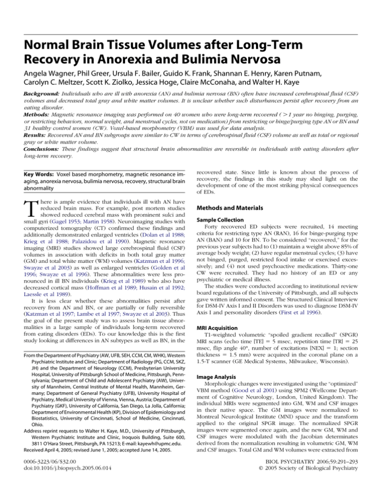
Normal Brain Tissue Volumes after Long-Term Recovery in Anorexia and Bulimia Nervosa Angela Wagner, Phil Greer, Ursula F. Bailer, Guido K. Frank, Shannan E. Henry, Karen Putnam, Carolyn C. Meltzer, Scott K. Ziolko, Jessica Hoge, Claire McConaha, and Walter H. Kaye Background: Individuals who are ill with anorexia (AN) and bulimia nervosa (BN) often have increased cerebrospinal fluid (CSF) volumes and decreased total gray and white matter volumes. It is unclear whether such disturbances persist after recovery from an eating disorder. Methods: Magnetic resonance imaging was performed on 40 women who were long-term recovered ( ⬎1 year no binging, purging, or restricting behaviors, normal weight, and menstrual cycles, not on medication) from restricting or binge/purging type AN or BN and 31 healthy control women (CW). Voxel-based morphometry (VBM) was used for data analysis. Results: Recovered AN and BN subgroups were similar to CW in terms of cerebrospinal fluid (CSF) volume as well as total or regional gray or white matter volume. Conclusions: These findings suggest that structural brain abnormalities are reversible in individuals with eating disorders after long-term recovery. Key Words: Voxel based morphometry, magnetic resonance imaging, anorexia nervosa, bulimia nervosa, recovery, structural brain abnormality recovered state. Since little is known about the process of recovery, the findings in this study may shed light on the development of one of the most striking physical consequences of EDs. T Methods and Materials here is ample evidence that individuals ill with AN have reduced brain mass. For example, post mortem studies showed reduced cerebral mass with prominent sulci and small gyri (Gagel 1953; Martin 1958). Neuroimaging studies with computerized tomography (CT) confirmed these findings and additionally demonstrated enlarged ventricles (Dolan et al 1988; Krieg et al 1988; Palazidou et al 1990). Magnetic resonance imaging (MRI) studies showed large cerebrospinal fluid (CSF) volumes in association with deficits in both total gray matter (GM) and total white matter (WM) volumes (Katzman et al 1996; Swayze et al 2003) as well as enlarged ventricles (Golden et al 1996; Swayze et al 1996). These abnormalities were less pronounced in ill BN individuals (Krieg et al 1989) who also have decreased cortical mass (Hoffman et al 1989; Husain et al 1992; Laessle et al 1989). It is less clear whether these abnormalities persist after recovery from AN and BN, or are partially or fully reversible (Katzman et al 1997; Lambe et al 1997; Swayze et al 2003). Thus the goal of the present study was to assess brain tissue abnormalities in a large sample of individuals long-term recovered from eating disorders (EDs). To our knowledge this is the first study looking at differences in AN subtypes as well as BN, in the From the Department of Psychiatry (AW, UFB, SEH, CCM, CM, WHK), Western Psychiatric Institute and Clinic; Department of Radiology (PG, CCM, SKZ, JH) and the Department of Neurology (CCM), Presbyterian University Hospital, University of Pittsburgh School of Medicine, Pittsburgh, Pennsylvania; Department of Child and Adolescent Psychiatry (AW), University of Mannheim, Central Institute of Mental Health, Mannheim, Germany; Department of General Psychiatry (UFB), University Hospital of Psychiatry, Medical University of Vienna, Vienna, Austria; Department of Psychiatry (GKF), University of California, San Diego, La Jolla, California; Department of Environmental Health (KP), Division of Epidemiology and Biostatistics, University of Cincinnati, School of Medicine, Cincinnati, Ohio. Address reprint requests to Walter H. Kaye, M.D., University of Pittsburgh, Western Psychiatric Institute and Clinic, Iroquois Building, Suite 600, 3811 O’Hara Street, Pittsburgh, PA 15213; E-mail: kayewh@upmc.edu. Received April 4, 2005; revised June 1, 2005; accepted June 14, 2005. 0006-3223/06/$32.00 doi:10.1016/j.biopsych.2005.06.014 Sample Collection Forty recovered ED subjects were recruited, 14 meeting criteria for restricting type AN (RAN), 16 for binge-purging type AN (BAN) and 10 for BN. To be considered “recovered,” for the previous year subjects had to (1) maintain a weight above 85% of average body weight; (2) have regular menstrual cycles; (3) have not binged, purged, restricted food intake or exercised excessively; and (4) not used psychoactive medications. Thirty-one CW were recruited. They had no history of an ED or any psychiatric or medical illness. The studies were conducted according to institutional review board regulations of the University of Pittsburgh, and all subjects gave written informed consent. The Structured Clinical Interview for DSM-IV Axis I and II Disorders was used to diagnose DSM-IV Axis I and personality disorders (First et al 1996). MRI Acquisition T1-weighted volumetric “spoiled gradient recalled” (SPGR) MRI scans (echo time [TE] ⫽ 5 msec, repetition time [TR] ⫽ 25 msec, flip angle 40°, number of excitations [NEX] ⫽ 1; section thickness ⫽ 1.5 mm) were acquired in the coronal plane on a 1.5-T scanner (GE Medical Systems, Milwaukee, Wisconsin). Image Analysis Morphologic changes were investigated using the “optimized” VBM method (Good et al 2001) using SPM2 (Wellcome Department of Cognitive Neurology, London, United Kingdom). The individual MRIs were segmented into GM, WM and CSF images in their native space. The GM images were normalized to Montreal Neurological Institute (MNI) space and the transform applied to the original SPGR image. The normalized SPGR images were segmented once again, and the new GM, WM and CSF images were modulated with the Jacobian determinates derived from the normalization resulting in volumetric GM, WM and CSF images. Total GM and WM volumes were extracted from BIOL PSYCHIATRY 2006;59:291–293 © 2005 Society of Biological Psychiatry 292 BIOL PSYCHIATRY 2006;59:291–293 A. Wagner et al Table 1. Demographics CW n ⫽ 31 RAN n ⫽ 14 BAN n ⫽ 16 BN n ⫽ 10 Mean SD Mean SD Mean SD Mean SD p Group Differences 26.8 21.9 22.8 20.1 7.3 2.0 2.0 1.4 23.7 21.2 21.6 14.1 18.2 28.7 5.3 2.0 2.3 1.4 4.5 20.4 27.4 21.2 23.6 14.8 15.7 39.5 7.2 1.5 2.8 2.0 2.7 52.7 24.0 23.1 24.7 19.2 15.5 29.8 6.1 2.4 2.85 2.1 2.3 18.1 .307 .076 .016 ⬍.001 .140 .753 — — 2⬍4 2,3 ⬍ 1,4 — — Study Age (years) Current BMI (kg/m2) High Past BMI (kg/m2) Low Past BMI (kg/m2) Age of Onset (years) Length of Recovery (months) CW, healthy control women; RAN, recovered anorexic women, restricting type; BAN, recovered anorexic women, binge/purging type; BN, recovered bulimic women; BMI, body mass index. the modulated images. Modulated images were smoothed with a 12 mm Gaussian kernel prior to statistical analysis in SPM2. Statistical Analysis All data analyses were performed using SPSS 11.0 (SPSS Inc., Chicago, Illinois). For multiple group comparisons analysis of variance was used to test for differences across the groups followed by Tukey post hoc test when the overall difference was statistically significant. All image analysis group comparisons were performed using analysis of variance within SPM2 (p ⬍ .05 corrected for multiple comparisons using false discovery rate). Results Recovered subjects and CW were of similar age and body mass index (BMI) (Table 1). The average length of recovery ranged from 29.8 to 39.5 months. Groups had similar volumes for total (Table 2) GM, WM, and CSF as well as regional values (data not shown). There was a small but not significant decline of total GMV with age, while total WMV and total CSF volume showed a small but not significant increase in all recovered subjects (age/GMV: r ⫽ ⫺.193, p ⫽ .232; age/ WMV: r ⫽ .167, p ⫽ .304; age vs. CSF: r ⫽ .097, p ⫽ .552) as well as in CW (age/ GMV: r ⫽ ⫺.160, p ⫽ .388; age/WMV: r ⫽ .139, p ⫽ .457; age/CSF: r ⫽ .229, p ⫽ .216). The findings remained the same after covarying for age, BMI, total brain volume and length of recovery (recovered subjects). However, the BN subgroup had an increase in GM insula volume (Z ⫽ 4,14; p ⫽ .03) when age was included as a nuisance variable. There was no relationship between total or regional GMV, WMV or CSF and co-morbid diagnoses (data not shown). Discussion Recovered AN and BN women were similar to each other and to CW, in total and regional GMV, WMV and CSF volume. Thus, this study suggests that structural brain abnormalities may be reversible after long-term recovery from an ED. To our knowledge, this is the first study to use rigorous criteria to define recovery status based upon the requirements of normal nutrition, stable weight, absence of medication, and normal menstrual cycle for at least one year. Not all studies have compared AN subjects to CW. Previous research studies found results consistent with the present study, while other studies reported persistent abnormalities. Golden et al (1996) studied individuals with AN after nutritional rehabilitation and detected normal total ventricular volumes compared to CW. Kingston found enlarged lateral ventricles and dilated sulci in AN compared to CW. However, subjects were rescanned after at least 10% weight gain (Kingston et al 1996) so that length of recovery was variable. Swayze rescanned individuals with AN after weight normalization, finding a decrease in total and regional CSF volumes as well as an increase in GMV and WMV compared to pre-scan (Swayze et al 2003). Degree of recovery may be critical for normalization of brain morphometry. However, there was no comparison to CW, or information about menstrual function or length of recovery. Lambe described an increase in GMV and WMV as well as a decrease in CSF volume in recovered compared to ill AN. Compared to CW, recovered AN had greater CSF volume and smaller GMV, but normal WMV (Lambe et al 1997). Katzman replicated these findings in a longitudinal study of a subset of 6 weight recovered AN subjects (Katzman et al 1997). Subjects in both of these studies were weight restored for at least 1 year and had started to menstruate or returned to normal menses at follow-up. More recently Neumarker found a significant volume difference of the lateral ventricles and the Sylvian fissure in AN compared to CW, which abated with weight restoration, whereas deficits of the mesencephalon and pons persisted (Neumarker et al 2000). Again length of weight restoration is unclear. All subjects in the present study also participated in positron emission tomography (PET) studies. For PET image analysis partial volume correction (PVC) factors were created by segmenting the SPGR MR image into brain and nonbrain regions, which created a binary image (Meltzer et al 1996, 1999). After Table 2. Distribution of Segmented Tissues CW n ⫽ 31 GMV (ml) WMV (ml) CSF (ml) TIV (ml) RAN n ⫽ 14 BAN n ⫽ 16 BN n ⫽ 10 Mean SD Mean SD Mean SD Mean SD p 729.3 393.6 313.4 1436.3 45.4 35.3 55.6 109.2 749.9 383.6 309.3 1442.8 53.0 26.0 61.9 115.1 740.2 403.1 342.2 1485.4 55.6 42.3 42.9 107.5 770.2 402.7 334.1 1507.0 54.8 25.7 47.5 106.6 .155 .400 .231 .224 Group Differences CW, healthy control women; RAN, recovered anorexic women, restricting type; BAN, recovered anorexic women, binge/purging type; BN, recovered bulimic women; GMV, gray matter volume; WMV, white matter volume; CSF, cerebrospinal fluid; TIV, total intracranial volume. www.sobp.org/journal A. Wagner et al smoothing, regions of interest (ROI) were placed on the binary image to determine the fraction of nonbrain within each ROI. PVC values were similar for CW and recovered subjects (Bailer et al 2004). Thus 2 different methods showed similar conclusions (Good et al 2002). While emaciation and malnutrition are known to be associated with altered brain morphometry abnormalities in AN, the physiology underlying changes in brain structure remains elusive. Because brain morphometry abnormalities appear to be reversible, they may be related to fluid shifts, perhaps caused by altered hormonal function. For example, Katzman reported that urinary free cortisol levels in ill AN were positively correlated with total CSF volume and inversely correlated with GMV (Katzman et al 1996). Free serum cortisol levels of CW and recovered subjects in this study were similar (CW 16.1 ⫾ 7.0, all REC: 17.1 ⫾ 6.2, p ⫽ .994). Other studies suggest that low triiodthyronine is associated with ventricular size in AN and BN (Krieg et al 1989). In addition, decreased serum proteins could result in decreased colloidal osmotic pressure and a shift of fluid from the intravascular space into the subarachnoid spaces (Heinz et al 1977). In summary, it is possible that alterations in brain volume in the ill state are a consequence of starvation-induced changes in fluid compartments which are reversed by improved nutrition. The study is limited by its cross-section design. However, subjects met DSM-IV criteria and had elevated EDI-2 scores (not shown) when ill. Moreover, AN subjects had a history of low BMI similar in magnitude to that of ill subjects with altered brain volumes in past studies. There was a selection bias as only subjects whose MRs showed a high quality of brain tissue segmentation were included. In addition, subjects may not be representative of all who recover. While we depend on selfreports of the participants regarding their state of recovery, their normal plasma -hydroxybutyrate levels (Bailer et al, in Press) support the likelihood of normal nutritional status. In summary, these data indicate that brain tissue abnormalities might be reversible in ED patients after long-term recovery. We want to thank Eva Gerardi for the manuscript preparation. We are indebted to the participating subjects for their contribution of time and effort in support of this study. Funding for this work was provided by the National Institute of Mental Health (NIMH) MH46001, MH42984 and K05MH01894, and grant support from Erwin-Schrödinger-ResearchFellowship of the Austrian Science Fund (J2188 and J2359-B02) to UFB. Bailer UF, Frank GK, Henry SE, Price JC, Meltzer CC, Weissfeld L, et al (in Press): Altered brain serotonin 5-HT1A receptor binding after recovery from anorexia nervosa measured by positron emission tomography and [11C]WAY100635. Arch Gen Psychiatry. In press. Bailer UF, Price JC, Meltzer CC, Mathis CA, Frank GK, Weissfeld L, et al (2004): Altered 5-HT2A receptor binding after recovery from bulimia-type anorexia nervosa: relationships to harm avoidance and drive for thinness. Neuropsychopharmacology 29:1143–1155. Dolan RJ, Mitchell J, Wakeling A (1988): Structural brain changes in patients with anorexia nervosa. Psychol Medicine 18:349 –353. First MB, Gibbon M, Spitzer RL, Williams JBW (1996): Users guide for the structured clinical interview for DSM-IV Axis I disorders- research version BIOL PSYCHIATRY 2006;59:291–293 293 (SCID-I, version 2.0, February 1996 FINAL VERSION). New York: Biometrics Research Department, New York State Psychiatric Institute. Gagel O (1953): Magersucht, Vol 5. Berlin: Springer. Golden NH, Ashtari M, Kohn MR, Patel M, Jacobson MS, Fletcher A, et al (1996): Reversibility of cerebral ventricular enlargement in anorexia nervosa, demonstrated by quantitative magnetic resonance imaging. J Pediatr 128:296 –301. Good CD, Johnsrude IS, Ashburner J, Henson RNA, Friston KJ, Frackowiak RS (2001): A Voxel-Based Morphometric Study of Ageing in 465 Normal Adult Human Brains. Neuroimage 14:21-36. Good CD, Scahill RI, Fox NC, Ashburner J, Friston KJ, Chan D, et al (2002): Automatic Differentiation of Anatomical Patterns in the Human Brain: Validation with Studies of Degenerative Dementias. NeuroImage 17:29 – 46. Heinz ER, Martinez J, Haenggeli A (1977): Reversibility of Cerebral Atrophy in Anorexia Nervosa and Cushing’s Syndrome. J Comput Assist Tomogr 1:415– 418. Hoffman GW, Ellinwood EHJ, Rockwell WJ, Herfkens RJ, Nishita JK, Guthrie LF (1989): Cerebral atrophy in bulimia. Biol Psychiatry 25:894 –902. Husain MM, Black KJ, Doraiswamy PM, Shah SA, Rockwell WJ, Ellinwood EHJ, et al (1992): Subcortical Brain Anatomy in Anorexia and Bulimia. Biol Psychiatry 31:735–738. Katzman DK, Lambe EK, Mikulis DJ, Ridgley JN, Goldbloom DS, Zipursky RB (1996): Cerebral gray matter and white matter volume deficits in adolescent girls with anorexia nervosa. J Pediatri 129:794 – 803. Katzman DK, Zipursky RB, Lambe EK, Mikulis DJ (1997): A longitudinal magnetic resonance imaging study of brain changes in adolescents with anorexia nervosa. Arch Pediatr Adolesc Med 151:793–797. Kingston K, Szmukler G, Andrewes D, Tress B, Desmond P (1996): Neuropsychological and structural brain changes in anorexia nervosa before and after refeeding. Psychol Med 26:15–28. Krieg JC, Lauer C, Pirke KM (1989): Structural brain abnormalities in patients with bulimia nervosa. Psychiatry Res 27:39 – 48. Krieg JC, Pirke KM, Lauer C, Backmund H (1988): Endocrine, metabolic, and cranial computed tomographic findings in AN. Biol Psychiatry 23:377– 387. Laessle RG, Krieg JC, Fichter MM, Pirke KM (1989): Cerebral atrophy and vigilance performance in patients with anorexia and bulimia nervosa. Neuropsychobiology 21:187–191. Lambe EK, Katzman DK, Mikulis DJ, Kennedy SH, Zipursky RB (1997): Cerebral gray matter volume deficits after weight recovery from anorexia nervosa. Arch Gen Psychiatry 54:537–542. Martin F (1958): Pathologie des aspects neurologiques et psychiatriques des quelques manifestations carentielles avec troubles digestifs et neuroendocriniens: etude des alterations du systeme nerveux central dans deux cas d’ anorexie survenue chez la jeune fille (dite anorexie mentale). Acta Neurologica Belgica 52:816 – 830. Meltzer CC, Kinahan P, Nichols TE, Greer PJ, Comtat C, Cantwell MN, et al (1999): Comparative evaluation of MR-based-partial-volume correction schemes for PET. J Nucl Med 40:2053–2065. Meltzer CC, Zubieta JK, Links JM, Brakeman P, Stumpf MJ, Frost JJ (1996): MR-based correction of brain PET measurements for heterogeneous gray matter radioactivity distribution. J Cereb Blood Flow Metab 16:650 – 657. Neumarker KJ, Bzufka WM, Dudeck U, Hein J, Neumarker U (2000): Are there specific diabilities of number processing in adolescent patients with Anorexia nervosa? Evidence from clinical and neurpsychological data when compared to morphometric measures from magnetic resonance imaging. Eur Child Adolesc Psychiatry 9:111–121. Palazidou E, Robinson PS, Lishman WA (1990): Neuroradiological and neuropsychological assessment in anorexia nervosa. Psychol Med 20:521– 527. Swayze VW, Andersen A, Arndt S, Rajarethinam R, Fleming F, Sato Y, et al (1996): Reversibility of brain tissue loss in anorexia nervosa assessed with a computerized Talairach 3-D proportional grid. Psychol Med 26:381–90. Swayze VW II, Andersen AE, Andreasen NC, Arndt S, Sato Y, Ziebell S (2003): Brain tissue volume segmentation in patients with anorexia nervosa before and after weight normalization. Int J Eat Disord 33:33– 44. www.sobp.org/journal
