RabMAb Advantages A guide to advantages of Abcam’s Rabbit Monoclonal Antibodies
advertisement
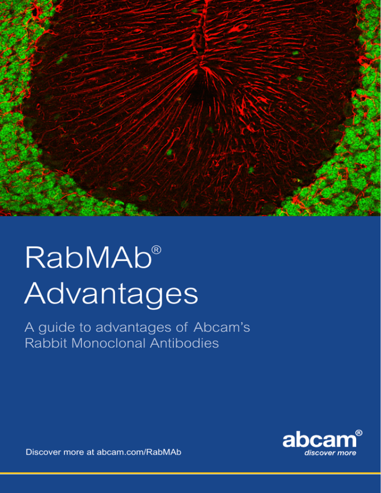
RabMAb Advantages ® A guide to advantages of Abcam’s Rabbit Monoclonal Antibodies Discover more at abcam.com/RabMAb Table of Contents About RabMAb® technology..................................................................................................3 What are the RabMAb advantages?.....................................................................................4 What do these advantages mean to you?.............................................................................4 RabMAb technology vs. Mouse MAb technology .................................................................5 RabMAb Advantages 1. Low background............................................................................................................6 2. Ideal for post-translational modification detection.........................................................7 3. Excellent for IHC usage................................................................................................8 4. High affinity...................................................................................................................9 5. High specificity............................................................................................................10 6. Diverse/novel epitope recognition...............................................................................11 7. Fully validated in multiple applications........................................................................12 8. Ideal for use on mouse samples.................................................................................13 Featured RabMAb products................................................................................................14 “The RabMAb products I received are specific to an immunogen with no crossreactivity to other peptides or proteins and possess higher affinity than mouse monoclonal antibodies.” Dr. Pankaj D. Mehta, Institute for Basic Research in Developmental Disabilities (NY) Cover: IHC-P image of NeuN (green) and GFAP (red) double staining on mouse cerebellum sections using anti-NeuN Rabbit Monoclonal (ab177487, 1/5000 DF) and anti-GFAP Chicken Polyclonal (ab4674, 1/1500 DF). The sections were deparaffinized and subjected to heat mediated antigen retrieval using citric acid. The primary antibody was detected using Goat anti-rabbit IgG conjugated to Alexa Fluor® 488 (ab150097, 1/500 DF) and Goat anti-chicken IgY conjugated to Alexa Fluor® 594 (ab150176, 1/500 DF) 2 Discover more at abcam.com/RabMAb About RabMAb® Technology Abcam RabMAb technology is a unique and proprietary method for developing monoclonal antibodies from rabbits rather than the conventional method of starting with mice. This results in delivering antibodies with the high affinity of a rabbit polyclonal and the high specificity of a monoclonal. Fig. 1 - An overview of RabMAb® development The basic principle for making antibodies using RabMAb technology is the same as for mouse monoclonals. Proprietary rabbit fusion partner cells can fuse to rabbit B-cells to create the Rabbit Hybridoma cells. Hybridomas are then screened to select for clones with specific and sensitive antigen recognition and the antibodies are characterized using a variety of methods (Fig. 1). Antibodies generated with RabMAb technology provide better antigen recognition in comparison to traditional murine antibodies (1, 2). This stems from the uniqueness of the rabbit immune system and it has been widely observed that compared to mice and other rodents, rabbits: > Mount immune responses against a broader range of compounds, including various murine proteins > Lower immune dominance towards carrier proteins > Undergo more somatic gene conversion > Possess longer and more heterogeneous CDR3 sequences for increased sequence variation The combination of these properties results in a wider range of antibodies and therefore a better chance of obtaining a functional antibody that will work in a variety of applications (3). 1. Rabbit monoclonal antibodies: a comparative study between a novel category of immunoreagents and the corresponding mouse monoclonal antibodies. Rossi S, [Am J Clin Pathol. 2005 Aug;124(2):295-302] 2. Rabbit monoclonal antibodies show higher sensitivity than mouse monoclonals for estrogen and progesterone receptor evaluation in breast cancer by immunohistochemistry. Rocha R, [Pathol Res Pract, 2008;204(9):655-62. Epub 2008 Jun 18] 3. Therapeutic Monoclonal Antibodies: From Bench to Clinic (Book) 2009 Discover more at abcam.com/RabMAb 3 What are the RabMAb® advantages? Antibodies developed using RabMAb technology provide the combined benefits of superior antigen recognition of the rabbit immune system with the specificity and consistency of a monoclonal antibody, bringing you the highest quality antibody possible. Published results from independent laboratories comparing rabbit monoclonal and mouse monoclonal antibodies have found that RabMAb primary antibodies offer increased sensitivity with similar or better specificity than comparable mouse monoclonal antibodies. Some unique benefits of RabMAb® technology: > > > > Diverse epitope recognition Improved immune response to small epitopes High specificity and affinity Improved response to mouse antigens What do these advantages mean to you? High quality antibodies for a wide range of applications The rabbit immune system generates antibody diversity and optimizes affinity by mechanisms that are more efficient than those of mice and other rodents. This increases the possibility of obtaining a functional antibody that will work in a variety of applications. RabMAb primary antibodies are even ideal for most demanding applications such as IHC on formalin-fixed, paraffin-embedded (FFPE) tissue. Novel antibodies for previously hard-to-generate targets With a RabMAb primary antibody’s diverse epitope recognition and improved immune response to non-immunogenic targets (e.g. mouse antigens, small epitopes, small compounds/peptides), highly functional antibodies to novel targets can be developed. One example is development of high quality antibodies to dual phosphorylated sites. Extensive validation In addition to the benefits of the rabbit monoclonal technology, every RabMAb primary antibody is screened and tested in multiple applications (ELISA, WB, IHC, ICC/IF, IP and FCM) and species (Human, Mouse, Rat). Extensive validation and Abcam’s standard for high quality products, equates to the highest quality antibody possible. 4 Discover more at abcam.com/RabMAb RabMAb® technology vs. traditional Mouse Monoclonal technology Comparison ANTIGEN RECOGNITION RabMAb® technology Mouse MAb technology • High chance of success with range of antigens including small molecules and peptides • Limited immuno response • Good responses to rodent proteins • Small molecules/peptides often non-immunogenic • Very limited response to rodent antigens • Recognizes several • Recognizes limited number of epitopes (due to immunodominance) AFFINITY Picomolar (10-12 KD M) possible Nanomolar (~10-9 KD M) SPECIFICITY High Med-High APPLICATIONS Western blot, ELISA, Flow Cytometry, IP, IHC, ICC (excellent results in IHC) Western blot, ELISA, Flow Cytometry, IP, not always suitable for IHC, ICC. epitopes per protein antigen CDX2 Paraffin-embedded human colonic carcinoma tissue stained with CDX2 rabbit monoclonal (ab76541) and Vendor A’s CDX2 mouse monoclonal (both at 1:1000 dilution factor) RabMAb® Primary Antibody Mouse MAb RabMAb® Primary Antibody Mouse MAb RabMAb® Primary Antibody Mouse MAb E-CADHERIN Paraffin-embedded human breast carcinoma tissue stained with E-Cadherin rabbit monoclonal (ab40772) and Vendor A’s E-Cadherin mouse monoclonal (both at 1:50 dilution factor) HER2 / ErbB2 Paraffin-embedded human breast carcinoma tissue stained with HER2 / ErbB2 rabbit monoclonal (ab134182) and Vendor A’s HER2 / ErbB2 mouse monoclonal (both at 1:500 dilution factor) Discover more at abcam.com/RabMAb 5 1. Low background As monoclonals, RabMAb primary antibodies detect a single epitope and are therefore less likely to cross-react with other proteins. At the same time we have observed that RabMAb primary antibodies bind to their target with greater affinity, enabling higher signal to-noise ratio than mouse mAbs at a given concentration of antibody. The benefit of this is that RabMAb primary antibodies typically provide more specific and sensitive detection of their target protein with low background. c-Myc IHC on FFPE human colonic adenocarcinoma using anti-c-Myc rabbit monoclonal (ab32072). Histone H3 (phospho S10) IF on HeLa using anti-Histone H3 (phospho S10) rabbit monoclonal (ab32107). 6 AMPK alpha 1 (phospho S496) IF on HeLa cells using anti-PhosphoAMPK alpha 1 (pS496) rabbit monoclonal (ab92701). ERG IHC on human prostate cancer tissue on anti-ERG rabbit monoclonal (ab92513). MCM2 WB on (1) MCF-7, (2) Ramos, (3) SW480, (4) Molt-4, (5) Jurkat, and (6) HeLa cell lysates using anti-MCM2 rabbit monoclonal (ab108935). Ki67 (Alexa Fluor® 647) Flow cytometry on HeLa cells using anti-Ki67 (Alexa Flour 647) (red) (ab194724), rabbit IgG isotype control (black), unlabelled sample (blue). Discover more at abcam.com/RabMAb 2. Ideal for post-translational modification detection The ability of rabbit generated antibodies to recognize small epitopes translates to success with recognition of post-translational modifications (e.g. phosphorylation, methylation, acetylation, sumoylation). In addition, many small compounds and peptides do not elicit a good immune response in mice but do so in rabbits. Examples of Histone H3 modification specific RabMAb primary antibodies Methylation Acetylation 1 2 1 WB on C6 cell lysates using anti-Histone H3 (acetyl K56) rabbit monoclonal (ab76307). Lane 1: untreated, lane 2: treated with TSA. Phosphorylation 2 1 WB using anti-Histone H3 (di methyl K4) rabbit monoclonal (ab32356). Lane 1: HeLa cell lysate, lane 2: recombinant Histone H3 (nonmethylated). 2 WB on NIH 3T3 cell lysates using anti-Histone H3 (phospho S28) rabbit monoclonal (ab32388). Lane 1: untreated, lane 2: treated with FBS and Calyculin A. Examples of phospho site specific RabMAb primary antibodies Phosphotyrosine (pY) 1 Phosphoserine (pS) Phosphothreonine (pT) 1 2 1 2 3 4 WB on A431 cell lysates using anti-Phospho-EGFR (Phospho Y1110) rabbit monoclonal (ab68470). Lane 1: untreated, lane 2: treated with EGF. Dual Phospho (pT/pS) WB on NIH 3T3 cell lysate using Anti-c-Jun (phospho S63) rabbit monoclonal (ab32385). Lanes 1 and 3: untreated, lanes 2 and 4: treated with UV or Anisomycin. Discover more at abcam.com/RabMAb 1 2 2 WB on 293 cell lysate using anti-eIF4EBP1 (phospho T37) rabbit monoclonal (ab75767). Lane A1 untreated, lane 2: treated with insulin. WB on 293T cell lysate using anti-Phospho-S6K (pT421/ pS424) rabbit monoclonal (ab32525). Lane 1: untreated, lane 2: treated with EGF. 7 3. Excellent for IHC usage RabMAb technology offers increased sensitivity with no loss of specificity, making antibodies ideal for demanding applications like IHC on FFPE tissues. RabMAb primary antibodies permit higher working dilutions (5 - 10X on average) and can be used with various tissue fixations, such as FFPE at minimal level of pretreatment. Additionally, when used along with a mouse monoclonal, one can perform dual staining with two monoclonal antibodies for high quality double staining on the same tissue sample. HER2 RabMAb primary antibody IHC comparison HER2 / ErbB2 RabMAb® primary antibody 3 ng / mL Rabbit Polyclonal Antibody (Vendor A) Mouse Monoclonal Antibody (Vendor B) 20 ng / mL 30 ng / mL A comparison of HER2 / ErbB2 rabbit monoclonal against leading commercially available HER2 / ErbB2 rabbit polyclonal (Vendor A) and mouse monoclonal (Vendor B) on FFPE human breast carcinoma tissue. Recommended IHC protocol and dilution factor were used for each case, the antibody concentration used listed below for each stain. Examples of double staining using RabMAb primary antibodies CK8 & Ki67 IHC on human urinary bladder cancer tissue using anti-CK8 rabbit monoclonal (ab53280) with Fast Red and anti-Ki67 Mouse MAb with DAB. 8 HER2 / ErbB2 & ER Alpha AIF & Ki67 IHC on human breast cancer tissue using anti-HER2 / ErbB2 rabbit monoclonal (ab134182) with Fast Red and anti-ER Alpha Mouse MAb with DAB. IHC on human cervical carcinoma tissue using anti-AIF rabbit monoclonal (ab32516) with Fast Red and anti-Ki67 Mouse MAb with DAB. Discover more at abcam.com/RabMAb 4. High affinity Antibody affinity is typically represented by the equilibrium dissociation constant (KD), a ratio of koff/kon, between the antibody and its antigen, where lower KD value suggests higher affinity relationship (1). While most therapeutic monoclonal antibodies have a KD value in the nanomolar range (KD =10-9 M), RabMAb primary antibodies consistently demonstrate higher affinity. With the KD values which can often reach the picomolar level (KD =10-12 M), effectively eliminating the need for further affinity maturation (2). Affinity comparison by KD value against popular therapeutic antibodies RabMAb primary antibody KD (M) ER 1.28 x 10⁻¹² ID1 2.82 x 10⁻¹² C29 9.57 x 10⁻¹¹ TNF-alpha 1.25 x 10⁻¹¹ IL-1-beta 1.99 x 10⁻¹⁰ Marketed Therapeutic MAb KD (M) Herceptin 5.0 x 10⁻⁹ Rituxan 8.0 x 10⁻⁹ Synagis 1.0 x 10⁻⁹ Remicade 2.0 x 10⁻¹⁰ Avastin 5.0 x 10⁻¹⁰ Affinity Meter: RabMAb® primary antibodies vs. Typical MAbs Antibody affinity comparison by KD value for RabMAb primary antibodies vs. typical MAbs. Measurement based on KD measurement analysis of over 850 RabMAb primary antibodies. RabMAb primary antibody displays antibody affinity level in the picomolar range compare to typical MAbs which are in the nano molar range. 1. Applications And Engineering Of Monoclonal Antibodies. David J. King, 2007. 2. Anti-peptide antibody screening: Selection of high affinity monoclonal reagents by a refined surface plasmon resonance technique. Pope ME, [J Immunol Methods. 2009 Feb 28;341(1-2):86-96. doi: 10.1016/j.jim.2008.11.004. Epub 2008 Nov 28] Discover more at abcam.com/RabMAb 9 5. High specificity The RabMAb technology is capable of delivering antibodies that are highly specific and able to distinguish between very similar proteins or sequences. While mouse mAbs can also be highly specific, subtle changes in epitopes are often not recognizable by the mouse immune system. On the contrary, the rabbit immune system is capable of recognizing subtle differences, enabling the generation of antibodies against epitopes that distinguish one protein from another. The example below demonstrates the high specificity of an anti-Progesterone RabMAb primary antibody, that does not cross-react with closely related analogues of progesterone. Identity % Reactivity Progesterone 100 17-aHydroxyprogesterone 0.901 Cortisol 0.035 Desoxycorticosterone 1.59 20a Dihydroprogesterone 0.016 20b Dihydroprogesterone 0.02 Pregnenolone 0.764 Pregnenolone-3-SO4 0.426 RabMAb technology delivers superior specificity for the generation of antibodies which recognize subtle changes in epitopes, such as those created upon protein cleavage. The specificty of various PARP RabMAb® primary antibodies A comparison of Poly (ADP-ribose) polymerase (PARP-1) specific rabbit monoclonals. Each PARP-1 rabbit monoclonals specifically recognizes specific cleavage site and with no cross-reactivity to non-specific cleavage sites. 10 Discover more at abcam.com/RabMAb 6. Diverse/novel epitope recognition The rabbit’s immune system develops affinity in ways that are different to the mouse. These differences are largely attributed to the rabbit’s lower immune dominance and larger B-cell repertoire. The benefit is a wider epitope recognition during antibody development. The comparison of rabbit and murine immune responses below shows that the rabbit antisera recognizes a wider range of epitopes than the mouse by western blot analysis. Our experience in antibody development has demonstrated that the rabbit immune system will generally yield a wider range of antibodies recognizing unique epitopes. Immune response comparison: Rabbit vs. Mouse 5 mice and 3 rabbits MOUSE 1 2 3 RABBIT 1 2 3 Rabbits recognize more epitopes 1. Molecular-weight marker 2. Fibronectin 3. Protease digested fibronectin Immunized with human fibronectin Polyclonal sera Western Analysis The comparison of rabbit and murine immune responses below shows that the rabbit antisera recognizes a wider range of epitopes than the mouse by western blot In addition, many major relevant human protein targets are highly conserved between mouse and human. Therefore, these proteins tend to be less immunogenic when using mouse or rat as a host. With its unique mechanism of immune diversification and affinity maturation, rabbit antibodies can be produced to a wider range of epitopes to enable the discovery of more novel antibody targets. Discover more at abcam.com/RabMAb 11 7. Fully validated in multiple applications Every RabMAb primary antibody is tested in multiple applications (WB, IHC, ICC/IF, IP and FCM) and multiple species (Human, Mouse and Rat) before release. For IHC, each RabMAb primary antibody is tested on mulitiple FFPE human tissue array for more accurate verification of antibody sensitivity and localization. Validation results for Caspase-3 (Pro) RabMAb® primary antibody Validation data for Caspase-3 (Pro) rabbit monoclonal (ab32150) on A) WB on HeLa cell lysate, B) IHC on FFPE human cervical carcinoma tissue, C) FCM on Jurkat cells and D) ICC on HeLa cells. “We have spent the last year buying AGR2 antibody from various companies and sometimes from the same company with different lot numbers, searching for an antibody that does not cross react with AGR3 and therefore is specific for AGR2, does not give us background when developing western blots, does not give us background in negative control when looking at IF staining in tissue, and of course works well for both western blot and IHC. We have finally found it with AGR2 RabMAb product from Abcam!” Researcher, University of Southern California 12 Discover more at abcam.com/RabMAb 8. Ideal for use on mouse samples Use of mouse mAbs on mouse tissues can be problematic and require complex protocols due to cross-reactivity and high background from anti-mouse secondary antibodies. With the rabbit as a host, the RabMAb technology can produce a monoclonal antibody to mouse targets which are less immunogenic in mouse or rat. With RabMAb primary antibodies, one can use monoclonal antibodies in mouse models without the issue of nonspecific signal due to native mouse Ig cross-reactivity. Every RabMAb primary antibody has been tested for species cross-reactivity in western blot before release, using both human and mouse samples. RabMAb® validations on mouse tissues by IHC EGFR Phospho (pY1092) IHC on FFPE mouse intestine using anti-EGFR (phospho Y1092) rabbit monoclonal (ab40815). GFAP IHC on FFPE mouse brain using antiGFAP rabbit monoclonal (ab68428). Cytokeratin 8 IHC on FFPE mouse colon using anti-Cytokeratin 8 rabbit monoclonal (ab53280). CD3 gamma IHC on FFPE mouse spleen using anti-CD3 gamma rabbit monoclonal (ab134096). Discover more at abcam.com/RabMAb MCM2 IHC on FFPE mouse intestine using antiMCM2 rabbit monoclonal (ab108935). p120 Catenin IHC on FFPE mouse large intestine using anti-p120 Catenin rabbit monoclonal (ab92514). 13 Featured RabMAb® Products NeuN antibody [EPR12763] (ab177487) WB using anti-NeuN rabbit monoclonal on Lane 1: Human fetal brain, Lane 2: Mouse brain, Lane 3: Rat brain. . IHC-P using anti-NeuN rabbit monoclonal on mouse cerebellum tissue. Reactivity: Hu, Ms, Rt, Sh, Gt, Ct, Dg, Zf, Mtt Applications: WB, ICC/ IF, IHCFoFr, IHC-Fr, IHC-P Amount: 100 µl ATM (phospho S1981) antibody [EP1890Y] (ab81292) WB using anti-ATM (phospho S1981) rabbit monoclonal on HEK293 cell lysates. Lane 1: untreated, lane 2: doxorubicin treated. IHC using anti-ATM (phospho S1981) rabbit monoclonal on FFPE human endometrial carcinoma tissue. Reactivity: Hu Applications: WB, IHC-P, ICC/IF, IP Amount: 100 µl beta Catenin antibody [E247] (ab32572) WB using anti-beta Catenin rabbit monoclonal on whole cell lysate of U2OS cells. 14 IHC using anti-beta Catenin rabbit monoclonal on FFPE human cervical carcinoma tissue. Reactivity: Hu, Ms, Rt, Hm, Mk Applications: WB, IHC-F, IHC-P, ICC/IF, IP Amount: 100 µl Discover more at abcam.com/RabMAb c-Myc antibody [Y69] (ab32072) IF using anti-c-Myc rabbit monoclonal on HeLa cells. IHC using anti-c-Myc rabbit monoclonal on FFPE Human colonic adenocarcinoma tissue. Reactivity: Hu, Ms, Rt Applications: WB, IHC-P, ICC/IF, IP Amount: 100 µl Reactivity: Hu, Ms Applications: WB, IHC-F, IHC-P, ICC/IF Amount: 100 µl Reactivity: Hu, Ms, Rt Applications: WB, IHC-F, IHC-P, ICC/IF, IP, FCM Amount: 100 µl EGFR (phospho Y1092) antibody [EP774Y] (ab40815) WB using anti-EGFR (phospho Y1092) rabbit monoclonal on A431 cell lysates. Lane 1: EGF untreated, lane 2: EGF treated. IHC using anti-EGFR (phospho Y1092) rabbit monoclonal on FFPE human cervical carcinoma tissue. alpha Tubulin antibody [EP1332Y] (ab52866) WB using anti-alpha Tubulin rabbit monoclonal on HeLa cell lysate at 10 µg. IF using anti-alpha Tubulin rabbit monoclonal on human embryonic kidney cells. Discover more at abcam.com/RabMAb 15 Alexa Fluor® is a registered trademark of Life Technologies. Alexa Fluor® dye conjugates contain(s) technology licensed to Abcam by Life Technologies RabMAb® is a registered trademark of Abcam. Discover more at abcam.com/RabMAb Copyright © 2015 Abcam, All rights reserved. E-322
