The Effects of pH and Osmolarity Conditions on the Type... fim ABSTRACT
advertisement
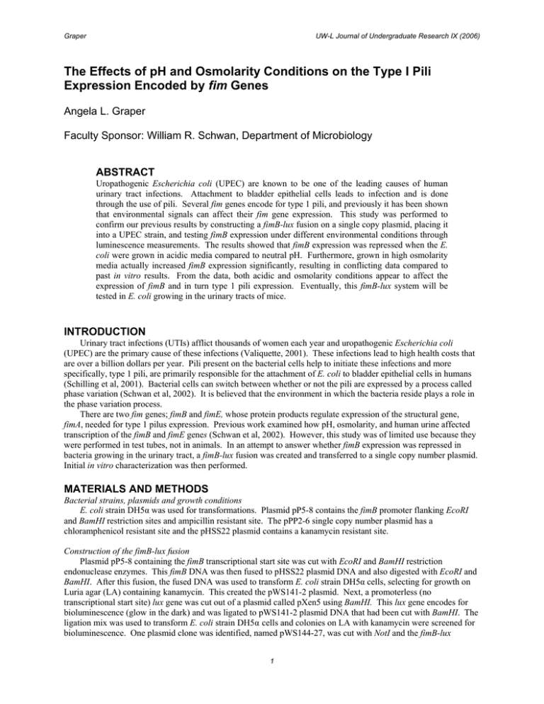
Graper UW-L Journal of Undergraduate Research IX (2006) The Effects of pH and Osmolarity Conditions on the Type I Pili Expression Encoded by fim Genes Angela L. Graper Faculty Sponsor: William R. Schwan, Department of Microbiology ABSTRACT Uropathogenic Escherichia coli (UPEC) are known to be one of the leading causes of human urinary tract infections. Attachment to bladder epithelial cells leads to infection and is done through the use of pili. Several fim genes encode for type 1 pili, and previously it has been shown that environmental signals can affect their fim gene expression. This study was performed to confirm our previous results by constructing a fimB-lux fusion on a single copy plasmid, placing it into a UPEC strain, and testing fimB expression under different environmental conditions through luminescence measurements. The results showed that fimB expression was repressed when the E. coli were grown in acidic media compared to neutral pH. Furthermore, grown in high osmolarity media actually increased fimB expression significantly, resulting in conflicting data compared to past in vitro results. From the data, both acidic and osmolarity conditions appear to affect the expression of fimB and in turn type 1 pili expression. Eventually, this fimB-lux system will be tested in E. coli growing in the urinary tracts of mice. INTRODUCTION Urinary tract infections (UTIs) afflict thousands of women each year and uropathogenic Escherichia coli (UPEC) are the primary cause of these infections (Valiquette, 2001). These infections lead to high health costs that are over a billion dollars per year. Pili present on the bacterial cells help to initiate these infections and more specifically, type 1 pili, are primarily responsible for the attachment of E. coli to bladder epithelial cells in humans (Schilling et al, 2001). Bacterial cells can switch between whether or not the pili are expressed by a process called phase variation (Schwan et al, 2002). It is believed that the environment in which the bacteria reside plays a role in the phase variation process. There are two fim genes; fimB and fimE, whose protein products regulate expression of the structural gene, fimA, needed for type 1 pilus expression. Previous work examined how pH, osmolarity, and human urine affected transcription of the fimB and fimE genes (Schwan et al, 2002). However, this study was of limited use because they were performed in test tubes, not in animals. In an attempt to answer whether fimB expression was repressed in bacteria growing in the urinary tract, a fimB-lux fusion was created and transferred to a single copy number plasmid. Initial in vitro characterization was then performed. MATERIALS AND METHODS Bacterial strains, plasmids and growth conditions E. coli strain DH5α was used for transformations. Plasmid pP5-8 contains the fimB promoter flanking EcoRI and BamHI restriction sites and ampicillin resistant site. The pPP2-6 single copy number plasmid has a chloramphenicol resistant site and the pHSS22 plasmid contains a kanamycin resistant site. Construction of the fimB-lux fusion Plasmid pP5-8 containing the fimB transcriptional start site was cut with EcoRI and BamHI restriction endonuclease enzymes. This fimB DNA was then fused to pHSS22 plasmid DNA and also digested with EcoRI and BamHI. After this fusion, the fused DNA was used to transform E. coli strain DH5α cells, selecting for growth on Luria agar (LA) containing kanamycin. This created the pWS141-2 plasmid. Next, a promoterless (no transcriptional start site) lux gene was cut out of a plasmid called pXen5 using BamHI. This lux gene encodes for bioluminescence (glow in the dark) and was ligated to pWS141-2 plasmid DNA that had been cut with BamHI. The ligation mix was used to transform E. coli strain DH5α cells and colonies on LA with kanamycin were screened for bioluminescence. One plasmid clone was identified, named pWS144-27, was cut with NotI and the fimB-lux 1 Graper UW-L Journal of Undergraduate Research IX (2006) containing piece of DNA was ligated to NotI digested pPP2-6. The ligated DNA was used to transform DH5α cells, selecting for colonies on LA that were bioluminescent and resistant to chloramphenicol. One clone resulted, named pWS145-5, which was used for in vitro characterization. In vitro bioluminescence assays The E. coli strain with the pWS145-5 plasmid, containing a fimB-lux fusion, was grown in LB media with variations of pH and osmolarity. Luminescence assays were then performed to measure the effects of the growth environment on the fimB gene when the bacteria were propagated in a test tube. Strains DH5α/pWS145-5 and DH5α/pWS145-38 were grown in LB with a pH of 5.5 with or without 400 mM NaCl (high salt) compared to a neutral pH with or without 400 mM NaCl. The results were tabulated by dividing the RLU relative luminescence units by the OD600 reading of the culture. RESULTS Examination of the fimB-lux fusion at different pHs and osmolarities. In order to determine whether the pH affected the transcription of the fimB gene, a fusion of the fimB promoter with the lux gene was generated. The fimB-lux fusion was effectively cloned into a single-copy number plasmid, resulting in pWS145-5 and pWS145-38 and then transformed into E. coli strain DH5α. Luminescence measurements were taken when the cells were in mid-log phase, which was determined by optical density. Higher luminescence activity was observed when cells were grown in media of pH 7.0 for both single-copy number plasmids compared to pH 5.5 (FIG. 1). When both strains were grown in media of pH 7.0 with the addition of 400 mM NaCl, expression of the fimB gene was higher with 400 mM NaCl compared to the assays noted above. Furthermore, an examination of growth media at pH 5.5 with 400 mM NaCl also showed higher levels of expression as compared to the no salt tester. 16000 14000 Luminescene (RLU/s) 12000 10000 8000 6000 4000 2000 0 7.0 7.0+ 5.5 5.5+ Growth Conditions FIG. 1. Luminescence assays of strains DH5α/pWS145-5 (left bar) and DH5α/pWS145-38 (right bar) cells grown in LB at pH 5.5 or 7.0 ± 400 mM NaCl with means and ± standard deviations shown. Each data point represents at least three separate runs. 2 Graper UW-L Journal of Undergraduate Research IX (2006) DISCUSSION The effects of pH and osmolarity on the expression of the fimB gene were studied through the use of the lux gene by measuring luminescence. Transcription of the fimB gene tested was repressed under acidic conditions but actually increased under osmolarity conditions with NaCl acting as the osmolyte. Previous work done by Schwan et al. (2) had shown that both acidic conditions as well as osmolarity conditions repressed the fimB gene when using βgalactosidase to measure the activity. The lux gene may be affected differently under osmolarity conditions resulting in the conflicting results. The lux gene may need to be studied on its own, in order to determine if it is affected differently than the fimB gene normally would by itself, to obtain accurate results when used in vivo studies. With conflicting results from the study done with β-galactosidase, the actual effect of osmolarity conditions on the fimB gene would have to be determined prior to in vivo studies for correct results. ACKNOWLEDGEMENTS This work was supported in part by an undergraduate research grant from the University of Wisconsin-La Crosse to A.L.G., a NIH Area grant to W.R.S. and Xenogen Corp. for the pXen5 plasmid. REFERENCES Schilling, J. D., M. A. Mulvey, S. J. Hultgren. 2001. Structure and function of Escherichia coli type 1 pili: new insight into the pathogenesis of urinary tract infections. J Infect. Disease. 183(Suppl. 1):36-40. Schwan, W .R., J. L. Lee, F. A. Lenard, B. R. Matthews, and M. T. Beck. 2002. Osmolarity and pH growth conditions regulate fim gene transcription and type 1 pilus expression in uropathogenic Escherichia coli. Infect. Immun. 70:1391-1402. Valiquette, L. 2001. Urinary tract infections in women. Can J Urol. 8(Suppl. 1):6-12. 3
