The Effectiveness of Recombinant Fibroblast Human Cells Timothy A. Bloomquist
advertisement
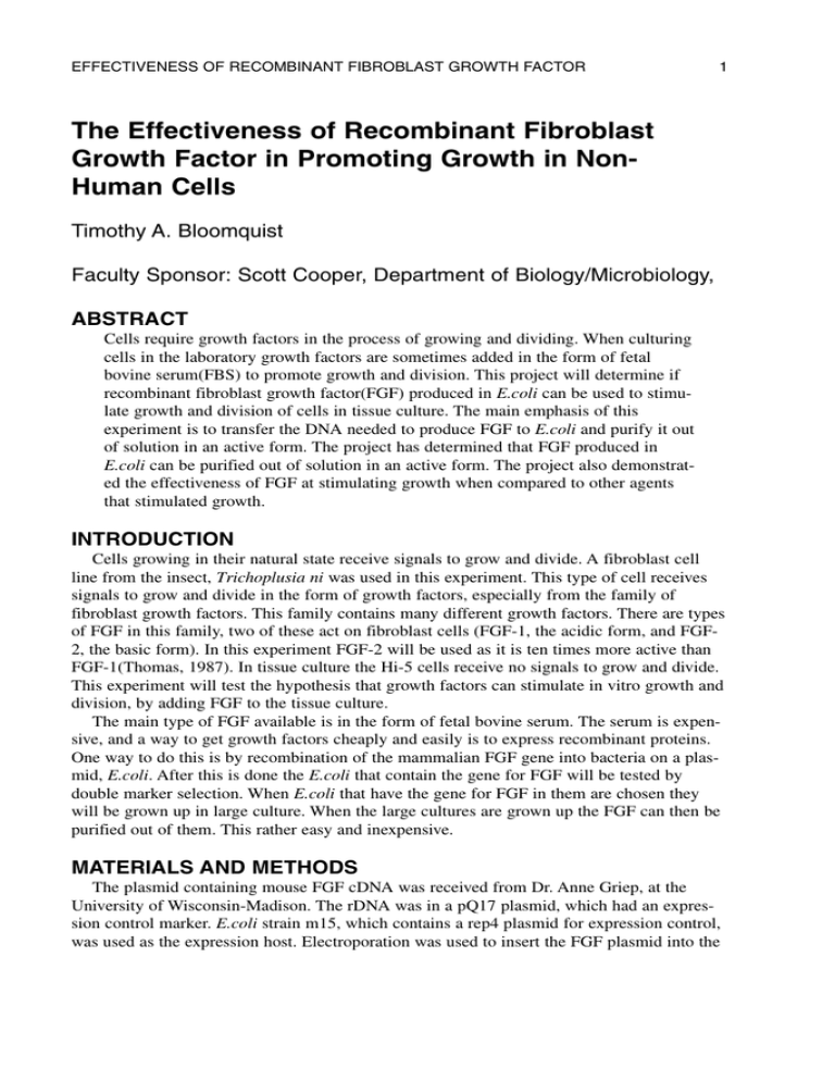
EFFECTIVENESS OF RECOMBINANT FIBROBLAST GROWTH FACTOR 1 The Effectiveness of Recombinant Fibroblast Growth Factor in Promoting Growth in NonHuman Cells Timothy A. Bloomquist Faculty Sponsor: Scott Cooper, Department of Biology/Microbiology, ABSTRACT Cells require growth factors in the process of growing and dividing. When culturing cells in the laboratory growth factors are sometimes added in the form of fetal bovine serum(FBS) to promote growth and division. This project will determine if recombinant fibroblast growth factor(FGF) produced in E.coli can be used to stimulate growth and division of cells in tissue culture. The main emphasis of this experiment is to transfer the DNA needed to produce FGF to E.coli and purify it out of solution in an active form. The project has determined that FGF produced in E.coli can be purified out of solution in an active form. The project also demonstrated the effectiveness of FGF at stimulating growth when compared to other agents that stimulated growth. INTRODUCTION Cells growing in their natural state receive signals to grow and divide. A fibroblast cell line from the insect, Trichoplusia ni was used in this experiment. This type of cell receives signals to grow and divide in the form of growth factors, especially from the family of fibroblast growth factors. This family contains many different growth factors. There are types of FGF in this family, two of these act on fibroblast cells (FGF-1, the acidic form, and FGF2, the basic form). In this experiment FGF-2 will be used as it is ten times more active than FGF-1(Thomas, 1987). In tissue culture the Hi-5 cells receive no signals to grow and divide. This experiment will test the hypothesis that growth factors can stimulate in vitro growth and division, by adding FGF to the tissue culture. The main type of FGF available is in the form of fetal bovine serum. The serum is expensive, and a way to get growth factors cheaply and easily is to express recombinant proteins. One way to do this is by recombination of the mammalian FGF gene into bacteria on a plasmid, E.coli. After this is done the E.coli that contain the gene for FGF will be tested by double marker selection. When E.coli that have the gene for FGF in them are chosen they will be grown up in large culture. When the large cultures are grown up the FGF can then be purified out of them. This rather easy and inexpensive. MATERIALS AND METHODS The plasmid containing mouse FGF cDNA was received from Dr. Anne Griep, at the University of Wisconsin-Madison. The rDNA was in a pQ17 plasmid, which had an expression control marker. E.coli strain m15, which contains a rep4 plasmid for expression control, was used as the expression host. Electroporation was used to insert the FGF plasmid into the 2 BLOOMQUIST E.coli. The recombinant E.coli were grown on LB plates that contained ampicillin and kanamycin. Expression of FGF was preformed by growing up individual overnight cultures of the recombinant E.coli in LB broth containing ampicillin and kanamycin. These cultures were then used to inoculate a 500mL culture. The 500mL culture was grown to an OD of 0.6. At this time ITPG was added to the culture for increased expression of FGF. This was then allowed to grow to an OD of 1. All the media from the culture was then removed by centrifugation and a lysis buffer was added. The E.coli were then lysed with four short bursts of sonication. The cellular material was then centrifuged to remove extraneous protein. The solution was then applied to a DEAE-cellulous column. The elutant was then applied to a Heparin affinity column. To elute the FGF off the Heparin affinity column two washes of high concentration salt was used, a 0.5M NaCl wash and a 2M NaCl elution. The FGF was eluted with the 2M NaCl buffer. A dialysis was done to remove the salt using PBS buffer. The activity of FGF was tested by adding it to the tissue culture media of Hi5 insect fibroblast cells. The activity of FGF was compared to the activity of FBS. The tissue culture was set up with a control group which received no FGF or FBS, two FGF groups , and a FBS group. RESULTS The tissue cultures were allowed to grow to confluence. Then at this time the total cell number and total cellular protein was determined. The cell counts were determined by counting and the total protein was determined by Bradford assay. The total cell counts for each of the groups is listed in Figure 1. The total protein concentrations of each of the groups is listed in Figure 2. Cell Number Average Cell Counts Control FBS Test Groups Figure 1 Average Cell Counts FGF EFFECTIVENESS OF RECOMBINANT FIBROBLAST GROWTH FACTOR 3 Cellular Protein (mg/mL) Average Total Cellullar Protein Control FBS Test Groups FGF Figure 2 Average Total Cellular Protein DISCUSSION That the E.coli grew in the presence of both ampicillin and kanamycin showed that a successful electroporation had taken place and that these E.coli now contained the rFGF plasmid. The cell counts show that an active FGF protein was produced by the E.coli. This was determined when the cell counts of the control group were compared to the FGF groups. These showed an increased number of cells had grown in the same amount of time. This proved that FGF can stimulate growth in Hi5 cells. The cell counts of the FGF groups were then compared to the FBS group. When this had been done the cell counts were relatively similar. Showing the FGF had stimulated the effects as FBS. This shows that the FGF works just as well as FBS at stimulating cell growth. When the total cellular protein was determined by Bradford assay the same conclusions were reached. Overall producing FGF in E.coli is comparable to commercial FBS. For the same desired effects are found. Now with the recombinant E.coli on hand one can produce adequate amounts of FGF. REFERENCES QIAGEN (1997). The QIAexpressionist Third Edition. Klagsbrun, M., Sasse, J., Sullivan, R. & Smith, J.A. Proc. Natl. Acad. Sci. USA. 83:22482452; 1986 Thomas, K.A. Fibroblast growth factors. FASEB J. 1:434-440; 1987 Vlodavsky, I., Folkman, J., Sullivan, R., Fridman, R., Ishai-Michaeli, R., Sasse, J. & Klagsbrun, M. Proc. Natl. Acad. Sci. USA. 84:2292-2296; 1987 4
