Label Free Mid-IR Photothermal Imaging of Bird Brain
advertisement
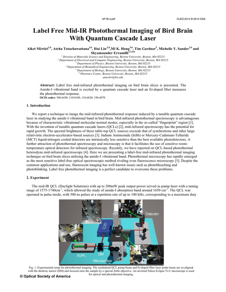
AF1B.4.pdf CLEO:2014 © 2014 OSA Label Free Mid-IR Photothermal Imaging of Bird Brain With Quantum Cascade Laser Alket Mërtiri1,6, Atcha Totachawattana2,6, Hui Liu,2,6,Mi K. Hong3,6, Tim Gardner5, Michelle Y. Sander2,6 and Shyamsunder Erramilli1,3,4,6 2 1 Division of Materials Science and Engineering, Boston University, Boston, MA 02215 Department of Electrical and Computer Engineering, Boston University, Boston, MA 02215 3 Department of Physics, Boston University, Boston, MA 02215 4 Department of Biomedical Engineering, Boston University, Boston, MA 02215 5 Department of Biology, Boston University, Boston, MA 02215 6 Photonics Center, Boston University, Boston, MA 02215 amertiri@bu.edu Abstract: Label free mid-infrared photothermal imaging on bird brain slices is presented. The Amide-I vibrational band is excited by a quantum cascade laser and an Er:doped fiber measures the photothermal response. OCIS codes: 300.6430, 110.0180, 110.6820, 190.4870 1. Introduction We report a technique to image the mid-infrared photothermal response induced by a tunable quantum cascade laser in studying the amide-I vibrational band in bird brain. Mid-infrared photothermal spectroscopy is advantageous because of characteristic vibrational molecular normal modes, especially in the so-called “fingerprint” region [1]. With the invention of tunable quantum cascade lasers (QCLs) [2], mid-infrared spectroscopy has the potential for rapid growth. The spectral brightness of these table-top QCL sources exceeds that of synchrotrons and other large relativistic electron-accelerator-based sources [3]. Indium Antimonide (InSb) or Mercury-Cadmium-Telluride (MCT) liquid-nitrogen cooled detectors are intrinsically less sensitive than the best available photodetectors. A further attraction of photothermal spectroscopy and microscopy is that it facilitates the use of sensitive roomtemperature optical detectors for infrared spectroscopy. Recently, we have reported on QCL-based photothermal heterodyne mid-infrared spectroscopy [4]. Here we are presenting a label-free mid-infrared photothermal imaging technique on bird brain slices utilizing the amide-I vibrational band. Photothermal microscopy has rapidly emerged as the most sensitive label-free optical spectroscopic method rivaling even fluorescence microscopy [5]. Despite the common applications and use, fluorescent imaging has well known issues such as photobleaching and photoblinking. Label free photothermal imaging is a perfect candidate to overcome these problems. 2. Experiment The mid-IR QCL (Daylight Solutions) with up to 200mW peak output power served as pump laser with a tuning range of 1575-1740cm-1, which allowed the study of amide-I absorption band around 1650 cm-1. The QCL was operated in pulse mode, with 500 ns pulses at a repetition rate of up to 100 kHz, corresponding to a maximum duty Fig. 1. Experimental setup for photothermal imaging. The modulated QCL pump beam and Er:doped fiber laser probe beam are co-aligned with the dichroic mirror (DM) and focused onto the sample by a special ZnSe objective. An inverted Nikon Eclipse Ti-U microscope is used for optical and photothermal imaging. © Optical Society of America AF1B.4.pdf CLEO:2014 © 2014 OSA Fig. 2. a) Illustration of the pump-probe setup for photothermal imaging of bird brain slices on CaF2 window. b) FTIR absorbance spectrum showing the amide-I and amide-II band. c) Optical image of the bird brain slice and d) is a photothermal image. cycle of 5%. Compared to fluorescence, where molecules can eventually be photobleached, the conversion of photon energy into heat is fast, occurring within a few picoseconds. The molecule can be repeatedly excited after that time scale. Our probe laser is an Er:doped fiber laser with 10mW of power at telecommunication wavelengths, which is chosen outside any vibrational band. The pump and probe beams are collinearly combined using a dichroic mirror (DM) and focused coaxially onto the sample by a zinc-selenide (ZnSe) focusing objective (NA=0.25). Losses at the beamsplitters and in the ZnSe objective lens resulted in an estimated QCL pump beam incident intensity on the sample of up to a maximum of ~ 1.2 ×10⁴ W/cm2. The transmitted probe beam is then collected by a lens and focused onto an InGaAs photodetector for monitoring the photothermal signal. To enhance sensitivity, the probe signal is detected using a conventional photodetector using a lock-in amplifier, phase locked to the modulation frequency of the pump beam. 3. Results Label-free photothermal image of the bird brain slice is shown in figure 2 and compared to the optical image. The samples were sliced thin and placed on top of a CaF2 window, which is transparent in the mid-infrared range. An illustration of the pump-probe Gaussian beams focused onto the sample is shown in figure 2a. The FTIR shows the absorbance peaks of both the amide-I and amide-II bands. Our pump laser is able to excite the amide-I band, which is used as our contrast feature in acquiring the photothermal image. Optical and photothermal images are compared and remarkable similarities are demonstrated. The photothermal image was taken in a raster-scan approach across the sample with 20µm steps. The pump laser was maintained at the peak of amide-I vibrational band and the probe was operated in cw-mode. The photothermal images are created as an interaction of the probe laser with the pump induced refractive index changes hence overcoming Abbe’s limitation on imaging structures less than a wavelength. Our results show the capability for sensitive label-free images that have the potential to achieve resolution below the diffraction limit [6]. References [1] P. R. Griffiths and J. A. de Haseth, Fourier Transform Infrared Spectrometry, 2nd ed. (Wiley, New York, 2007). [2] J. Faist, F. Capasso, D. L. Sivco, C. Sirtori, A. L. Hutchinson, and A. Y. Cho, “Quantum cascade laser,” Science 264, 553 (1994). [3] G. L. Carr, P. Dumas, C. J. Hirschmug, and G. P. Williams, “Infrared synchrotron radiation programs at the National Synchrotron Light Source,” Nuovo Cim. D 20, 375 (1998). [4] A. Mertiri, T. Jeys, V. Liberman, M. K. Hong, J. Mertz, H. Altug, and S. Erramilli, “Mid-infrared photothermal heterodyne spectroscopy in a liquid crystal using a quantum cascade laser,” Appl. Phys. Lett. 101, 44101 (2012). [5] S. E. Bialkowski, Photothermal Spectroscopy Methods for Chemical Analysis (Wiley, New York, 1996). [6] V. P. Zharov, “Far-field photothermal microscopy beyond the diffraction limit,” Opt. Lett. 28, 1314 (2003)
