THE SPECIAL SENSES , vision, hearing and equilibrium.
advertisement
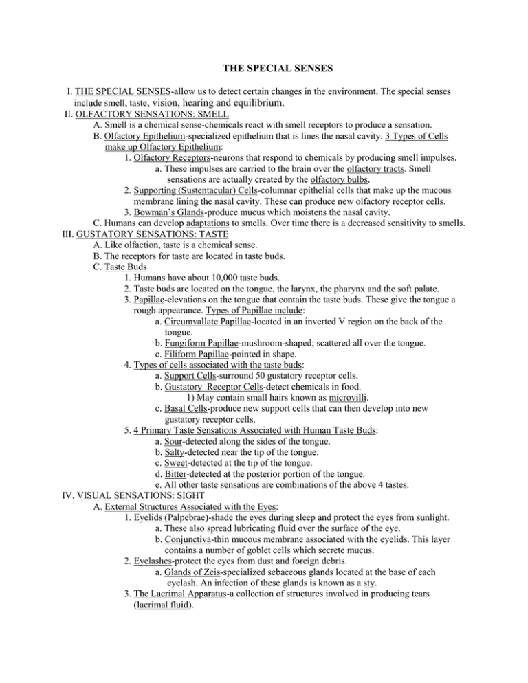
THE SPECIAL SENSES I. THE SPECIAL SENSES-allow us to detect certain changes in the environment. The special senses include smell, taste, vision, hearing and equilibrium. II. OLFACTORY SENSATIONS: SMELL A. Smell is a chemical sense-chemicals react with smell receptors to produce a sensation. B. Olfactory Epithelium-specialized epithelium that is lines the nasal cavity. 3 Types of Cells make up Olfactory Epithelium: 1. Olfactory Receptors-neurons that respond to chemicals by producing smell impulses. a. These impulses are carried to the brain over the olfactory tracts. Smell sensations are actually created by the olfactory bulbs. 2. Supporting (Sustentacular) Cells-columnar epithelial cells that make up the mucous membrane lining the nasal cavity. These can produce new olfactory receptor cells. 3. Bowman’s Glands-produce mucus which moistens the nasal cavity. C. Humans can develop adaptations to smells. Over time there is a decreased sensitivity to smells. III. GUSTATORY SENSATIONS: TASTE A. Like olfaction, taste is a chemical sense. B. The receptors for taste are located in taste buds. C. Taste Buds 1. Humans have about 10,000 taste buds. 2. Taste buds are located on the tongue, the larynx, the pharynx and the soft palate. 3. Papillae-elevations on the tongue that contain the taste buds. These give the tongue a rough appearance. Types of Papillae include: a. Circumvallate Papillae-located in an inverted V region on the back of the tongue. b. Fungiform Papillae-mushroom-shaped; scattered all over the tongue. c. Filiform Papillae-pointed in shape. 4. Types of cells associated with the taste buds: a. Support Cells-surround 50 gustatory receptor cells. b. Gustatory Receptor Cells-detect chemicals in food. 1) May contain small hairs known as microvilli. c. Basal Cells-produce new support cells that can then develop into new gustatory receptor cells. 5. 4 Primary Taste Sensations Associated with Human Taste Buds: a. Sour-detected along the sides of the tongue. b. Salty-detected near the tip of the tongue. c. Sweet-detected at the tip of the tongue. d. Bitter-detected at the posterior portion of the tongue. e. All other taste sensations are combinations of the above 4 tastes. IV. VISUAL SENSATIONS: SIGHT A. External Structures Associated with the Eyes: 1. Eyelids (Palpebrae)-shade the eyes during sleep and protect the eyes from sunlight. a. These also spread lubricating fluid over the surface of the eye. b. Conjunctiva-thin mucous membrane associated with the eyelids. This layer contains a number of goblet cells which secrete mucus. 2. Eyelashes-protect the eyes from dust and foreign debris. a. Glands of Zeis-specialized sebaceous glands located at the base of each eyelash. An infection of these glands is known as a sty. 3. The Lacrimal Apparatus-a collection of structures involved in producing tears (lacrimal fluid). a. Lacrimal Glands-secrete lacrimal fluid through the lacrimal ducts onto the surface of the eyeball. b. Lacrimal fluid is composed of: water, salts, mucus and lysozyme (an enzyme that kills bacteria). 1) This fluid acts to lubricate, moisten and protect they eyeball. B. External Parts of the Eyeball 1. The Cornea-nonvascular covering that surrounds the iris (colored portion of the eye) of the eye. The cornea is involved in focusing light on the lens. a. Corneal Transplants 2. The Sclera-the white portion of the eye. 3. The Pupil-a hole in the center of the iris. Light passes into the eye through the pupil. a. Muscles can adjust the size of the pupil. C. Internal Parts of the Eyeball 1. The Choroid-lines the internal surface of the sclera. This layer provides nutrients to the posterior surface of the retina. 2. The Ciliary Body which consists of: a. The Ciliary Processes-which secrete a fluid known as aqueous humor. b. The Ciliary Muscle-smooth muscle that alters the shape of the lens for near or far vision. 3. The Retina-the inner coat of the eyeball a. This layer lines the posterior portion of the eyeball and it is highly vascular. b. Parts Associated with the Retina: 1) The Optic Disc-the site where the optic nerve exits the eyeball. 2) Rods-photoreceptors that detect black and white light. These allow us to see shape and movement. 3) Cones-allow for color vision. 4) The Macula lutea-the center of the posterior portion of the retina. 5) The Fovea Centralis-a small depression in the center of the macula lutea that contains only cones. This region provides resolution to our vision. 6) The Blind Spot-this is also another name for the optic disc. This portion of the retina lacks rods and cones. c. Detached Retina-usually caused by some type of trauma. Can lead to blindness. 4. The Lens-located posterior to the pupil and iris. It is primarily protein in composition. a. It is held in place by the suspensory ligaments and it is involved in focusing the object that one is observing. b. Cataracts-a loss of transparency of the lens. Can by caused by aging, trauma, exposure to UV light, certain medications. Can be repaired via surgery. 5. Chambers within the eyeball: a. The Anterior Cavity-space anterior to the lens. This area is filled with a watery fluid known as aqueous humor. b. The Posterior Cavity-space posterior to the lens. This cavity is filled with a liquid known as vitreous humor. 1) Intraocular pressure-pressure within the eyeball. This is usually caused by the aqueous humor and vitreous humor 2) Glaucoma-excessively high intraocular pressure in the eyeball. It can cause degeneration of the retina which may lead to blindness. This can be repaired via surgery. V. AUDITORY SENSATIONS AND EQUILIBRIUM A. The ear plays a major role in providing auditory sensations and in allowing us to maintain our balance. B. External Parts of the Ear 1. The Auricle (Pinna)-composed of elastic cartilage, forms the outer ear itself. The auricle is composed of the helix and the lobule. 2. The External Auditory Meatus (Canal)-tube that extends from the auricle to the tympanum (eardrum). This meatus runs through the temporal bone. a. Ceruminous glands-line the external auditory meatus. These are the wax glands that secrete cerumen (earwax). This earwax prevents dust and debris from collecting in the external auditory meatus. C. Internal Parts of the Ear 1. The Tympanic Membrane (Eardrum)-a thin layer of tissue between the external auditory meatus and the inner ear. This structure helps to move sound waves deep into the ear. a. Perforated Eardrum-hole in the tympanic membrane. It is characterized by extreme pain. Is usually caused by trauma. 2. The Eustachian or Auditory Tube-connects the middle ear with the nasopharynx. This tube opens during swallowing and yawning. This tube helps to regulate pressure in the middle ear. 3. The Auditory Ossicles-3 bones within the middle ear. These are held in place by ligaments. a. They are all involved in moving sound waves in to the inner ear. b. The Malleus, Incus and Stapes are the three auditory ossicles. 4. The Semicircular Canals-3 of these, they contain otoliths which are involved in providing us with our balance and equilibrium. 5. The Cochlea-resembles a snail’s shell. This structure contains the Spiral Organ which houses receptors that receive sound waves. This is the organ of hearing. a. Cochlear Implants VI. DISORDERS/MEDICAL TERM TO KNOW: A. Otitis media-infection of the inner ear, usually caused by bacteria. B. Conjunctivitis-inflammation of the conjunctiva, also known as Pinkeye. C. Otalgia-earache. D. Tinnitus-a ringing in the ears. E. Vertigo-sensation of spinning. F. Macular degeneration-progressive degeneration of the retina and the macula lutea. This is the main cause of blindness in those over the age of 65. Currently there is no cure for this illness. G. Exopthalmos-bulging eyeballs. Is a common sign of hyperthyroidism.
