Chapter 1 Steroid Chemistry and Steroid Hormone Action
advertisement
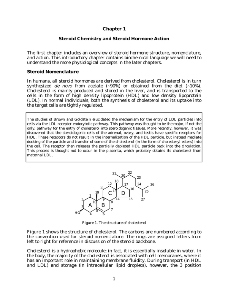
Chapter 1 Steroid Chemistry and Steroid Hormone Action The first chapter includes an overview of steroid hormone structure, nomenclature, and action. This introductory chapter contains biochemical language we will need to understand the more physiological concepts in the later chapters. Steroid Nomenclature In humans, all steroid hormones are derived from cholesterol. Cholesterol is in turn synthesized de novo from acetate (~90%) or obtained from the diet (~10%). Cholesterol is mainly produced and stored in the liver, and is transported to the cells in the form of high density lipoprotein (HDL) and low density lipoprotein (LDL). In normal individuals, both the synthesis of cholesterol and its uptake into the target cells are tightly regulated. The studies of Brown and Goldstein elucidated the mechanism for the entry of LDL particles into cells via the LDL receptor endocytotic pathway. This pathway was thought to be the major, if not the only, pathway for the entry of cholesterol into steroidogenic tissues. More recently, however, it was discovered that the steroidogenic cells of the adrenal, ovary, and testis have specific receptors for HDL. These receptors do not result in the internalization of the HDL particle, but instead mediate docking of the particle and transfer of some of the cholesterol (in the form of cholesteryl esters) into the cell. The receptor then releases the partially depleted HDL particle back into the circulation. This process is thought not to occur in the placenta, which probably obtains its cholesterol from maternal LDL. 21 18 12 11 1 2 3 HO A 4 C 19 10 5 9 B 13 8 14 20 22 23 24 17 D 16 15 26 25 27 7 6 Figure 1. The structure of cholesterol Figure 1 shows the structure of cholesterol. The carbons are numbered according to the convention used for steroid nomenclature. The rings are assigned letters from left to right for reference in discussion of the steroid backbone. Cholesterol is a hydrophobic molecule; in fact, it is essentially insoluble in water. In the body, the majority of the cholesterol is associated with cell membranes, where it has an important role in maintaining membrane fluidity. During transport (in HDL and LDL) and storage (in intracellular lipid droplets), however, the 3 position 1 Endocrine -- Dr. Brandt Chapter 1. Steroid Chemistry and Steroid Hormone Action hydroxyl is modified to increase the hydrophobicity of the molecule still further, by esterification with a fatty acid (Figure 2). O O Figure 2. A cholesteryl ester. As we will see in greater detail in the next chapter, all steroid hormones are derived from cholesterol. The production of the hormones involves a number of precise modifications to the cholesterol structure, with different series of modifications occurring in different pathways. These modifications include attack at the 11, 16, 17, 18, 19, 20, and 21 positions, conversion of the 3-hydroxyl to a ketone, and isomerization of the 5-6 double bond to the 4-5 position (Figure 3). A number of additional modifications, not shown here, occur during conversion of the steroid hormones to inactive metabolites. Each set of alterations to the steroid backbone alters the affinity of the steroid for a given steroid receptor. 21 20 18 11 19 17 16 3 O HO 5 4 6 ∆5 Figure 3. Locations of attacks on the cholesterol backbone during conversion to steroid hormones. In organic chemistry, sites of unsaturation (i.e. double bonds) are often referred to using the Greek letter ∆. Thus, steroids containing the 5-6 double bond, such as cholesterol, are designated ∆5 steroids; those with a 4-5 double bond are called ∆4 steroids. As we will see in Chapter 9, Vitamin D 3 and its metabolites are also derived from cholesterol. The numbering used for the metabolites of Vitamin D is the same as that used for all of the other cholesterol metabolites; however, some aspects of the nomenclature can be somewhat confusing because during the synthesis of Vitamin D3 the 9-10 bond of the B-ring is cleaved and the A-ring is flipped around the 6-7 bond. The active derivative of Vitamin D3 , 1α,25-dihydroxy-Vitamin D 3 , is produced 2 Endocrine -- Dr. Brandt Chapter 1. Steroid Chemistry and Steroid Hormone Action by successive hydroxylations at the 25 and 1 positions. Figures 1-3 are representations of the steroid backbone that ignore most of the stereochemistry. Figure 4 shows cholesterol with a little more attention to the stereochemical details. For our purposes, this representation contains more detail than we are usually going to need, but it does illustrate a few important points. The 3-hydroxyl, the 18- and 19-methyl groups and the side-chain attached to the 17carbon all project above the plane of the fused ring system. Substituents located above the ring plane are denoted β. Substituents located below the ring plane (such as the hydrogens shown in Figure 4 at the 3 and 17 positions) are denoted α. The difference between α and β is often the difference between an active and an inactive compound. CH3 CH3 β HO α H H Figure 4. Stereochemical projection of cholesterol. Note that most of the hydrogens are omitted for clarity. The names we will use for the biologically relevant steroids imply the correct stereochemistry unless specifically noted otherwise (e.g. in order to be cholesterol the steroid must have a 3β-hydroxyl, and the branched 8-carbon side-chain at the 17β position). However, there is a chemical nomenclature for each steroid that uniquely denotes the structure for that compound. This nomenclature is based on describing the modifications to one of four possible backbones (Figure 5). Cholestane (C27) Pregnane (C21) Androstane (C19) Estrane (C18) Figure 5. The basic backbone structures of the physiological steroids. All physiological steroids are derivatives of one of these four backbone structures. The differences among these basic structures are relatively minor. The differences between cholestane (which has 27 carbons), pregnane (which has 21 carbons), and 3 Endocrine -- Dr. Brandt Chapter 1. Steroid Chemistry and Steroid Hormone Action androstane (which has 19 carbons) are limited to the length of the side-chain at the 17β position. Estrane differs from androstane in that it lacks the 19 methyl group. To generate the names of the actual compounds, it is simply necessary to decide which backbone is correct, and then make a note of the modifications. For example the chemical name of cholesterol is 5-cholestene-3β-ol. In other words it is a C27 steroid (cholestane) with a 5-6 double bond (5-cholestene), and a 3β-hydroxyl (3β-ol). This same nomenclature is used for all physiological steroids. Further examples of this nomenclature are given in Table I. Table I. Steroid Nomenclature Examples Backbone Cholestane Pregnane Androstane Estrane Trivial Name Chemical Name Cholesterol 5-cholestene-3β-ol Cortisol Aldosterone Progesterone Pregnenolone 4-pregnene-11β,17α,21-triol-3,20-dione 4-pregnene-11β,21-diol-3,18,20-trione 4-pregnene-3,20-dione 5-pregnene-3β-ol-20-one Androstenedione Testosterone DHEA Dihydrotestosterone 4-androstene-3,17-dione 4-androstene-17β-ol-3-one 5-androstene-3β-ol-17-one androstane-17β-ol-3-one Estradiol Estrone Estriol 1,3,5(10)-estratrien-3,17β-diol 1,3,5(10)-estratrien-3-ol-17-one 1,3,5(10)-estratrien-3,16α,17β-triol During the initial purification and characterization of the adrenal steroids in the 1930s, the structures of the compounds were not known, and, in fact, it was not known that these compounds were steroids. The researchers involved gave the compounds identifying letters for reference purposes. The table gives the code letters most commonly associated with the steroids. This table is of more than simply historical interest, since these letter codes are still often used as abbreviations in the literature. The structures of all of these steroids are shown in Chapter 2. Compound B E F S Current Name corticosterone cortisone cortisol (hydrocortisone)* deoxycortisol *In the Merck Index, and some other reference books cortisol is listed under the name hydrocortisone. 4 Endocrine -- Dr. Brandt Chapter 1. Steroid Chemistry and Steroid Hormone Action The Major Classes of Steroid Hormones In human endocrine physiology, there are three major classes of steroid hormones: glucocorticoids, mineralocorticoids, and the sex steroids. The following discussion is intended only to point out a few of the structural differences among these types; the function of some of these steroids (glucocorticoids and mineralocorticoids) are discussed in more detail in later chapters. 1) Glucocorticoids: steroid hormones that affect energy metabolism (among a large variety of other actions). The primary glucocorticoid in humans is cortisol (Figure 6). 21-hydroxyl Cortisol Aldosterone 20-ketone 18-aldehyde OH 11β-hydroxyl OH O O O HO HO OH 17α position not modified 3-ketone 17α-hydroxyl O O ∆4 Figure 6. The structures of the primary human glucocorticoid steroid hormone, cortisol (4-pregnene-11β,17α,21-triol-3,20-dione), and of the primary human mineralocorticoid, aldosterone (4-pregnene-11β,21-diol-3,18,20-trione). Cortisol is a 21-carbon steroid, a pregnane. The modifications shown in Figure 6 are all required for activity. For example, conversion of the 11β-hydroxyl to a ketone yields cortisone, an inactive metabolite of cortisol. The steroid that lacks the 17αhydroxyl, corticosterone, has 70% lower glucocorticoid activity in humans, although it is the major glucocorticoid in rats. 2) Mineralocorticoids: steroid hormones that affect electrolyte balance. The primary human mineralocorticoid, aldosterone, is also shown in Figure 6. Note that it is structurally very similar to cortisol, except that it lacks the 17α-hydroxyl group, and has an aldehyde at the 18-methyl. The 18-aldehyde is critical for mineralocorticoid activity; the sole difference between corticosterone and aldosterone is the 18aldehyde, but aldosterone has 200 times higher mineralocorticoid activity than corticosterone. 3) Sex steroids: steroid hormones that affect sexual development and reproductive functioning. There are three types of sex steroids in humans: progestins, androgens, and estrogens. Representative structures of these hormones are shown in Figure 7. The human progestin is progesterone, which is a 21-carbon (pregnane) 3-keto ∆4 steroid like cortisol and aldosterone. In addition to its hormonal function, progesterone is also a precursor to the other hormonal steroids, and therefore it has fewer modifications from the basic steroid backbone. 5 Endocrine -- Dr. Brandt Chapter 1. Steroid Chemistry and Steroid Hormone Action 17β-hydroxyl Progesterone 17β-hydroxyl Testosterone O O Estradiol OH Missing 19 carbon O OH HO ∆4 ∆4 Aromatic A-ring Figure 7. The structures of the sex steroids: the progestin progesterone (4-pregnene3,20-dione), the androgen testosterone (4-androstene-17β-ol-3-one), and the estrogen estradiol (1,3,5(10)-estratriene-3,17β-diol). Humans use several androgens; the structure shown is that of testosterone. Testosterone, like progesterone, aldosterone, and cortisol, is a ∆4 steroid. However, it lacks the 2-carbon side-chain attached to the 17 position, making it a 19-carbon steroid (an androstane). The side-chain has been replaced by a 17β-hydroxyl. The stereochemistry at this position has important consequences for receptor binding: the 17-ketosteroid, androstenedione, has a much lower affinity for the receptor, and epitestosterone (17α-hydroxy-testosterone) is inactive. The estrogens are unique among the steroid hormones in that they have an aromatic A-ring (i.e. the phenol-like structure). This requires the loss of the 19-methyl; thus estrogens are 18-carbon (estrane) steroids. The nomenclature for estradiol, the most potent human estrogen is also shown in Figure 7. The aromatic ring has three double bonds (estratriene). By convention, the double bond positions are given using only the lower numbered carbon, with the assumption that the bond goes from there to the next higher numbered carbon. However, this is not the case for one of the double bonds in estradiol, which goes from the 5 to the 10 carbon, hence the “5(10)” in the name. As with testosterone, the 17-ketosteroid, estrone, has a much lower affinity for the receptor, and 17α-hydroxy-estradiol is inactive. You may remember from organic chemistry that in aromatic ring structures, the double bonds are delocalized around the ring; the position of the double bonds mentioned is that used for the purposes of nomenclature. Chemically, it would be equivalent to state that the double bonds are at positions 2, 4, and 10(1), but for reasons having to do with the rules of nomenclature, this is not used when describing the structure. Because of the delocalization of the double bond, aromatic rings are often presented with a circle (as at left, below) instead of with discrete bonds (center and right, below). These structures are identical in meaning, and are generally used interchangeably. OH HO OH HO OH HO 6 Endocrine -- Dr. Brandt Chapter 1. Steroid Chemistry and Steroid Hormone Action Steroid Hormone Mechanism of Action Steroids, like all hormones, act to transmit information. However, the information being transmitted is not contained within the actual hormone molecule; instead, the hormone acts as a signal to activate (or deactivate) cellular processes. The precise nature of these processes depend on the cell type. Different types of hormones use different mechanisms to affect cellular functioning. The following discussion applies to steroids and related hormones; other lecturers and later chapters discuss other types of hormones and their mechanisms. A great deal has been learned about steroid hormone action recently as a result of improved techniques. All of the well characterized steroid hormone receptors are intracellular proteins. The following general description of steroid hormone action via these intracellular receptors is illustrated in Figure 8. The steroid hormone enters the cell, probably by passive diffusion, and binds the receptor. The binding of hormone triggers a hormone-dependent activation of the receptor (a conformational change, and possibly phosphorylation). The activated receptor interacts with DNA and with other nuclear proteins, resulting in a change in the rate of mRNA transcription. The mRNA is then translated into new proteins which either have direct effects or return to the nucleus to alter the rate of transcription of other genes; in combination, these changes yield the biological effects associated with the steroid hormone. (Note the “±” next to the DNA; the effect of the activated receptor, and of any new proteins returning to the nucleus may be an increase or a decrease in the rate of transcription.) S Cell S R R Key: R Steroid S S ± R S R R ± Unoccupied Receptor S R Steroid Receptor Complex S R Activated Steroid Receptor Complex mRNA Nucleus New Proteins EFFECTS Figure 8. Steroid hormone mechanism of action. In Figure 8 the unoccupied receptor is shown as being located both in the cytoplasm and in the nucleus. Most of the available evidence suggests that the receptors are predominantly present within the nucleus in both the presence and absence of hormone. The older model, in which the binding of hormone was thought to result in a translocation of the receptor from the cytoplasm to the nucleus, is probably incorrect, at least for the majority of the receptor types. However, due to the conflicting data concerning this issue, I have presented both models in this drawing. 7 Chapter 1. Steroid Chemistry and Steroid Hormone Action Endocrine -- Dr. Brandt It should be noted that there is evidence for cell surface receptors for some of the steroid hormones, and for some steroids that had not previously been considered to be hormones. In some experiments, responses to hormonal administration have been observed on short time scales (i.e. a few seconds to a few minutes). These responses are too rapid to be mediated by alterations in gene transcription, and may be mediated by cell surface receptors working through one of the classical second messenger pathways. Although most of the current evidence suggests that the major actions of the steroid hormones are mediated by the intracellular receptors, this may be due, at least in part, to the fact that the cell surface receptors have not yet been isolated and characterized. The steroid hormone receptors are generally present in small quantities (a few hundred to a few thousand molecules per cell). They have high affinity for their ligands (dissociation constant usually less than 1 nanomolar). They function as ligand activated transcription factors, specifically activating a small number of genes (less than 50, and possibly less than 10 genes per cell). The Steroid Hormone Receptor Superfamily All of the characterized steroid hormone receptors are members of a large evolutionarily-related superfamily of proteins. The family also includes receptors for thyroid hormone and some metabolites of Vitamins A and D. This family of proteins is also called the nuclear receptor superfamily, because the actions of these proteins are thought to occur also a result of gene transcription modulatory effects exerted within the nucleus. Members of the superfamily are thought to be present in most if not all multicellular animals. Table II shows the names of some of the human protein known to be members of the superfamily, the size of the receptor protein (in amino acids), and the chromosomal location of the gene. The receptor genes are widely scattered throughout the genome (note that the androgen receptor is on the X chromosome!), and vary in size from 427 amino acids for the Vitamin D receptor to 984 amino acids for the mineralocorticoid receptor. Note the Type I and Type II glucocorticoid receptors. The GR Type I is listed as the mineralocorticoid receptor; this is only partially true, because it also binds glucocorticoids with high affinity (in fact, with 10-fold higher affinity than GR Type II receptors), and probably mediates glucocorticoid action in some tissues. This illustrates an important point: there is no label on the cloned gene; the name of the ligand is not spelled out in the nucleotide sequence! The names of the receptors are related to their major observed functions, but under some circumstances it is possible for a receptor to respond to other ligands, both physiological and nonphysiological. Type II lists thyroid hormone twice. These are the two thyroid hormone receptor genes; in fact, there are actually at least four different thyroid hormone receptor proteins as a result of differential splicing of the gene transcripts to form the final mRNA. The presence of multiple forms of the receptor protein is probably also the case for most if not all of the other receptors as well. The receptor sizes listed in 8 Chapter 1. Steroid Chemistry and Steroid Hormone Action Endocrine -- Dr. Brandt Tables II and III are for one representative isoform of the gene product. Table II. Members of the Steroid Hormone Receptor Superfamily Receptor Size (amino acids) Chromosome Thyroid hormone-α Thyroid hormone-β 490 456 17 3 Vitamin D 427 12 Retinoic Acid-α Retinoic Acid-β Retinoic Acid-γ 462 448 454 17 3 12 Retinoid-X-α Retinoid-X-β Retinoid-X-γ 462 533 454 9 6 1 Estrogen-α Estrogen-β 595 477 6 14 Glucocorticoid (GR Type II) 777 5 Mineralocorticoid (GR Type I) 984 4 Androgen 919 X Progesterone 934 11 A total of at least six different genes code for receptors for the morphogen retinoic acid. It is thought that the physiological ligand for the Retinoic Acid Receptors is all-trans-retinoic acid, while that of the Retinoid-X Receptors is 9-cis-retinoic acid (Figure 9). Neither form of retinoic acid is usually considered to be a classical hormone, but the control of retinoic acid levels and the scope of their physiological function are currently poorly understood. COOH all trans-retinoic acid 9-cis-retinoid acid COOH Figure 9. The two known physiologically active forms of retinoic acid. A number of other members of the receptor superfamily have been identified in humans; however, the physiological ligands of these “orphan receptors” are not known. These orphan receptors are all thought to act as transcription factors. Some may be constitutively active, and some may have their activity modulated by mechanisms other than ligand binding (e.g. phosphorylation). In some cases, the proteins are known to be ligand-activated, but the ligands are either unknown or only been tentatively identified. For example, all three of the known peroxisome proliferator-activated receptor (PPAR) gene products bind some xenobiotics, but have not been found to bind any endogenous 9 Endocrine -- Dr. Brandt Chapter 1. Steroid Chemistry and Steroid Hormone Action compounds in physiological concentration ranges. Features of the Steroid Receptor Proteins The receptor amino acid sequences are deduced from the sequence of the cloned receptor genes. By comparing the sequences of receptors for different hormones and the sequences of receptors for the same hormone from different species, and by experiments using the cloned receptors, it has been discovered that the receptor proteins contain three main regions (Figure 10). Variable domain DNA binding 1 421 Hormone Binding 486 528 777 Figure 10. Schematic of the glucocorticoid receptor (GR Type II). One region, a short segment of about 70 amino acids, is the part of the protein that specifically binds DNA, and is the most highly conserved part of the protein (see Table II). An approximately 250 amino acid region at or near the C-terminus of the protein is the hormone binding domain. Finally, there is a third region at the Nterminus (the variable domain) that is by far the least conserved, in either length or amino acid sequence. This last region is responsible for mediating some of the transcriptional effects of the protein. Table III shows the percent amino acid sequence identity of the different regions relative to the corresponding part of the glucocorticoid Type II receptor. Table III. Features of Selected Receptor Proteins DNA Binding Domain Receptor Glucocorticoid (GR Type II) Mineralocorticoid (GR Type I) Progesterone Androgen Estrogen-α Thyroid hormone-β Retinoic acid-α Vitamin D Size of Size % variable (amino Identity domain acids) 421 66 100 603 66 94 567 559 180 102 88 24 66 66 66 67 66 66 10 90 91 53 47 45 42 Hormone Binding Domain Size (amino acids) 250 251 % Identity 255 243 250 225 265 236 55 52 30 17 15 <15% 100 57 Chapter 1. Steroid Chemistry and Steroid Hormone Action Endocrine -- Dr. Brandt Notice the differences in length of the variable domain (this domain has very little sequence similarity among the different receptors). Also, notice the similarities of the size and the sequence identities of the DNA binding domain. The most divergent receptors still have greater than 40% identity in the DNA binding domain; due to this high degree of conservation, this region is used as a marker for identifying members of the superfamily. The receptors are divided into three sub-families by similarities of protein sequence and by some functional aspects: The GR Type I and II, progesterone, and androgen receptors form one family; the estrogen receptor forms a family of least two genes for the same ligand (as well as some closely related orphan receptors); and the Vitamin D, thyroid hormone, and the two types of retinoic acid receptors comprise a third family. Molecular Mechanism of Steroid Hormone Receptor Action It has not yet been possible to determine the three-dimensional structure of one of the steroid hormone receptors, and there are many gaps in our understanding of mechanism by which the interaction of the steroid hormone with its receptor elicits a biological effect. The following discussion presents a working model for steroid hormone action at the molecular level. The model is certainly incomplete, is somewhat simplified, and may be incorrect in some details, but represents a picture of the current consensus opinion. The receptors for retinoic acid, thyroid hormone, and Vitamin D appear to be tightly associated with DNA in both the presence and absence of hormone. For the thyroid hormone receptor, in particular, the effect of hormone appears change the nature of the receptor activity from a negative effect to a positive effect (see Chapter 7). In contrast, the receptors for the steroid hormones are, at most, loosely associated with the DNA (or the chromatin) in the absence of hormone. In addition, ligand-free steroid receptors are probably bound to several other proteins, including hsp90 (hsp = heat shock protein, 90 refers to the fact that this particular heat shock protein has a molecular weight of 90,000) and hsp70. The hsp complex is thought to both stabilize the receptor and prevent the unoccupied receptor from affecting gene transcription. The hsp complex proteins dissociate from the receptor upon ligand binding. The receptors for retinoic acid, thyroid hormone, and Vitamin D are thought not to form these complexes. The model shown in Figure 11 uses the estrogen receptor as an example; all of the steroid hormone receptors are thought to work by similar mechanisms, although the ligands are different. If an antagonist (shown in Figure 11 as 4-hydroxy-tamoxifen, but the same principle applies to all steroid hormone antagonists) binds to the receptor, nothing happens. This “nothing” can be profound therapeutically, because as long as an antagonist is bound to the receptor, the receptor cannot bind an agonist, and therefore a hormone, even if present, has no effect. On the other hand, if an agonist binds (shown as estradiol in Figure 11), the receptor undergoes an event called “activation” or “transformation”. The nature of this event is not yet 11 Chapter 1. Steroid Chemistry and Steroid Hormone Action Endocrine -- Dr. Brandt clear, but it probably involves a conformational change within the hormone binding domain. The activated receptor binds a specific type of enhancer DNA sequence called a hormone response element (HRE). (Note that an HRE is a region of DNA just like any other except for the specific sequence -- the real DNA double helix does not have a box labeled HRE!) The receptor dimer can also interact with other transcription factors (shown in Figure 11 as irregular polygons with various patterns). The transcription factors that comprise this complex are not yet fully characterized, and probably vary in different cell types and/or for different genes. It is thought that the presence of the activated receptor stabilizes the complex formed by the other transcription factors and stimulates the binding of RNA polymerase, and as a result, initiation of RNA synthesis. Unoccupied Receptor N OH ER 4-hydroxy Tamoxifen Estradiol HO HO HO ER Antagonist Bound Receptor OH OH O ER Agonist Bound Receptor HO HRE ER* Transformed Receptor RNA Polymerase TATA mRNA Figure 11. Model for steroid hormone receptor-mediated initiation of transcription. Agonist binding of results in dissociation of other proteins, a conformational change, and interaction with the transcription machinery, while binding of antagonist does not affect transcription directly. The full length receptor proteins are somewhat difficult to work with. However, individual domains are often more tractable than the full length protein. While the three-dimensional structure of the complete receptor protein has not yet been determined, the structure of the isolated DNA-binding domain has been solved for several receptors. In all cases this highly conserved region of the protein has been shown to fold around two zinc ions into a structure that can bind specific sequences of DNA with high affinity. Like the DNA-binding domain, the hormone binding domain for some receptors has been produced by recombinant DNA technology; this part of the protein appears to exhibit all of the features required for ligand discrimination. In the last few years, hormone-binding domain structures have been solved for several of the receptors. The model in Figure 11 implies that steric hindrance by the antagonist results in a different conformational change from that induced by an agonist ligand. This is based on comparison of estradiol-bound and antagonist-bound estrogen receptor-α hormone binding domain structures, and is currently believed to be a general phenomenon for the steroid receptor superfamily proteins. 12 Chapter 1. Steroid Chemistry and Steroid Hormone Action Endocrine -- Dr. Brandt Appropriate Cellular Response The key event in the development of higher organisms, such as humans, is the differentiation of pluripotent embryonic cells into specific cell types. Maintenance of the differentiated phenotype requires appropriate response (or non-response) to the correct hormonal stimuli. In most, although not all cases, this requires differential response by different cells to the same levels of the same circulating hormones. How can two cells exposed to the same hormones respond differently? A complete answer to this question would require an entire textbook. The following is a list containing a few general examples illustrating some of the mechanisms that cells use to allow the correct response to a given stimulus. These examples apply specifically to the steroid hormone receptors, but similar phenomena have been observed for all types of hormones. 1) Presence of the receptor: Most cells do not respond to the steroid hormone progesterone because they lack the progesterone receptor. This is the simplest method of responding “correctly”: for a cell without the receptor for a hormone, that hormone does not exist, regardless of the circulating levels of that hormone. In some cases, the amount of receptor may be important in regulating the response to the hormone also -- in general, a cell with a higher level of receptor has a stronger response than a cell with a lower level. 2) Transcription factor competition: The steroid receptors do not initiate transcription themselves; instead they bind other transcription factors that are required for binding the RNA polymerase. Often these other transcription factors are present in small amounts, and may become limiting. In some cases this inhibitory effect does not require DNA binding by the receptor. For example, a cell exposed to a high level of glucocorticoids may not be able to respond concurrently to other hormones, because too many of the required transcription factors are bound to the glucocorticoid receptor. 3) Competition for the HRE: The DNA sequences recognized by the receptors are fairly similar. In some experiments the thyroid hormone receptor has been shown to act as a estrogen receptor “antagonist”, by binding to the Estrogen Response Element. For structural reasons, the thyroid hormone receptor cannot activate transcription at the ERE, and its presence prevents the estrogen receptor from binding. 4) Heterodimer formation: In Figure 11 the estrogen receptor is shown binding to DNA as a homodimer: i.e. two molecules of estrogen receptor interact with each other and bind the DNA. Most available evidence indicates that the receptors must dimerize (or possibly form even larger complexes) in order to exhibit physiologically relevant affinity for DNA. Current evidence suggests that the receptors may form both homodimers and heterodimers depending conditions within the cell. This is clearly the case for the Vitamin D, retinoic acid, and thyroid hormone receptors, which form heterodimers with each other. Receptor heterodimer formation allows the expression of a gene to be under the simultaneous control of two different hormones. In addition, the two estrogen receptor gene products probably form heterodimers. Three complexes could therefore be formed (i.e. homodimers formed 13 Chapter 1. Steroid Chemistry and Steroid Hormone Action Endocrine -- Dr. Brandt from estrogen receptor-α and estrogen receptor-β, and the αβ heterodimer). These complexes probably have different effects on different genes; a cell could tailor its response to estrogen by producing only one form, or by altering the relative amounts of the two forms produced. 5) Multiple forms of the receptor: There are two genes for thyroid hormone receptors. Each gene produces at least two different proteins due to differential splicing of the exons during mRNA maturation. As a result there are at least four different “thyroid hormone receptors”; in fact, one of these proteins is incapable of binding hormone. By controlling the relative amounts of the different forms of the receptor a cell can vary its response to a given level of thyroid hormone. Similar phenomena have been observed for several other members of the steroid receptor superfamily. 6) Phosphorylation: Most, if not all of the receptors have been shown to be phosphorylated. In some cases this inactivates the receptor; in others it may be required for full activity. Since both protein kinases and phosphatases are under the control of other hormones, modulation of the phosphorylation state of the receptor allows the response to a steroid hormone to be blocked, attenuated, or enhanced by other hormones. 7) Alteration of the hormone: An additional mechanism by which a cell can modulate its response to a hormone is for the cell to modify the hormone. This can take two forms: conversion of an inactive compound into an active hormone, or inactivation of an active hormone prior to receptor binding. An example of the latter is elaborated in the Clinical Correlation section (below). The important concept to remember at this point is that the response to a given hormone depends on a number of other factors. The response cannot be considered in isolation; it depends on the cell type, on the presence or absence of other proteins, and on the levels of other hormones to which the cell can respond, or to which the cell has recently responded. Clinical Correlation Diseases, and especially genetic disorders, involve defects in normal biochemical pathways. One example of a defect in steroid hormone action is a rare disorder called “Syndrome of Apparent Mineralocorticoid Excess.” AME is characterized by hypertension and other symptoms of high mineralocorticoid levels, but presents with low levels of aldosterone (and all other physiological mineralocorticoids). Individuals ingesting large amounts of licorice have similar symptoms, as do individuals given carbenoxolone (an anti-ulcer drug, now rarely used due to its hypertensive side effects). In order to understand the basis for the disorder, it is necessary to consider a few facts regarding mineralocorticoid physiology. In in vitro experiments, cortisol, the major human glucocorticoid, binds the mineralocorticoid receptor with equal affinity to that of aldosterone, the major human mineralocorticoid. (As mentioned in an earlier section, the “mineralocorticoid receptor” is also called the “Type I glucocorticoid receptor”, because the Type I GR has a higher affinity for cortisol 14 Endocrine -- Dr. Brandt Chapter 1. Steroid Chemistry and Steroid Hormone Action than does the Type II GR.) In normal individuals, the concentration of cortisol free in circulation is always much (~100-fold) higher than that of aldosterone. You might predict from these facts that the mineralocorticoid receptor is misnamed, and never responds to aldosterone. However, there is a specific response to aldosterone. How can these observations be explained? There must be some mechanism to allow mineralocorticoid-responsive cells to screen out the vast excess of cortisol so that they can specifically respond to aldosterone. Since the usual method of doing this (i.e. having a specific receptor for the desired ligand) doesn’t apply, the cells must do something else. A few years ago this “something else” was discovered: mineralocorticoid-responsive tissues were found to contain an enzyme that inactivates cortisol, but not aldosterone. For both GR Type I and Type II, steroid ligands that contain a ketone at the 11 position, such as cortisone, are inactive. An enzyme, 11β-hydroxysteroid dehydrogenase (11β-HSD), reversibly interconverts cortisol and cortisone. Aldosterone, unlike cortisol, contains an 18-aldehyde; in solution, this forms a cyclic hemiacetal with the 11β-hydroxyl (Figure 12). The cyclic hemiacetal form of aldosterone is not a substrate for 11β-HSD, and therefore aldosterone is not inactivated by 11β-HSD. OH HO O O Aldosterone (aldol form) OH O HO Nonenzymatic O O O Aldosterone (hemiacetal form) OH OH O O HO O OH 11β-HSD O OH O Cortisol Cortisone Figure 12. The 11β-hydroxysteroid dehydrogenase catalyzed inactivation of cortisol. The forms of the steroids within the box are the major forms of the steroids present in circulation. The enzyme 11β-hydroxysteroid dehydrogenase (11β-HSD) converts cortisol to an inactive metabolite, cortisone; aldosterone is not a substrate for this enzyme and is not inactivated. Syndrome of Apparent Mineralocorticoid Excess appears to be caused by reduced or absent 11β-HSD activity. As a result, cortisol is not inactivated by the 11β-HSD, and is able to bind to the mineralocorticoid receptor; therefore, mineralocorticoidresponsive tissues sense a high level of “mineralocorticoid”. Aldosterone (and all other classical mineralocorticoid hormone) levels are very low due to negative feedback. However, the negative feedback has no effect on cortisol levels (which are controlled by a separate mechanism). Treatment with carbenoxolone (an inhibitor of 11β-HSD) or excessive licorice ingestion (licorice contains compounds related to carbenoxolone) has somewhat similar effects to those observed in patients with this syndrome. 15 Chapter 1. Steroid Chemistry and Steroid Hormone Action Endocrine -- Dr. Brandt Why does an individual with AME respond as if the levels of mineralocorticoid were high? After all, aldosterone levels are unmeasurably low! The reason is that the cell doesn’t know (or care) that we call cortisol a “glucocorticoid” -- the cell simply responds to the high levels of a circulating compound that is capable of binding to and activating the GR Type I (mineralocorticoid receptor). This is an important concept: activation of the receptor by a compound results in all of the consequences of hormone action at that receptor, regardless of what we call the compound. Drugs such as synthetic agonists use this as their mode of action. In some cases, drugs may affect hormone action unintentionally; the hypertension induced by the antiulcer drug carbenoxolone (as a result of its inhibition of 11β-HSD) is this type of a side effect. Why is the 11β-HSD mechanism required (i.e. why isn’t there a mineralocorticoid receptor that doesn’t bind cortisol)? The simple answer is we don’t know. A separate mineralocorticoid response may be a relatively late evolutionary development. There is some evidence that the fetus uses 11β-HSD to reduce its exposure to cortisol until relatively late in fetal life (when cortisol is required for development, especially in the lungs). It is possible that the differential functioning of the two receptor types in some tissues is an evolutionary outgrowth of this ability to selectively inactivate cortisol. In some tissues, such as sweat glands, there is evidence that cortisol mediates the sodium retention response. It appears, in fact, the separate actions of mineralocorticoids and glucocorticoids are largely confined to a few selected tissues (e.g. kidney, colon, and parotid, and a few small areas of the brain responsible for salt appetite), and that, with those exceptions, the primary adrenal hormone for physiological purposes is cortisol. (This is even more apparent in adults, who have much lower requirements for mineralocorticoids than do children; in fact, adrenalectomized adults treated with cortisol may not require mineralocorticoid supplementation.) In tissues lacking 11β-HSD but containing Type I receptors (e.g. heart, liver, and hippocampus), cortisol appears to exert differential effects via interaction with high (Type I) and low (Type II) affinity receptors; these effects may be mediated by Type I/Type II heterodimer formation or through different HREs activated by the different receptors. Thus, the designation “mineralocorticoid receptor” probably represents recognition of a special case; physiologically (other than in a few tissues) there are actually two forms of glucocorticoid receptor: Type I and II. Type I and II receptors can be differentially affected by synthetic agonists or antagonists, which is useful therapeutically. Steroid Hormone Superfamily Receptors and Cancer Several types of endocrine cancer have been described. These fall into three groups: hormonedependent (requiring the hormone for growth, and regressing in the absence of the hormone), hormone-responsive (not requiring the hormone, but increasing or decreasing growth rate in response to the hormone), and hormone-independent (the hormone has no effect on the rate of growth). Breast cancer is the most famous type of hormone-dependent cancer: about one-third of the 180,000 new cases detected annually in the United States are found to be estrogen-dependent at the time of diagnosis. The existence of hormone-dependent tumors in itself constitutes a role for 16 Chapter 1. Steroid Chemistry and Steroid Hormone Action Endocrine -- Dr. Brandt hormones and their receptors in the etiology and maintenance of at least some types of cancer. In addition, there is evidence that tumors derived from hormonally responsive tissue are usually, if not always, initially hormone-dependent. According to this hypothesis, the 65-70% of breast cancer cases that are not hormone-dependent upon detection have lost their requirement for estrogen. Based on this hypothesis, a large (16,000 women) clinical trial is currently underway, testing the efficacy of prophylactic treatment with tamoxifen (an estrogen receptor antagonist) in women at high risk for breast cancer. This trial is somewhat controversial, because there is some evidence that tamoxifen treatment during tumor initiation may select for more aggressive tumors, and because tamoxifen has been implicated in the induction of uterine cancer, as well as having other side effects. Cancer may be the result of expression of mutant forms and/or overexpression of wild-type forms of the receptors. The classical definition of an oncogene is a coding segment acquired by a tumorigenic retrovirus. By this definition v-erbA is the only known oncogene derived from the superfamily (v-erbA is a mutated form of one of the thyroid hormone receptor genes), and v-erbA is, in fact, only directly oncogenic in a few cell lines. By a wider definition, a number of the receptors can be designated as potentially proto-oncogenic. A number of hepatic carcinomas have been shown to overexpress retinoic acid receptor-β, a receptor not normally found in hepatic cells, suggesting a role for this receptor in carcinogenesis. A variant form of the estrogen receptor that lacks exon 5 (i.e. part of the hormone binding domain) has been described in a number of putatively estrogen receptor-negative but progesterone receptor-positive breast tumors (this is important because progesterone receptor gene expression is normally considered to require estrogen action). The Exon 5 (-) ER is constitutively active (albeit with only 10-15% of wild-type activity) in cultured cells, which may account for the presence of PR in the breast cancer tissue. Furthermore, a constitutively active ER would be expected, even in the absence of estrogen, to exhibit the mitogenic capabilities normally inherent in its hormonally responsive wild-type homologue. Constitutive activity, either as an activator or as a repressor, may be a common feature in carcinogenesis resulting from some receptor-related oncogene products. The v-erbA protein is a constitutive repressor. RAR-β is a constitutive repressor in the absence of retinoic acid; alternatively, retinoids have been shown to exhibit proliferative effects in epithelial cells, such as those from which hepatic tumors are usually derived. Acute promyelocytic leukemia was an essentially uniformly fatal disorder. A few years ago, the disease was discovered to be associated with a chromosomal translocation between chromosomes 17 and 15 that fuses the genes for RAR-α and a previously unknown nuclear protein called PML. The PML-RAR fusion appears to inhibit differentiation of the mutant cells. Treatment of APL patients with retinoids induces differentiation and cessation of growth of the tumor cells, and has resulted in a dramatically reduced fatality rate, with complete remission in a majority of cases. An additional potential mechanism for carcinogenesis involves the formation of covalent adducts resulting in constitutively active receptors. Long-term exposure of estrogen-responsive tissue to the estrogen metabolite 16α-hydroxy-estrone may result in irreversible covalent modification of the receptor. There is some evidence suggesting that some 16α-hydroxy-estrone-protein adduct appears to be tightly associated with the nucleus. If this protein is ER, and if the 16α-hydroxy-estrone-ER is constitutively active, this would suggest a mechanism for tumor induction. 16α-hydroxy-estrone has relatively low affinity for the estrogen receptor in vitro, but has been shown to be a potent estrogen in bioassays, and some correlation between 16α-hydroxy-estrone levels and breast cancer incidence has been observed. 17 Chapter 1. Steroid Chemistry and Steroid Hormone Action Endocrine -- Dr. Brandt References Evans (1988) “The steroid and thyroid hormone receptor superfamily”, Science 240, 889-894. Godowski & Picard (1989) “Steroid receptors: how to be both a receptor and a transcription factor”, Biochem. Pharmacol. 38, 3135-3143. Sluyser (1990) “Steroid/thyroid receptor-like proteins with oncogenic potential: a review”, Cancer Research 50, 451-458. Carson-Jurica, Schrader, & O’Malley (1990) “Steroid receptor family: structure and functions”, Endocr. Rev. 11, 201-220. Wahli & Martinez (1991) “Superfamily of steroid nuclear receptors: positive and negative regulators of gene expression.” FASEB J. 5: 2243-2249. Ortí, et al. (1992) “Phosphorylation of steroid hormone receptors.” Endocr. Rev. 13:105-128. Freedman (1992) “Anatomy of the steroid receptor zinc finger region.” Endocr. Rev. 13:129-145. Funder (1993) “Aldosterone action.” Ann. Rev. Physiol. 55:115-130. Rhodes & Klug (February, 1993) “Zinc fingers.” Scientific American 268: 56-65. Wurtz, et al. (1996) “A canonical structure for the ligand-binding domain of nuclear receptors.” Nature Struct. Biol. 3: 87-94. Katzenellenbogen, et al. (1996) “Tripartite steroid hormone receptor pharmacology: interaction with multiple effector sites as a basis for the cell- and promoterspecific action of these hormones.” Mol. Endocrinol. 10: 119-131. Parker & White (1996) “Nuclear receptors spring into action.” Nature Struct. Biol. 3: 113-115. Bamberger, et al. (1996) “Molecular determinants of glucocorticoid receptor function and tissue sensitivity to glucocorticoids.” Endocr. Rev. 17: 245-261. Revelli, et al. (1998) “Non-genomic actions of steroid hormones in reproductive tissues.” Endocr. Rev. 19: 3-17. 18
