Cell Division Overview
advertisement
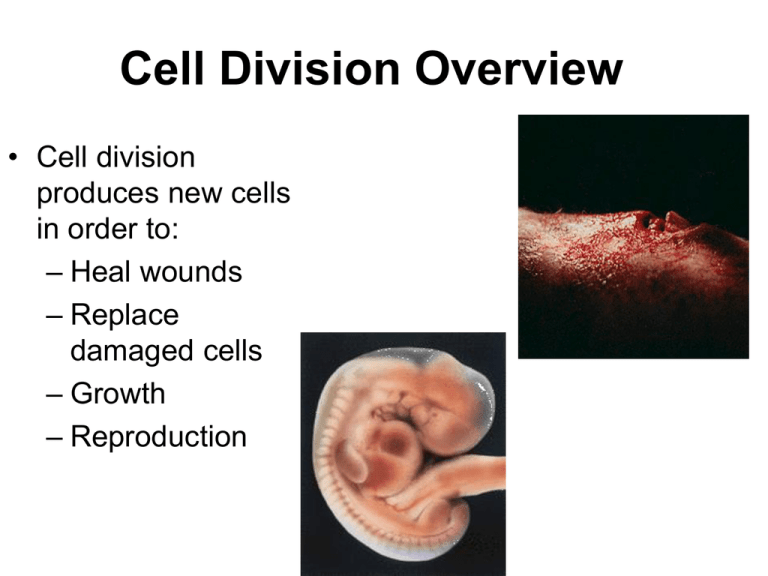
Cell Division Overview • Cell division produces new cells in order to: – Heal wounds – Replace damaged cells – Growth – Reproduction Overview of Cell Division • Before dividing, a copy of DNA (deoxyribonucleic acid) must first be made • DNA starts out in an string-like, uncondensed form called chromatin • Before cell division, DNA is condensed into short, linear chromosomes • The number of chromosomes in each cell depends on the organism: humans have 46 • Each chromosome is copied and the copy is called a sister chromatid • The sister chromatid is connected to the original DNA by a centromere • DNA is a double stranded molecule made of two single strands of nucleotides that are bonded together • The DNA molecule looks a lot like a twisted rope ladder • The “rungs” of the molecule are the bases: – – – – A (adenine) T (thymine) G (guanine) C (cytosine) • The bases across the “ladder” are connected in a specific way: – A always bonds with T – C always bonds with G • The connection is a hydrogen bond DNA Replication • DNA molecule separates at hydrogen bonds that hold bases together • The enzyme DNA polymerase adds the correct base to the now single strand of DNA • The covalent bond between sugars and phosphates is made • This results in two identical DNA molecules • Each new DNA molecule is half new and half from the old molecule “semiconservative” DNA is folded into structures called chromosomes during ____ _____. • Cell division Each chromosome is copied and the copy is called a ________ _________ • Sister chromatid Sister chromatids are connected to each other at the __________. • centromere Each strand of DNA is made up of smaller units called _________. • nucleotides Adenine always pairs with ___ • Thymine The enzyme ____ ___________adds the correct base to the now single strand of DNA • DNA polymerase Each new DNA molecule is half new and half from the old molecule and therefore is called ______________. • semiconservative The Cell Cycle and Mitosis For cells that divide by mitosis, there are 3 steps in the cell cycle: 1. interphase 2. mitosis 3. cytokinesis Interphase • Most of a cell’s life is spent in interphase • Normal functions are carried out • Three stages of interphase: – G1 –S – G2 http://cellsalive.com/cell_ cycle.htm Mitosis • The purpose of mitosis is to separate the sister chromatids so that each new cell has a complete set of chromosomes • PMAT http://cellsalive.com/mitosis.htm 5.4 Cell Cycle Control and Mutation Controls in the Cell Cycle • Checkpoints exist in the cell cycle • Cell determines if cell is ready to enter next part of cell cycle http://highered.mcgrawhill.com/olc/dl/120082/bio 34a.swf 5.1 What Is Cancer? • Cancer begins when the proteins that regulate the cell cycle don’t work, the cell divides uncontrollably – Mutations in the DNA can produce nonfunctioning proteins – Mutations can be inherited or induced by exposure to U.V. radiation or carcinogens that damage DNA and chromosomes • Unregulated cell division leads to a tumor, a mass of cells with no apparent function in the body • Cancer travels through the body by way of the lymphatic and circulatory systems (metastasis) • The lymphatic system collects fluids lost from capillaries • Lymph nodes are structures that filter lost fluids, called lymph • After they metastasize, cells gain access to the circulatory system and the heart, allow them to travel almost anywhere in the body Cancer: Uncontrolled cell growth • Tumor – Malignant vs benign • Metastasis • Types of cancer – Carcinoma (epithelials) • Melanoma (melanocytes) – Sarcoma (muscle/connective) – Osteogenic (bone) – Leukemia (blood forming organs) ↑ WBC’s – Lymphoma (lymphatic) • Malignant cells trigger angiogenesis Mutations to Cell-Cycle Control Genes • Proto-oncogenes: Normal genes on many different chromosomes regulate cell division • When mutated, they become oncogenes • Many organisms have proto-oncogenes, so many organisms can develop cancer Mutations to Cell-Cycle Control Genes • Proto-oncogenes carry instructions for building growth factors – Stimulate cell division when needed • Oncogenes overstimulate cell division •Suppressors are backup in case proto-oncogenes are mutated •They can also be mutated •Cells can then override the checkpoints From Benign to Malignant • Angiogenesis – growth of blood cells caused by secretions from cancer cells – Increases the blood supply to cancer cells: more oxygen and nutrients • Cancer cells can divide more • Tumors develop, sometimes filling entire organs From Benign to Malignant • Contact inhibition in normal cells prevents them from dividing all the time, which would force the new cells to pile up on each other • Anchorage dependence in normal cells keeps the cells in place From Benign to Malignant • Cancer cells divide too quickly and can leave the original site and enter the blood, lymph or tissues • Most cells divide a set number (60-70) of times, then they stop dividing • This usually limits benign tumors to small sizes • Cancer cells can divide indefinitely, as they are immortal through the manipulation of the enzyme telomerase Multiple Hit Model • Many changes, or hits, to the cancer cell are required for malignancy • Multiple hit model describes the process of cancer development • Mutations can be inherited and/or can stem from environmental exposures • Knowledge of cancer risk factors is important • Earlier detection and treatment of cancer greatly increase the odds of survival Detection Methods: Biopsy • Different cancers are detected by different methods, including high protein production possibly indicating a tumor • Biopsy, the surgical removal of cells, tissue, or fluid for analysis is performed • Under a microscope, benign tumors appear orderly and resemble other cells in the same tissue • Malignant tumors do not resemble normal tissue Treatment Methods: Chemotherapy • Chemicals that kill dividing cells are injected into the bloodstream during chemotherapy • Combinations of chemical agents are used since cancer cells grow resistant • Adverse effects on chemotherapy patients during treatment are numerous Treatment Methods: Radiation • High energy particles damage DNA in radiation therapy, so cells don’t divide • Radiation therapy is often administered in addition to chemotherapy • A patient is in remission if the patient is no longer suffering negative impacts from cancer after a given period The cell spends most of its life in ___________. • interphase The correct sequence of phases in mitosis is: • • • • Prophase Metaphase Anaphase Telophase Mitosis is followed by: • cytokinesis During which phase is DNA copied? • S phase Where are the 3 checkpoints for the cell cycle? • G1 • G2 • Metaphase Cancer that does not spread and therefore is considered not harmful • benign Normal genes on many different chromosomes that regulate cell division • Proto-oncogenes Identify 2 characteristics of normal cells that cancer cells do not exhibit • Anchorage dependance • Contact inhibition 5.6 Meiosis • • • • • • Sexual reproduction (Pro’s vs Con’s) Occurs within gonads (testes:ovaries) Meiosis produces sex cells – gametes (sperm:egg) Gametes have half the chromosomes (23) that somatic cells do (46) Meiosis reduces the number of chromosomes by one-half Fertilization of the male and female gamete will result in 46 chromosomes • Karyotype – There are 22 pairs of autosomes – There is one pair of sex chromosomes • The pairs of chromosomes (homologous pairs) carry the same genes Meiosis • During the S phase of interphase, the DNA is copied and the homologous chromosomes consist of sister chromatids • All four sister chromatids carry the same genes at the same locations, but not necessarily the exact same information Meiosis Meiosis • Meiosis is preceded by interphase (G1, S, G2) and followed by cell division • Meiosis consists of phases: – Meiosis I, in which the homologous pairs are separated – Meiosis II, in which the sister chromatids are separated Crossing Over and Random Alignment • There are millions of possible combinations of genes that each parent can produce because of: – Random alignment of homologous pairs (the way the homologs place themselves during metaphase I of meiosis) – Crossing over Crossing Over • When the homologous pairs are in prophase I of meiosis, they can exchange genetic information in the process of crossing over http://cellsalive.com/meiosis.htm Identify 3 differences between mitosis and meiosis • Somatic vs gametes • Divides once vs divides twice • Crossing over *somatic cells *divide once diploid *forms identical cells http://highered. mcgrawhill.com/sites/0 072437316/stu dent_view0/ch apter12/animat ions.html# *gametes *divide twicehaploid *forms different cells (crossing over)

