a genetic monitoring programme was part
advertisement
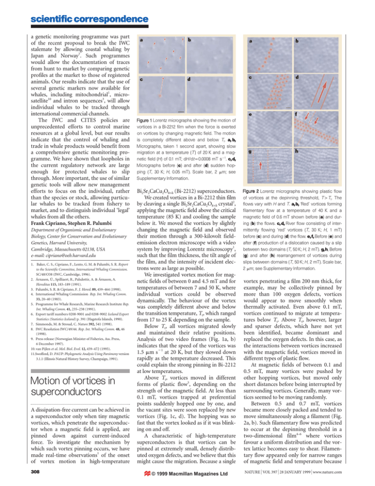
scientific correspondence a genetic monitoring programme was part of the recent proposal to break the IWC stalemate by allowing coastal whaling by Japan and Norway7. Such programmes would allow the documentation of traces from hunt to market by comparing genetic profiles at the market to those of registered animals. Our results indicate that the use of several genetic markers now available for whales, including mitochondrial1, microsatellite10 and intron sequences3, will allow individual whales to be tracked through international commercial channels. The IWC and CITES policies are unprecedented efforts to control marine resources at a global level, but our results indicate that the control of whaling and trade in whale products would benefit from a comprehensive genetic monitoring programme. We have shown that loopholes in the current regulatory network are large enough for protected whales to slip through. More important, the use of similar genetic tools will allow new management efforts to focus on the individual, rather than the species or stock, allowing particular whales to be tracked from fishery to market, and to distinguish individual ‘legal’ whales from all the others. Frank Cipriano, Stephen R. Palumbi Department of Organismic and Evolutionary Biology, Center for Conservation and Evolutionary Genetics, Harvard University, Cambridge, Massachusetts 02138, USA e-mail: cipriano@oeb.harvard.edu 1. Baker, C. S., Cipriano, F., Lento, G. M. & Palumbi, S. R. Report to the Scientific Committee, International Whaling Commission. SC/48/O38 (IWC, Cambridge, 1996). 2. Árnason, Ú., Spilliaert, R., Palsdottir, A. & Árnason, A. Hereditas 115, 183–189 (1991). 3. Palumbi, S. R. & Cipriano, F. J. Hered. 89, 459–464 (1998). 4. International Whaling Commission Rep. Int. Whaling Comm. 33, 20–40 (1983). 5. Programme for Whale Research, Marine Research Institute Rep. Int. Whaling Comm. 41, 235–238 (1991). 6. Export tariff numbers 0208-9001 and 0208-9002 Iceland Export Statistics (Statistics Iceland) p. 391 (Hagstofa Islands, 1990). 7. Simmonds, M. & Stroud, C. Nature 392, 541 (1998). 8. IWC Resolution IWC/49/44. Rep. Int. Whaling Comm. 48, 46 (1998). 9. Press release (Norwegian Minister of Fisheries, Ass. Press, 6 December 1997). 10. van Pijlen et al. Mol. Biol. Evol. 12, 459–472 (1995). 11.Swofford, D. PAUP: Phylogenetic Analysis Using Parsimony version 3.1.1 (Illinois Natural History Survey, Champaign, 1991). Motion of vortices in superconductors A dissipation-free current can be achieved in a superconductor only when tiny magnetic vortices, which penetrate the superconductor when a magnetic field is applied, are pinned down against current-induced force. To investigate the mechanism by which such vortex pinning occurs, we have made real-time observations1 of the onset of vortex motion in high-temperature 308 Figure 1 Lorentz micrographs showing the motion of vortices in a Bi-2212 film when the force is exerted on vortices by changing magnetic field. The motion is completely different above and below Tt. a, b, Micrographs, taken 1 second apart, showing slow migration at a temperature (T ) of 20 K and a magnetic field (H) of 0.1 mT; dH/dtǃ0.0008 mT sǁ1. c, d, Micrographs before (c) and after (d) sudden hopping (T, 30 K; H, 0.05 mT). Scale bar, 2 Ȗm; see Supplementary Information. Bi2Sr2CaCu2O8+Ȏ (Bi-2212) superconductors. We created vortices in a Bi-2212 thin film by cleaving a single Bi2Sr2CaCu2O8+Ȏ crystal2, applying the magnetic field above the critical temperature (85 K) and cooling the sample below it. We moved the vortices by slightly changing the magnetic field and observed their motion through a 300-kilovolt fieldemission electron microscope with a video system by improving Lorentz microscopy1, such that the film thickness, the tilt angle of the film, and the intensity of incident electrons were as large as possible. We investigated vortex motion for magnetic fields of between 0 and 4.5 mT and for temperatures of between 7 and 50 K, where individual vortices could be observed dynamically. The behaviour of the vortex was completely different above and below the transition temperature, Tt, which ranged from 17 to 25 K depending on the sample. Below Tt, all vortices migrated slowly and maintained their relative positions. Analysis of two video frames (Fig. 1a, b) indicates that the speed of the vortices was 1.5 Ȗm sǁ1 at 20 K, but they slowed down rapidly as the temperature decreased. This could explain the strong pinning in Bi-2212 at low temperatures. Above Tt, vortices moved in different forms of plastic flow3, depending on the strength of the magnetic field. At less than 0.1 mT, vortices trapped at preferential points suddenly hopped one by one, and the vacant sites were soon replaced by new vortices (Fig. 1c, d). The hopping was so fast that the vortex looked as if it was blinking on and off. A characteristic of high-temperature superconductors is that vortices can be pinned at extremely small, densely distributed oxygen defects, and we believe that this might cause the migration. Because a single © 1999 Macmillan Magazines Ltd Figure 2 Lorentz micrographs showing plastic flow of vortices at the depinning threshold, T>Tt. The flows vary with H and T. a, b, ‘Red’ vortices forming filamentary flow at a temperature of 40 K and a magnetic field of 0.6 mT shown before (a) and during (b) the flows. c, d, River flow consisting of intermittently flowing ‘red’ vortices (T, 30 K; H, 1 mT) before (c) and during (d) the flow. e, f, Before (e) and after (f) production of a dislocation caused by a slip between two domains (T, 50 K; H, 2 mT). g, h, Before (g) and after (h) rearrangement of vortices during slips between domains (T, 50 K; H, 2 mT). Scale bar, 2 Ȗm; see Supplementary Information. vortex penetrating a film 200 nm thick, for example, may be collectively pinned by more than 100 oxygen defects, vortices would appear to move smoothly when thermally activated. Even above 0.1 mT, vortices continued to migrate at temperatures below Tt. Above Tt, however, larger and sparser defects, which have not yet been identified, became dominant and replaced the oxygen defects. In this case, as the interactions between vortices increased with the magnetic field, vortices moved in different types of plastic flow. At magnetic fields of between 0.1 and 0.5 mT, many vortices were pushed by other hopping vortices, but moved only short distances before being interrupted by surrounding vortices. Generally, many vortices seemed to be moving randomly. Between 0.5 and 0.7 mT, vortices became more closely packed and tended to move simultaneously along a filament (Fig. 2a, b). Such filamentary flow was predicted to occur at the depinning threshold in a two-dimensional film4–6 where vortices favour a uniform distribution and the vortex lattice becomes easy to shear. Filamentary flow appeared only for narrow ranges of magnetic field and temperature because NATURE | VOL 397 | 28 JANUARY 1999 | www.nature.com scientific correspondence our samples were just between two and three dimensions. We also found two filaments that were joined4. For magnetic fields between 0.7 and 4.5 mT, a single filament dragged adjacent ones along, forming rivers7 (Fig. 2c, d) that intermittently changed their location and width. To investigate stronger interactions between vortices, we increased the temperature instead of the magnetic field, as we could not observe vortices individually above 4.5 mT owing to the overlap of their magnetic fields, and found a new form of plastic flow, which we designate distortedlattice flow. At 50 K, and with a magnetic field exceeding 1.5 mT, the vortex lattice was divided into domains that tended to move separately because of non-uniform pinning. Sometimes one domain was suddenly displaced slightly with respect to its neighbouring domains, producing edge dislocations (Fig. 2e, f); sometimes all of the vortices in one domain disappeared while the vortices rearranged during the slip, before reappearing as different lattice orientations were formed (Fig. 2g, h). Under these conditions, vortices generally proceeded in units of domains, sometimes with slips between domains or with changing lattice orientations. A. Tonomura*‡, H. Kasai*, O. Kamimura*, T. Matsuda*, K. Harada*‡, J. Shimoyama†‡, K. Kishio†‡, K. Kitazawa†‡ *Advanced Research Laboratory, Hitachi Ltd, Hatoyama, Saitama 350-0395, Japan e-mail: tonomura@harl.hitachi.co.jp †Department of Applied Chemistry, University of Tokyo, Tokyo 113-8656, Japan ‡CREST, Japan Science and Technology Corporation (JST), Kawaguchi, Saitama 332-0012, Japan 1. Harada, K. et al. Nature 360, 51–53 (1992). 2. Kotaka, Y. et. al. Physica C 235–240, 1529–1530 (1994). 3. Crabtree, G. W. & Nelson, D. R. Phys. Today 50(4), 38–45 (1997). 4. Grønbech-Jensen, N., Bishop, A. R. & Dominguez, D. Phys. Rev. Lett. 76, 2985–2988 (1996). 5. Olson, C. J., Reichhardt, C. & Nori, F. Phys. Rev. B 56, 6175–6194 (1997). 6. Higgins, M. J. & Bhattacharya, S. Physica C 257, 232–254 (1996). 7. Matsuda, T. et. al. Science 271, 1393–1395 (1996). Supplementary information is available on Nature’s World-Wide Web site (http://www.nature.com) or as paper copy from the London editorial office of Nature. Role of the giant panda’s ‘pseudo-thumb’ The way in which the giant panda, Ailuropoda melanoleuca, uses the radial sesamoid bone — its ‘pseudo-thumb’ — for grasping makes it one of the most extraordinary manipulation systems in mammalian evolution1–5. The bone has been reported to function as an active manipulator, enabling the panda to grasp bamboo stems between the NATURE | VOL 397 | 28 JANUARY 1999 | www.nature.com bone and the opposing palm2,6–8. We have used computed tomography, magnetic resonance imaging (MRI) and related techniques to analyse a panda hand. The three-dimensional images we obtained indicate that the radial sesamoid bone cannot move independently of its articulated bones, as has been suggested1–3, but rather acts as part of a functional unit of manipulation. The radial sesamoid bone and the accessory carpal bone form a double pincer-like apparatus in the medial and lateral sides of the hand, respectively, enabling the panda to manipulate objects with great dexterity. Schematic drawings based on computedtomography and three-dimensional reconstructed images (Fig. 1a–c) explain the grasping mechanism used by the giant panda. Three-dimensional data obtained from artificial grasping of a carcass hand show that the radial sesamoid bone does not abduct or adduct independently of the first metacarpal and the radial carpal bones, and that the accessory carpal bone does not move substantially in the gripping action. When the movement is compared for open and gripping hands, the radial sesamoid bone, the first metacarpal and the radial carpal are actually moulded into a single bone, and the accessory carpal bone and the ulna constitute a single functional unit. When the hand is opened, the radial sesamoid bone and the accessory carpal bone therefore protrude at different angles from the plane of the palm (Fig. 1a). The radial carpal bone forms an enlarged articulated surface to the distal end of the radius. In the gripping action, the five long phalanges are crooked (Fig. 1b) while the panda flexes the wrist joint (Fig. 1c). This wrist flexion means that the radial sesamoid bone is parallel to the accessory carpal bone, and the distal phalanges are parallel with the radius and ulna. This arrangement gives the panda a degree of opposability between the phalanges and the functional unit comprising the radial sesamoid bone and the accessory carpal bone (Fig. 1c). The radial sesamoid bone and the accessory carpal bone do not move independently of their articulated bones in the grasping action, but constitute a functional unit with the first metacarpal and the radial carpal, and the ulna, respectively. The panda has three functional units: the RRM complex (radial sesamoid – radial carpal – first metacarpal), the AU complex (accessory carpal – ulna), and the phalanges (Fig. 1a–c). The RRM complex flexes and the radial sesamoid bone becomes parallel with the accessory carpal bone, and the phalanges bend and hold things in the hollow of the hand during the grasping action. The phalanges make a pincer-like apparatus with the RRM complex in the medial part of the hand, and another with the AU complex in © 1999 Macmillan Magazines Ltd a b Phalanges Metacarpals Radial sesamoid First metacarpal Radial carpal Accessory carpal c Radius and ulna RRM complex d AU complex Figure 1 Schematic drawings of the grasping mechanism of the giant panda (medial view of right hand, with the proximal direction at the bottom). a, Hand open. b, Hand open but with the phalanges flexed. c, The grasping action (from a small palmar angle). The radial sesamoid and accessory carpal bones do not move independently of their articulated bones in the grasping action, but constitute two functional units: the RRM complex (see text) in the medial part of the hand, and the AU complex (see text) in the lateral part. Pincer-like structures are made by the phalanges and the RRM complex in the medial part, and by the phalanges and the AU complex in the lateral part. d, As c, but showing the muscles in the pincer-like structures on both sides of the grasped hand (arrows). the lateral part of the hand (Fig. 1a–c). It is this pair of ‘pincers’ that gives the panda its manual dexterity. The MRI images indicate that the abductor pollicis brevis and the opponens pollicis muscles serve as a cushion for objects grasped between the radial sesamoid bone and the first metacarpal. The two muscle bundles surround the objects, increase friction between the hand and the objects, and alter the size and shape of the area in the hand. In the lateral part of the palm, the abductor digiti quinti muscle is well developed between the accessory carpal bone, the fifth metacarpal and the phalanges2. We suggest that the muscle pads may also help the panda to receive and hold objects grasped by the AU complex (Fig. 1d). Our idea that the radial sesamoid bone does not function independently, but as part of the RRM functional complex, takes no account of the role of its abductor and adductor muscles. We suggest that the three functional units, and the double-pincer-like apparatus of which they are made, can be completely controlled only by the same muscular system that is found in other bear species. The wrist flexion and the manipulation of the double-pincer apparatus have 309
