AN ABSTRACT OF THE THESIS OF
AN ABSTRACT OF THE THESIS OF
Matthew Joel Cannon for the degree of Master of Science in Animal Science presented on September 29,1995
Title: Evaluation of Alterations in the Bovine Zona Pellucida Induced by Plasmin or Embryonic Plasminogen Activator.
Approved:
Redacted for Privacy
Alfred R. Menino, Jr.
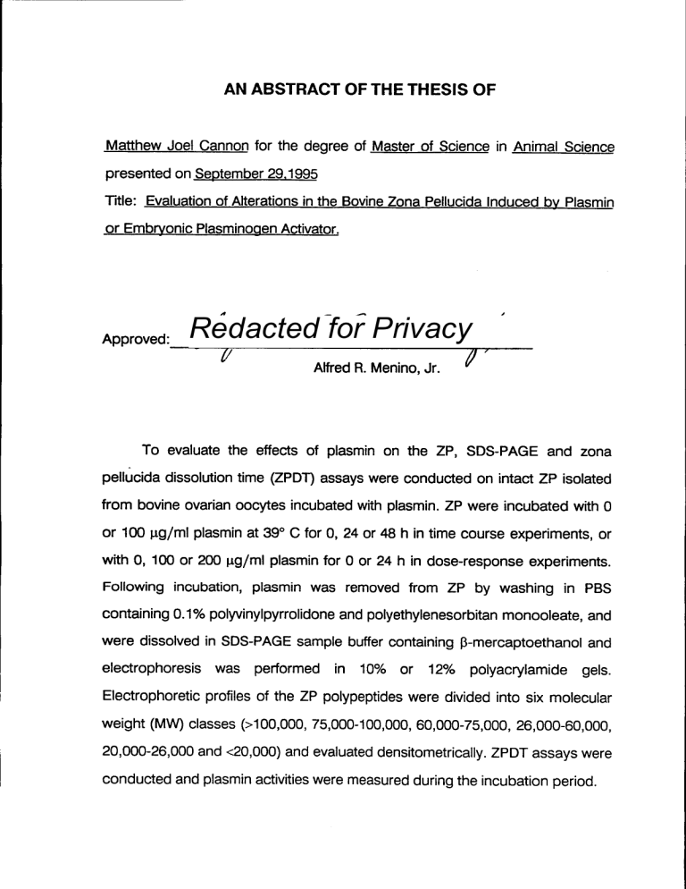
To evaluate the effects of plasmin on the ZP, SDS-PAGE and zone pellucida dissolution time (ZPDT) assays were conducted on intact ZP isolated from bovine ovarian oocytes incubated with plasmin. ZP were incubated with 0 or 100 µg /ml plasmin at 39° C for 0, 24 or 48 h in time course experiments, or with 0, 100 or 200 pg/m1 plasmin for 0 or 24 h in dose-response experiments.
Following incubation, plasmin was removed from ZP by washing in PBS
containing 0.1% polyvinylpyrrolidone and polyethylenesorbitan monooleate, and were dissolved in SDS-PAGE sample buffer containing f3-mercaptoethanol and electrophoresis was performed in
10% or
12% polyacrylamide gels.
Electrophoretic profiles of the ZP polypeptides were divided into six molecular weight (MW) classes (>100,000, 75,000-100,000, 60,000-75,000, 26,000-60,000,
20,000-26,000 and <20,000) and evaluated densitometrically. ZPDT assays were conducted and plasmin activities were measured during the incubation period.
SDS-PAGE of the bovine oocyte ZP resulted
in
resolution of two polypeptides with MW 76,000 and 65,000, respectively, and two minor polypeptides with MW 23,000 and 22,000 that migrated as a doublet. No
changes occurred in any MW classes in ZP incubated with 0 µg /ml plasmin for
0, 24 or 48 h, and ZPDT were not different (P>0.10). ZP treated with 100 µg/m1 plasmin exhibited decreases in the MW >100,000 and 20,000-26,000 classes at
24 and 48 h. ZPDT were lower (P<0.05) at 24 and 48 h in 100 µg /ml, and plasmin activities were decreased at 24 and 48 h compared to 0 h (P<0.05).
Bovine ZP treated with 100 or 200 µg/m1 plasmin exhibited a decrease (P<
0.10) in the polypeptides in the MW >100,000 class in 200 µg /ml, and increases
(P<0.10) in the MW 26,000-60,000 and <20,000 classes in 100 and 200 µg /ml compared to 0 µg /ml plasmin at 24 h. ZPDT were lower (P<0.05) in 100 or 200 t.t.
g/ml compared to 0 µg /ml plasmin at 24 h. ZPDT were also lower (P<0.05) for
ZP treated with 200 µg /ml plasmin compared to 100 µg /ml plasmin. Activity of both plasmin concentrations was decreased at 24 h (P<0.05).
In
bovine ZP treated
with
50 pg /ml
active or 4-(2-aminoethyl) benzenesulfonylfluoride-inactivated plasmin at 39° C for 0, 24 or 48 h, decreases in the MW >100,000 and 75,000-100,000 and increases in the 60,000-76,000 MW class occurred at 24 and 48 h in active plasmin. Solubility was increased at 24 and 48 h in active plasmin. Compared to 0 µg /ml plasmin, ZPDT were lower (P<
0.05) at 24 and 48 h in inactivated and active plasmin. Caseinolytic activity of active plasmin was lower (P<0.05) at 48 h compared to 0 and 24 h. Activity of inactivated plasmin was lower (P<0.05) at 0, 24 and 48 h compared to active plasmin, but activity of inactivated plasmin increased (P<0.05) at 24 and 48 h.
ZP were also treated with embryonic plasminogen activator. Day 7
embryos (day of estrus = Day 0) were collected non-surgically from superovulated cows and cultured in Ham's F -12+ 1.5% BSA at 39° C in 5% CO2
in air and conditioned medium was collected every 48 h and pooled. Embryo conditioned medium used to treat ZP contained plasminogen activating activity
equal to approximately 33 IU/m1 human urokinase. ZP treated with non-
conditioned or embryo-conditioned medium for 0 or 48 h at 39° C exhibited no changes in ZP polypeptide MW classes (P>0.10). ZPDT were not different between non-conditioned and embryo-conditioned medium at 0 and 48 h (P>
0.10). Addition of human plasminogen (100 pg/m1) to embryo-conditioned medium resulted in a decrease in the MW 75,000-100,000 class and an increase in the <20,000 class at 48 h (P<0.10). These results demonstrate that bovine plasmin
is capable of proteolytically degrading the bovine ZP, and that
embryonic plasminogen activator is capable of activating plasminogen in the presence of the ZP, thereby indirectly affecting the ZP through activation of plasminogen to plasmin. Changes in the specific MW classes and decreases in
ZPDT suggest that the plasminogen activator/plasmin system could exert proteolytic effects on the ZP during fertilization and hatching.
EVALUATION OF ALTERATIONS IN THE BOVINE ZONA
PELLUCIDA INDUCED BY PLASMIN OR EMBRYONIC
PLASMINOGEN ACTIVATOR
by
Matthew Joel Cannon
A THESIS submitted to
Oregon State University in partial fulfillment of the requirements of the degree of
Master of Science
Completed September 29, 1995
Commencement June 1996
Masters of Science thesis of Matthew Joel Cannon presented September 29,
1995.
APPROVED:
Redacted for Privacy
Major Professor Representing Animal dence
Redacted for Privacy
Redacted for Privacy
Dean of Graduate School
I understand that my thesis will become a part of the permanent collection of Oregon State University libraries. My signature below authorizes release of my thesis to any reader upon request.
Redacted for Privacy
Matthew Joel Cannon, Author
TABLE OF CONTENTS
INTRODUCTION
REVIEW OF THE LITERATURE
The Zona Pellucida
The Plasminogen Activator/ Plasmin System
The Plasminogen Activator / Plasmin System
In Reproduction
STATEMENT OF THE OBJECTIVES
EVALUATION OF ALTERATIONS IN THE BOVINE
ZONA PELLUCIDA INDUCED BY PLASMIN OR
EMBRYONIC PLASMINOGEN ACTIVATOR
Abstract
Introduction
Materials And Methods
Results
Discussion
Literature Cited
CONCLUSION
BIBLIOGRAPHY
1
4
4
12
18
30
32
47
65
70
32
34
37
74
76
Figure
1.
2.
3.
4.
5.
6.
7.
8.
9.
LIST OF FIGURES
Page
SDS-PAGE and densitometric scans of 250 bovine oocyte zonae pellucidae treated with 0 [ig/mIplasmin for a) 0, b) 24 or c) 48 h at 39° C 48
Bovine oocyte zonae pellucidae treated with 01.1g/m1 plasmin for 0 h at 39° C during zona pellucida dissolution time (ZPDT) assay at a) 0, b) 5 and c)10 minutes and d) at dissolution (200X) 50
(a) Zona pellucida dissolution times (ZPDT) for ZP treated with 0 or 100 pg/mIplasmin for 0, 24 or 48 h, and (b) recovered plasmin activities at 0, 24 or 48 h 51
SDS-PAGE and densitometric scans of 250 bovine oocyte zonae pellucidae treated with 100 lig/m1plasmin for a) 0, b)24 or c) 48 h at 39° C 52
SDS-PAGE and densitometric scans of 250 bovine oocyte zonae pellucidae treated with a) 0, b) 100 or c) 200 lig/mIplasmin for 0 h at 39° C 54
SDS-PAGE and densitometric scans of 250 bovine oocyte zonae pellucidae treated with a) 0, b) 100 or c) 200 gg/mIplasmin for 24 h at 39° C 55
(a) Zona pellucida dissolution times (ZPDT) for ZP treated with 0, 100 or 200 i_tg/mIplasmin for 0 or 24 h, and (b) recovered plasmin activities at 0 or 24 h 56
SDS-PAGE and densitometric scans of 250 bovine oocyte zonae pellucidae treated with 50 mg/mlactive plasmin for a) 0, b) 24 or c) 48 h at 39° C
58
SDS-PAGE and densitometric scans of 250 bovine oocyte zonae pellucidae treated with 50 .11g/m1
AEBSF-inactivated plasmin for a) 0, b) 24 or c) 48 h at 39° C C.
.
.
.59
Figure
10.
11.
12.
13.
(a) Zona pellucida dissolution times (ZPDT) for ZP treated with inactivated (50 pg/m1+AEBSF) or active
(50 gg/ml-AEBSF) plasmin for 0, 24 or 48 h, and b) recovered plasmin activities expressed as percent of active plasmin at 0 h
SDS-PAGE and densitometric scans of 250 bovine oocyte zonae pellucidae treated with non-conditioned medium for a) 0 and c) 48 h or embryo-conditioned medium for b) 0 and d) 48 h at 39° C
SDS-PAGE and densitometric scans of 250 bovine oocyte zonae pellucidae treated with embryo-conditioned medium containing 0 µg/m1 human plasminogen for a) 0 and c) 48 h or 100 lAg/mIhuman plasminogen for b) 0 and d) 48 h at 39° C
Zona pellucida dissolution times (ZPDT) for ZP treated with nonconditioned or embryo-conditioned medium for 0 or 48 h.
Page
60
62
63
64
EVALUATION OF ALTERATIONS IN THE BOVINE ZONA
PELLUCIDA INDUCED BY PLASMIN OR EMBRYONIC
PLASMINOGEN ACTIVATOR
INTRODUCTION
Preimplantation embryonic development in mammalian species involves a complex set of processes that begin with fertilization, and that end with the implantation or attachment of the developing embryo to the uterus. During most of this period the embryo is surrounded by a protein coat called the zona pellucida (ZP). The ZP is an acellular glycoprotein matrix that serves multiple functions in the preimplantation period of development. During fertilization, the
ZP functions as a sperm receptor and also induces the acrosome reaction in fertilizing sperm cells, allowing them to penetrate the ZP (Wassarman,
1988).
Immediately following fertilization, the ZP undergoes an event known as "zona hardening", which makes it impenetrable by further sperm, thereby serving as a block to polyspermy (Hoodbhoy and Talbot, 1994). The ZP also maintains the
integrity and conserves the microenvironment of the developing
embryo
(Betteridge and Flechon, 1988). At some point however, the embryo must shed the ZP for normal development to proceed. Prior to implantation, the embryo escapes from the ZP in a process known as hatching (Betteridge and Flechon,
1988).
Plasminogen activators (PA) are serine proteases that are capable of activating the tissue zymogen plasminogen to the broad-spectrum serine
protease plasmin (Dano et al., 1985). There are two main types of PA in
mammalian species, each having a different physiologic function: t-PA, which is mainly involved in lysis of fibrin clots in the circulatory system; and u-PA, which is primarily involved in extracellular matrix degradation during tissue migration, and is known to be associated with a number of invasive or migrating cell types
(Dano et al., 1985; Saksela and Rifkin, 1988). PA are also known or suspected to be involved in a number of reproductive processes. In the male, both types of
PA are involved in different processes in spermatogenesis (Lacroix et al., 1981;
Tolli et al., 1995), and in the female PA are involved in ovulation (Beers, 1975;
Canipari and Strickland, 1985; Ny et al., 1985).
The PA/plasmin system may also be involved
in other aspects of
reproduction. Fertilization is a process that up until recently was felt to be dependent on the protease acrosin for sperm to penetrate the ZP. However, it has recently been shown that fertilization can occur without acrosin (Baba et al.,
1994). Since both sperm and oocytes are known to secrete PA, and addition of plasminogen to in vitro fertilization media increases fertilization rates, this suggests that the PA/plasmin system may facilitate penetration of the ZP by sperm during fertilization (Huarte et al., 1993).
The mechanisms used by the embryo to hatch have long been a source of debate. Early on it was proposed that the embryo caused the rupture of the ZP through hydrostatic pressure generated by the expanding blastocoel combined with proteolytic weakening of the ZP (McLaren, 1970). Also, it was observed that the ZP of uterine embryos exhibited changes in solubility prior to hatching, suggesting that a zonalytic agent in the uterus catalyzed a "sublysis" of the ZP, which weakened it and facilitated hatching (Domon et al., 1973). Subsequently it was observed that the embryo secretes a trypsin-like protease prior to hatching, and that activity of this enzyme is found associated with the ZP following
2
hatching, with the greatest intensity at the point of rupture (Perona and
Wassarman, 1986). It is known that the embryo secretes PA (Stick land et al.,
1976; Fazleabas et al., 1983), is capable of activating plasminogen to plasmin
(Menino and Williams, 1987; Menino et al., 1989) and that embryos which produce more PA are more likely to hatch (Kaaekuahiwi and Menino, 1990).
Also, culture of embryos in medium containing plasminogen results in increased
solubility of the ZP. These results suggest that the PA/plasmin system is
involved in the hatching process.
Implantation is a process which involves the invasion of the uterine cell layers by cells from the developing embryo. This process has been described as very similar to tissue invasion by metastatic tumors, which is known to involve proteases, particularly PA (Strickland, 1980; Dano et al., 1985). PA secretion is correlated to the invasive period of development of the trophoblast (Strickland et al., 1976), and is localized to the regions of the trophoblast which are invading the uterine tissue (Sappino et al., 1989). The trophoblast also express a PA receptor on the cell surface, but only during the invasive period (Zini et al.,
1992). Also, trophoblast cells that are incapable of removing inactive PA-inhibitor complexes from their surface are incapable of implanting (Herz et al., 1992). It therefore seems that implantation is another event in which the PA/plasmin system is in some way involved.
Based on the evidence supporting the involvment of the PA/plasmin system in fertilization and early embryo development, the objective of this thesis was to evaluate changes in zona pellucida integrity following exposure to plasmin or the embryonic plasminogen activator.
3
REVIEW OF THE LITERATURE
The Zona Pellucida
In mammalian species, the female gamete, the oocyte, is surrounded by an acellular glycoprotein coat at the time of ovulation. This glycoprotein coat, the zona pellucida (ZP), serves functional roles in both the oocyte as well as the early embryo. The (ZP) is composed of multiple glycoprotein components
(Wassarman, 1988) that are products of developmentally regulated genes
(Dean, 1991) and which display antigenic cross-reactivity (Maresh and Dunbar,
1987) and biochemical similarities (Wassarman, 1988) across species. Although the ZP may serve more as yet unknown functions, components of the ZP have been demonstrated to be involved in fertilization as sperm receptors, in induction of the acrosome reaction, and in the post-fertilization block to polyspermy
(reviewed by Wassarman, 1988). Also, the ZP is essential in maintaining the physical integrity of the early embryo up to the fully-compacted late-morula stage and
has been suggested
to play a role in conserving the microenvironment of the perivitelline space in some species (Betteridge and
Flechon, 1988).
Growth and development of the oocyte occurs in conjunction with growth and development of the ovarian follicle during folliculogenesis. Folliculogenesis will not be extensively reviewed; it will suffice to say that follicle development proceeds from the primordial stage (the least mature) through the primary, secondary, and tertiary follicle stages, and finally to the Graafian,
or pre-
4
ovulatory follicle. These stages involve dynamic histological and biochemical changes in the follicle which are much more complex than development of the oocyte.
In most mammalian species studied (the rabbit being an exception), the
ZP is completely formed at the time of ovulation. However, it has been observed that oocytes isolated from primordial ovarian follicles are bounded only by a plasma membrane, and do not display a ZP (Chiquoine, 1960). Histologically the first evidence of formation of the ZP is seen on oocytes contained in late primary follicles. The ZP is not formed uniformly around the oocyte, but first appears as discontinuous portions around the oocyte, which later fuse to form a continuous glycoprotein coat. As the oocyte and follicle continue to grow and develop, the
ZP begins to thicken and take on a uniform appearance. By the time the follicle reaches the tertiary stage, the ZP is completely formed (Hafez, 1993).
Since the oocyte is encased within an ovarian follicle and the follicle cells are intimately associated with the oocyte, this observation led to some early contention as to the cellular origin of the ZP (Haddad and Nagai, 1977). It was suspected from initial morphological and histochemical studies that the source of the ZP was the follicle cells immediately surrounding the oocyte (reviewed by
Wassarman, 1988). However, in studies in which oocytes from primordial follicles were denuded of follicle cells and placed in culture, it was observed that the oocytes were capable of secreting the ZP glycoproteins, whereas cultured follicle cells were not (Bleil and Wassarman, 1980). Further, in pulse-chase autoradiographic studies in which mice were injected with radioactive fucose, it has been observed that the radioactive label appears first in the ZP adjacent to the oocyte plasma membrane, and subsequently progresses towards the outer surface of the ZP as the oocyte grows (Haddad and Nagai, 1977; Flechon et al.,
1984).
5
Expression of the mRNA coding for components of the ZP has also been
used to investigate not only the site of synthesis of the ZP, but also the
developmental stages at which the ZP components are expressed. Synthesis of the ZP glycoproteins is not only limited to the oocyte, but expression of the genes coding for the ZP is developmentally regulated to only those oocytes in the growth phase. In 1986, Ringuette and coworkers created a murine oocyte cDNA library, and cloned a cDNA coding for a component of the mouse ZP, ZP-
3. This cDNA was then used to demonstrate that mRNA for the ZP-3
glycoprotein is only expressed in the oocyte (Ringuette et al., 1986). This cDNA was subsequently used to demonstrate that ZP-3 is expressed only in growing mouse oocytes, and not in resting mouse oocytes (Ringuette et al., 1988).
These researchers further showed that another component of the mouse ZP,
ZP-2, is coordinately expressed with ZP-3, and that expression is developmentally restricted to oocytes in the growth phase (Liang et al., 1990).
One notable feature of the ZP is the differences between the inner and outer surfaces. Scanning electron micrographs reveal that the outside surface of the ZP is not a solid surface, but has the mesh-like appearance of a sponge.
This is different from the inside surface, which is more uniform in appearance
with no mesh-like fenestrations (reviewed by Hafez, 1993). Flechon and
coworkers have suggested that the fenestrated appearance of the outer surface of the ZP is a consequence of the increase in the outer diameter of the ZP due to
accretion of new material from the inside (Flechon et
al., 1984). These observations suggested that the ZP is not an impermeable barrier, and are reinforced by studies which show that the ZP is permeable to macromolecules such as ferritin (molecular weight (MW) 480,000 to 1,000,000) (Hastings et al.,
1972) and immunoglobulins (Sellens and Jenkinson, 1975).
6
Biochemically, the mammalian ZP is made up of a relatively small number of different glycoproteins. Due to the ease of isolation of large numbers of oocyte ZP, the most extensively studied ZP are the porcine and murine ZP.
However, biochemical and immunological similarities have been demonstrated between ZP from these species and rat, hamster, rabbit, cat, dog, squirrel monkey, and human ZP (Maresh and Dunbar, 1987; Wassarman, 1988).
The murine ZP is composed of three glycoproteins, ZP1, ZP2, and ZP3, with MW of 200,000, 120,000 and 83,000, respectively, as determined by sodium dodecyl sulfate polyacrylamide gel electrophoresis (SDS-PAGE). All of these glycoproteins are acidic, with pl between 4.1 and 5.5. Under native conditions
ZP2 and ZP3 are monomers, while ZP1 is a dimer. In the presence of reducing reagents on SDS-PAGE, ZP1 has an apparent MW of 120,000 and comigrates with ZP2, suggesting that the dimer is held together by intermolecular disulfide bonds. Migration of ZP3 is slightly retarded, suggesting that the native form possesses intramolecular disulfide bonds (Wassarman, 1988).
SDS-PAGE of the porcine ZP under reducing conditions yields four glycoproteins, ZP1, ZP2, ZP3, and ZP4, of molecular weight 80,000-90,000,
60,000-65,000, 55,000, and 20,000-25,000, respectively (Dunbar and Raynor,
1980; Dunbar et al., 1980; Hedrick and Wardrip, 1986). Like the murine ZP glycoproteins, the porcine ZP components are acidic, with pl between 5.2 and
5.6 pl units. Under non-reducing conditions, SDS-PAGE of the porcine ZP reveals only two glycoproteins, with molecular weights of 80,000-90,000 and
55,000. Since the sum of the molecular weights of ZP2 and ZP4 equal that of
ZP1, this suggests that ZP2 and ZP4 are derived from ZP1 due to reduction of intermolecular disulfide bonds (Yurewicz et al.1987; Hedrick and Wardrip,
1987a). Also, the amino acid and carbohydrate compositions of ZP2 and ZP4 equal ZP1, and ZP1 is immunologically related to ZP2 and ZP4. ZP2 and ZP4 are
7
not immunologically related (Wassarman, 1988). Together these observations suggest that ZP1 and ZP3 are the actual native porcine ZP glycoproteins.
Interestingly, there are actually two forms of porcine ZP3, designated as
ZP3a and ZP36. Deglycosylation of ZP3 yields two different polypeptides of MW
35,000 and 38,000 (Yurewicz et al., 1987) and antigenic studies reveal that ZP3a and ZP36 are immunologically distinct (Timmons et al., 1987). These findings
suggest that there are two structurally and immunologically distinct ZP3
glycoproteins, which are presumably encoded by different genes (Wassarman,
1988). Collectively, these data suggest that the intact porcine ZP is composed of three distinct glycoproteins: ZP1 (82,000), ZP3a (55,000), and ZP36 (55,000)
(Sacco et al., 1989).
Electrophoretic analysis of the bovine ZP under reducing conditions by
Gwatkin and coworkers (1980) resulted in three glycoprotein components, with molecular weights of 100,000, 50,000, and 25,000. However, an electrophoretic comparison of bovine ZP under reducing and non-reducing conditions by
Menino and Wright (1980) resulted in a single glycoprotein band of MW 73,000 under non-reducing conditions. Under reducing conditions, electrophoresis of the bovine ZP resulted in two major bands with molecular weights of 15,000 and
22,000. This led these authors to conclude that the bovine ZP is composed of a single protein, made up of four subunits, two of MW 15,000 and two of MW
22,000, held toether by intermolecular disulfide bonds (Menino and Wright,
1980). More recently two dimensional gel electrophoresis of the bovine ZP under non-reducing conditions resulted in the resolution
of three glycoprotein
components with varying ranges of pl: 94,000-110,000, pl= 7.0-8.3, 82,000-
94,000, pl= 4.3-4.8 and 70,000-76,000, pl= 5.8-7.3 (Florman and First, 1988).
Electrophoretic analysis of the bovine ZP under reducing conditions resulted in
8
resolution of three major glycoprotein bands with MW approximately 100,000,
80,000, and 65,000, and two minor bands with MW 21,000 and 22,000 (Cannon and Menino, 1995).
Electrophoretic mobilities and isoelectric points of the ZP components can
be significantly altered by various agents which are capable of removing
oligosaccharides from the glycoproteins. Digestion of murine ZP with endo-0-Nacetylglucosaminidase F (Endo F), which removes high-mannose- and complextype, asparagine-linked (N-linked) oligosaccharides, results in an increase in electrophoretic mobilities and an increase in isoelectric points of all three murine
ZP glycoproteins. Also, alkaline 0-elimination, which removes serine/threoninelinked (0-linked) oligosaccharides, results in increased electrophoretic mobilities of ZP2 and ZP3. Treatment with neuraminidase, which releases terminal sialic acid residues from oliogosaccharides, results in decreases in apparent molecular weight of the murine ZP components by 6,000-7,000 each, and increases in isoelectric points of as much as 1.1 pl units (Wassarman et al.,
1988). These observations indicate that the murine ZP glycoproteins contain both N- and 0-linked, high-mannose oligosaccharide chains, as well as terminal sialic acid residues on the oligosaccharides.
Similarly, chemical deglycosylation of porcine ZP glycoproteins results in decreases in apparent molecular weights of all ZP components (Wassarman et al., 1988). Yurewicz and coworkers (1991) demonstrated that ZP3 is heavily glycosylated with variably sized N- and 0-linked sulfated polylactosamines. Later it was demonstrated that the 0-linked oligosaccharides of porcine ZP3 are clustered within limited domains, rather than being widely dispersed along the polypeptide backbone (Yurewicz et al., 1992). These researchers concluded that this clustering may represent an essential determinant of biological activity of the ZP glycoproteins (Yurewicz et al., 1992).
9
10
The mammalian ZP performs a number of biological functions in the oocyte and the early embryo. The first of these is the role of sperm receptor.
Murine ZP3 has been shown to be the primary sperm receptor. It has been shown that purified ZP3 will bind to intact mouse sperm, but not to acrosomereacted sperm, and acrosome reacted sperm are incapable of binding to ZP
(Wassarman, 1988). Further, it has been reported that a ZP receptor on the inner acrosomal membrane binds to ZP2 to maintain binding of acrosome reacted sperm to the ZP. Thus, ZP2 seems to act as a secondary sperm receptor in the mouse (Mori et al., 1995). In the pig, sperm receptor activity has been shown to be associated with ZP3a (Sacco et al., 1989).
The ZP glycoproteins are also responsible for inducing the acrosome reaction in sperm following sperm binding. Purified murine ZP3 is capable of inducing the acrosome reaction in free-swimming mouse sperm (Wassarman,
1988). Bovine ZP glycoproteins will induce the acrosome reaction in capacitated, but not non-capacitated sperm (Florman and First, 1988).
It has been demonstrated that the oligosaccharide side chains found on the glycoprotein components of the ZP are necesary for sperm receptor activity.
Sacco and coworkers (1989) treated porcine ZP3 glycoproteins with antisera raised against either native or deglycosylated porcine ZP. Boar sperm were then
exposed to these glycoproteins prior to use in ZP binding assays. These
researchers observed that sperm exposed to ZP glycoproteins treated with antisera against native ZP were incapable of binding to ZP. Sperm that were exposed to ZP glycoproteins treated with antisera against deglycosylated ZP retained the ability to bind to ZP. This led to the conclusion that sperm binding of the ZP was dependent on the oligosaccharides present on the ZP glycoproteins
(Sacco et al., 1989). Subsequently, it was shown that 0-glycans isolated from purified porcine ZP3a are capable of inhibiting sperm binding to the ZP, and
11 these 0-glycans compete with ZP3a in sperm-ligand binding assays (Yurewicz et al., 1991). This suggests that sperm binding to the ZP, which occurs at fertilization, is
mediated by ZP3a
in the pig,
and that
the 0-linked oligosaccharides on ZP3a are necesary for sperm binding.
Following sperm binding and fertilization, an event known as the cortical granule reaction takes place. Cortical granules are membrane-bound organelles in the oocyte which contain a variety of proteolytic enymes. During the cortical granule reaction, the cortical granules fuse with the oocyte plasma membrane and release their contents into the perivitelline space (Hoodbhoy and Talbot,
1994). ZP glycoproteins isolated from zygotes exhibit different properties than those isolated from oocytes (Hedrick et al., 1987). Enzymes released during the cortical granule reaction are capable of modifying ZP glycoproteins. In the mouse, ZP3 loses the ability to bind sperm and induce the acrosome reaction.
Also, ZP2 is proteolytically modified to a form called ZP2f, which contains small peptide fragments which remain covalently attached to the glycoprotein by disulfide bonds. Treatment of ZP2f with reducing reagents will release the peptide fragments and yield a MW 90,000 glycoprotein (Wassarman, 1988). In the pig, the number of MW 90,000 minor glycoproteins seen on SDS-PAGE gels decreases substantially following fertilization. At the same time, sperm receptor activity of the ZP decreases (Hatanaka et al., 1992). In both cases, ZP from fertilized oocytes are more resistant to solubilization by detergents, strong proteases, or acidic solutions, demonstrating the "zona hardening" effect of the cortical granule reaction (Wassarman, 1988; Hatanaka et al.
1992). This suggests that the proteases released by the cortical granules modify the ZP, removing the sperm receptor activity and making the ZP more resistant to further sperm penetration, thereby leading to a block to polyspermy (Hatanaka et al., 1992).
12
It is evident that the ZP is a complex structure which serves a number of important functions in the oocyte and the early embryo. Proteolytic modification of the ZP during the cortical granule reaction is necesary to ensure that the oocyte is not fertilized by more than one sperm, thereby allowing development to proceed normally. However, this is not the only proteolytic event involving the
ZP. Proteolysis of the ZP is involved in penetration of the ZP by sperm (Dean,
1992), and may also be involved in shedding of the ZP by the developing embryo, a process known as hatching (Perona and Wassarman, 1986). In particular the plasminogen activator/plasmin proteolytic system has been implicated to be involved in both of these processes (Huarte et al., 1993; Menino and Williams, 1987).
The Plasminogen Activator/Plasmin System
Kaplan (1944) and Christensen (1945) first described an inactive form of a fibrinolytic enzyme, designated as profibrinolysin or plasminogen, and demonstrated that this enzyme could be converted to an active protease called fibrinolysin or plasmin.
Following these early reports,
the presence of
substances able to activate plasminogen to plasmin was demonstrated in tissue and tissue extracts, blood, and urine. These plasminogen-activating substances were later found to be proteolytic enzymes, and it was shown that conversion of plasminogen to plasmin occurred through limited proteolysis of plasminogen
(reviewed by Dano et al., 1985).
Plasminogen activators (PA) are now known to be serine proteases which use plasminogen as their primary substrate. Two main types of structurally
13 similar yet functionally and immunologically distinct PA, which are products of different genes, have been characterized in the vertebrate: urokinase-type PA
(u-PA), originally isolated from urine; and tissue-type PA (t-PA) originally isolated from tissue extracts. Additionally, serine proteases other than u-PA and t-PA have been shown to be capable of activating plasminogen. These include serum kallikrein, blood coagulation factors XI and XII, and very high molecular weight plasminogen-activating proteases characterized in various tumor cell lines (Dano et al., 1985; Saksela and Rifkin, 1988).
PA from non-vertebrate sources have also been identified. Strains of E.coli
have been reported to contain membrane-bound serine proteases capable of activating plasminogen. Streptokinase is a bacterial protein capable of activating plasminogen, however the mechanism of action is completely different from the vertebrate PA. It acts by complexing with plasminogen, and this complex then has plasminogen activating capability. Amino acid sequence analysis shows that it has homology with serine proteases, but it lacks the active site histidine (Dano et al., 1985).
The group of proteases known as the serine proteases, of which plasmin, u-PA, and t-PA are all members, are involved in a diverse range of cellular and physiological functions, including blood clotting and clot lysis, pro-hormone processing, complement activation, fertilization, and ovulation (Strickland, 1980;
Dano et al., 1985;). The serine proteases are divided into two families which are structurally very different, but which share the same active site and mechanism of action: the subtilisin family, and the trypsin family, which includes plasmin and
PA (Branden and Tooze, 1991).
Serine proteases all share a common amino acid motif in their active sites.
Aspartic acid, histidine, and serine residues comprise the active site, and together they form a system which is refered to as the charge-transfer relay
14 system, more commonly called the catalytic triad. The serine proteases use this system to catalyze the hydrolysis of peptide bonds in polypeptides and proteins
(Kraut, 1977). The specificity of the serine proteases for different substrates, however, is due to features other than the active site. To describe the specificity of a protease designates the position of amino acid residues in the substrate relative to the peptide bond to be cleaved:
-A4-A3-A2-Al -A' 1-A'2-A'3-
-A4-A3-A2-A1-0H + H-A'1-A'2-A'3-
The serine proteases catalyze the hydrolysis of peptide bonds in very similar ways, but they differ strikingly in their specificity for the amino acid residue at position Al (Creighton, 1993).
Plasminogen is the zymogen form of the serine protease plasmin. This inactive proenzyme is produced in the liver, and is present in plasma concentration of approximately 180 fig/m1.
It
at a has been reported
that approximately 40% of the total plasminogen in an organism is extravascular.
Plasminogen has been found in a variety of locations, including follicular fluid, uterine fluid, saliva, and the basal layers of the epidermis (Dano et al., 1985).
Native plasminogen exists as a single polypeptide chain with a molecular weight of 92,000. This form of plasminogen is termed Glu-plasminogen because the amino terminal residue is glutamine. Glu-plasminogen can by proteolytically modified to a form called Lys-plasminogen by cleavage of the Lys76-Lys77 bond by plasmin. This form of plasminogen has lysine at the amino terminus, and has a molecular weight 8,000 lower than Glu-plasminogen. Plasminogen activation is the result of the specific cleavage of the Arg560-Va1561 peptide bond by PA.
15
GLu- and Lys-plasminogen are converted to Glu- and Lys-plasmin, respectively, which are capable of converting Glu-plasminogen to Lys-plasminogen, and Gluplasmin to Lys-plasmin. Active plasmin is almost exclusively Lys-plasmin
(Christman et al., 1977; Dano et al., 1985).
Plasmin has a broad trypsin-like specificity, and hydrolyzes proteins at lysyl and arginyl bonds (Robbins et al., 1973; Dano et al., 1985). Active plasmin is composed of two polypeptide chains linked by a disulfide bond. The light chain, or B-chain, is derived from the carboxyl terminus and has a MW of approximately 25,000. It contains the active site and has amino acid sequence homology with other serine proteases. The heavy chain, or A-chain, is derived from the amino terminus, and has a MW of approximately 60,000. The A-chain is catalytically inactive, and contains five cysteine-rich, triple disulfide structural
domains called kringles. Kringles are considered essential for binding to
extracellular matrix-like proteins. Both plasminogen and plasmin display affinities
for fibrin, as well as for other extracellular matrix molecules. Binding of
extracellular matrix molecules may occur via the kringle domains, and may serve to immobilize the protease and protect it from inactivation (Saksela and Rifkin,
1988).
Activation of plasminogen to plasmin is carried out by PA, which are also serine proteases belonging to the trypsin family. The two types of PA, u-PA and t-PA, are structurally very similar and activate plasminogen in the same manner.
However, they are functionally, immunologically and biochemically distinct, and are products of different genes.
t-PA has been identified in plasma, saliva, tears, seminal plasma, uterine fluid, in small amounts in urine, and in varying amounts in all tissues. t-PA is a basic glycoprotein with a MW of approximately 70,000 and pl =8. t-PA can be found in two forms, a single chain form (sct-PA) and a two-chain form (tct-PA),
16 the latter being the proteolytically active form. Conversion of sct-PA to tct-PA is catalyzed by plasmin, tissue kallikrein, and blood coagulation factor Xa. tct-PA contains a heavy chain, or A-chain, with a MW of 40,000, and a light chain, or Bchain, with a MW of 30,000, linked by disulfide bonds. The B-chain contains the
active site, with the catalytic triad of His322, Asp371, and Ser478 being
homologous to the active sites found in other serine proteases. The A-chain contains other domains which relate to the physiological activities of t-PA. Within the A-chain are a fibronectin-like finger domain, an epidermal growth factor-like domain, and two kringle domains. The fibronectin-like finger domain and the second krigle domain are responsible for the high affinity of t-PA for fibrin.
The major physiological function of t-PA is in thrombolysis. This is most evident due to the fact that binding of t-PA to fibrin enhances enzymatic activity and results in increased conversion of plasminogen to plasmin. Also, t-PA but not u-PA is present in vascular endothelium, and the concentration of t-PA in the blood is greatly increased following venous occlusion (Dano et al., 1985).
u-PA was first discovered in urine, but was later found in small amounts in blood plasma and seminal plasma. u-PA was also found to be produced by a wide variety of neoplastic tumors and other migrating cell types. u-PA is a basic glycoprotein with MW of approximately 50,000, and pl between 8.4 and 9.7. Like t-PA, it exists in two forms, a single chain form (scu-PA) and a two-chain form
(tcu-PA). tcu-PA is the active enzyme, and conversion from scu-PA to tcu-PA is catalyzed by plasmin. tcu-PA contains a heavy chain or B-chain, with a MW of
30,000, and a light chain or A-chain, with a MW of 20,000, linked by a single disulfide bond. The A-chain contains a single kringle domain near the carboxyl terminal, and an epidermal growth factor-like domain at the amino terminal.
There is no fibronectin-like finger domain corresponding to that found in t-PA.
The B-chain contains the active site, where His204, Asp255 and Ser356
17 comprise the catalytic triad. These are in homologous positions to other serine proteases of the trypsin family. u-PA can also be further modified by plasmin to a molecule with a MW of 33,000. This form of u-PA contains a B-chain identical to the B-chain of the MW 50,000 form, and a truncated version of the A-chain which contains the C-terminal 21 amino acids. This form of u-PA lacks the epidermal growth factor-like domain and the kringle domain, but retains full enzymatic activity (Dano et al., 1985; Saksela and Rifkin, 1988).
Physiologically, the major importance of u-PA seems to be extracellular matrix degradation during tissue remodelling. For example, u-PA has been detected in decidual cells, in cells of the involuting mammary gland, and in invasive cells of the trophoblast (Sherman, 1980). Further, invasive tumor cells and cells which have been transformed by oncogenic viruses have been shown to secrete large amounts of u-PA (Dano et al., 1985).
The role of u-PA in degradation of extracellular matrix components and tissue remodelling is supported by the existence of a high affinity receptor for u-
PA which does not bind t-PA. This receptor binds u-PA via the epidermal growth factor-like domain. tcu-PA bound to the receptor retains full catalytic activity. The receptor has been found on several invasive cell types, including macrophages, monocytes, and several malignant cell types, as well as on sperm cells (Vassal' et al., 1984; Huarte et al., 1987). Binding of u-PA to the receptor localizes PA activity to the cell surface, and most likely facilitates the migration of invasive cells. In cells secreting u-PA and expressing the receptor, this mechanism would act in an autocrine fashion to facilitate cell migration. Cells not secreting u-PA but expressing the receptor may also bind u-PA from other sources, thereby utilizing u-PA mediated proteolytic machinery in cell migration (Huarte et al.,1987).
There is also evidence that PA can proteolytically modify other substrates.
Neurons in the developing rat dorsal root ganglia secrete u-PA, and u-PA
18 stimulates the proliferation of Schwann cells in a dose-dependent fashion. This mitogenic effect is observed in the absence of plasminogen, which suggests that the substrate for PA in this system is not plasminogen (Baron-Van Evercooren,
1987). u-PA has also been shown to cleave a MW 66,000 protein in the
pericellular matrices of human lung fibroblasts (Keski-Oja and Vaheri, 1982).
These observations demonstrate that although plasminogen is the primary substrate, PA is capable of catalyzing limited proteolysis of other substrates.
Male Reproduction
In the male, PA is thought to be involved in spermatogenesis by facilitating movement of the germ cells from the basement membrane of the seminiferous tubule, through the tight junctions between Sertoli cells to gain entry to the adluminal compartment of the seminiferous tubule. PA has also been suggested to be involved in release of the sperm cells into the lumen of the seminiferous tubule, or spermiation. Lacroix and coworkers (1977) observed that enriched
cultures of Sertoli cells secreted PA, and that the production of PA was
stimulated by follicle stimulating hormone (FSH) and dibutyryl cyclic adenosine monophosphate (dbcAMP). Subsequently, Lacroix and coworkers (1981) found that during spermiation in the rat (which occurs during stages VII and VIII of the cycle of the seminiferous epithelium) PA activity in the seminiferous tubules was much greater than at any other stage of the cycle of the seminiferous epithelium.
This stage of the cycle of the seminiferous epithelium in the rat also corresponds
to the time of movement of Sertoli cell
processes to surround leptotene spermatocytes. This suggests that PA is involved in both spermiation and remodeling of the Sertoli cells during spermatogenesis (Lacroix et al., 1981).
Organ culture of rat seminiferous tubule and culture of purified Sertoli cells demonstrates that under basal conditions Serto li cells produce u-PA, whereas in response to FSH or dbcAMP stimulation, u-PA production decreases and t-PA production increases (Nettle et al., 1986; To lli et al., 1995). Examination of Sertoli cell plasma membranes also supports the data that
two types of PA are
synthesized by the Serto li cell, one being associated with the plasma membrane and the other secreted (Marzowski et al., 1985). Investigation of the types of PA present in cells of the seminiferous tubule and the specific times at which they are present revealed that u-PA is present during stages VII-VIII of the cycle, whereas t-PA is present during stages VII-XIII (Vihko et al., 1988). Collectively these data seem to support the current hypothesis that the two types of PA play different roles in spermatogenesis. u-PA is thought to be involved with tissue
remodeling during the release of pre-leptotene spermatocytes from the
basement membrane of the seminiferous tubule, encasement of leptotene spermatocytes within Sertoli cell processes and spermiation. t-PA is felt to be
involved with opening of Sertoli
cell
tight junctions during migration of
spermatocytes from the basal to the adluminal compartment of the seminiferous tubule (To Ili et al. 1995).
19
Female Reproduction
Ovulation
PA is involved in rupture of the Graafian follicle at the time of ovulation.
Prior to the time of ovulation, the follicle wall is made up of two cell layers; the stratum granulosa, which is interior to the second layer, the theca folliculi. The majority of the follicle is made up of a fluid filled-cavity called the antrum. Fluid within the antrum (follicular fluid) has been shown to contain plasminogen in approximately the same concentration as that found in blood (Beers, 1975). It has also been shown that plasmin is capable of reducing the tensile strength of the follicle wall and that PA is present in the follicular fluid and follicle wall.
Granulosa cells collected from follicle walls during different stages of the estrous cycle exhibited peak PA activity prior to ovulation (Beers, 1975). In short term culture of cells collected from pre-ovulatory and non-ovulatory follicles, only cells from pre-ovulatory follicles exhibited PA activity (Strickland and Beers, 1976).
Both types of PA have been detected within the follicle. In one study, granulosa cells were shown to produce t-PA, whereas thecal cells produced only u-PA (Canipari and Strickland, 1985). However, other data suggest that the granulosa cells produce both u-PA and t-PA (Ny et al., 1985). Further, PA production by these cells seems to be effected by the pituitary gonadotropins follicle stimulating hormone (FSH) and luteinizing hormone (LH). Canipari and
Strickland (1985) observed that granulosa cells produce increased amounts of t-
PA under stimulation by FSH, whereas u-PA secretion by the thecal cells was not altered by FSH. However, LH stimulated u-PA production by thecal cells.
Alternatively, Ny and coworkers (1985) found that FSH and LH stimulated t-PA production by the granulosa cells, but u-PA activity was not affected. Both
20
21 studies observed that FSH stimulated t-PA production by granulosa cells, and that LH stimulation resulted in some form of increased PA activity.
In intact follicles, PA activity increases in response to treatment of animals with FSH and LH (Reich et al., 1985). These authors also found that injection of serine protease inhibitors into the ovary will inhibit ovulation (Reich et al., 1985).
PA activity in the walls of follicles that have ovulated is higher than that found in the walls of growing and mature follicles, and the highest PA activity is found at the area of the rupture (Somokovitis et al., 1988). These observations suggest that as the time of ovulation approaches increased gonadotropin stimulation of the granulosa and thecal cells stimulates production of PA. The increased PA
activity leads to increased production of plasmin, leading to
proteolytic degradation and weakening of the follicle wall, which facilitates ovulation.
Fertilization
During mammalian fertilization, sperm bind to the oocyte zona pellucida via the primary sperm receptor component of the ZP. This binding of the sperm
to the ZP triggers fusion of the sperm plasma membrane and the outer acrosomal membrane, an event known as the acrosome reaction. The
acrosome reaction results in the release of the lytic enzymes in the acrosome, of which acrosin is the most abundant, as well as exposure of lytic enzymes associated with the inner acrosomal membrane (reviewed by Dean et al., 1992).
It is felt that at the time of the acrosome reaction, acrosin acts locally to degrade the ZP glycoprotein matrix allowing the sperm to penetrate the ZP.
Acrosin has been shown to be capable of limited proteolysis of specific
22 components of the ZP (Dunbar et al., 1985). Also, it is felt that proacrosin may have some role to play in sperm binding to the ZP and retention of acrosomereacted sperm on the ZP (Jones, 1990). Further, ZP glycoproteins increase the rate of conversion of pro-acrosin to acrosin, as well as increasing the activity of acrosin (Lo Leggio et al., 1994).
Recently it has been shown that acrosin is not essential for penetration of the ZP at fertilization. Baba and coworkers (1994) have demonstrated that transgenic mice, in which acrosin is made non-functional through targeted mutagenesis of the acrosin gene, are still fertile. In vitro fertilization studies comparing sperm from wild-type and mutant mice revealed no differences in fertilization rate, however sperm penetration of the ZP was slower in the mutant mice (Baba et al., 1994). This suggests that although acrosin is probably involved in proteolysis of the ZP during sperm penetration, other proteases are most likely involved since acrosin is not absolutely essential. Also, various trypsin
inhibitors are cablable of markedly reducing sperm binding and
penetration of the ZP at fertilization (Saling, 1981). These observations seem to suggest the involvement of trypsin-like serine proteases besides acrosin in penetration of the ZP by fertilizing sperm.
It
has been suggested that the PA/plasmin system
is involved in fertilization (Huarte et al., 1993). Ovulated oocytes produce t-PA (Huarte et al.,
1985, 1993). In vitro fertilization of oocytes isolated from follicles with high t-PA content in the follicular fluid resulted in a higher percent of successfully fertilized oocytes than fertilization of oocytes from follicles in which t-PA in the follicular fluid was low (Deutinger et al., 1988).
Ejaculated spermatazoa possess the u-PA receptor and exhibit u-PA activity (Huarte et al., 1987), and binding of plasminogen to cell surfaces has been shown to enhance the conversion of plasminogen to plasmin (Miles and
23
Plow, 1985). Both spermatazoa and oocytes bind plasminogen, and both are capable of converting plasminogen to plasmin (Huarte et al., 1993). Further, in the presence of plasminogen, sperm binding to the ZP and fertilization in vitro are enhanced, suggesting that the PA/plasmin system does play a role in fertilization. However, it is not known whether enhanced fertilization in the presence of plasminogen is due to the increase in sperm binding to the ZP, or to enhanced ZP penetration by the sperm, or both (Huarte et al., 1993).
Hatching
Prior to implantation, the mammalian embryo must shed the
zona pellucida, the glycoprotein matrix which surrounds it throughout preimplantation development. This process, known as hatching, must occur in order for the trophectoderm to make contact with the endometrium during the implantation
process. Mechanical and biochemical systems are hypothesized to be responsible for the ability of the embryo to hatch. First, expansion of the
blastocoel, the fluid-filled cavity of the early embryo, near the time of hatching generates mechanical force on the ZP in the form of hydrostatic pressure. This pressure stretches and thins the ZP, which may result in a weakening of the ZP, allowing the embryo to rupture the ZP and escape as the blastocoel continues to expand (McLaren, 1970; Hurst and MacFarlane, 1981; Coates and Menino,
1994). Second, proteolysis of the ZP may occur, resulting in overall structural weakening and facilitating ZP rupture by the expanding blastocoel (Domon et al.,
1973; Perona and Wassarman, 1986).
24
It has been proposed that both mechanical and biochemical systems act
in concert to facilitate weakening and rupture of the ZP during hatching
(McLaren, 1970). Proteolytic activity is found in the uterus at the time of hatching
(Surani, 1975). However, the protein content of the ZP does not change as time of hatching approaches, suggesting that these proteases do not catalyze overall proteolytic degradation of the ZP (Bergstrom, 1972). [The rabbit is a notable exception. In rabbits, the embryo does not hatch, but rather the entire ZP is proteolytically degraded and removed prior to implantation. (Kirchner, 1972)]
Embryos hatch both in vivo and in vitro in the presence of protease inhibitors
(Fazleabas et al., 1983; Coates and Menino, 1994). Also, as the blastocoel expands, the ZP becomes thin and distorted, suggesting that the mechanical pressure generated by the embryo is the primary factor responsible for hatching
(Coates and Menino, 1994).
Proteolysis of the ZP during hatching does seem to occur however. While attempting to develop a more sensitive assay to detect subtle changes in the ZP
during embryonic development, Domon and coworkers (1973) observed
changes in the solubility of the ZP of embryos in the uterus prior to any thinning or rupture of the ZP. This led the authors to suggest the existence of a zonalytic agent which catalyzed a "sublysis" of the ZP at the time of hatching. It was unknown if this zonalytic agent was of uterine or embryonic origin.
Proteases of embryonic origin have been shown to be involved in the hatching processes of non-mammalian species, including echinoderms (Barret et al., 1971), amphibians (Carroll and Hedrick, 1974; Urch and Hedrick, 1981), and teleosts (Schoots, 1981). Proteolytic agents produced by the mammalian embryo have been suggested to be of importance in implantation, but may also be of importance for their involvement in the hatching process. Perona and
Wassarman (1986) described a trypsin-like protease in the mouse which was
25 associated with cells of the mural trophectoderm. This enzyme, which they called strypsin, was found to be associated with the ZP following hatching, with the strongest activity at the opening through which the blastocyst had emerged.
Subsequently, Sawada and coworkers (1990) showed that strypsin is secreted in increasing amounts over the course of hatching. These authors also showed that trypsin inhibitors are capable of decreasing the percentage of embryos
hatching, without any adverse effects otherwise on the physiology of the
embryo.
Embryos which are invasive when transplanted to ectopic sites
(a phenomena attributed to embryonic proteases) may be non-invasive in their normal uterine environment. This coupled with the observation that the uterus produces inhibitors of serine proteases under the influence of progesterone
(Mullins et al., 1980; Fazleabas et al., 1982) suggests that the embryo has a specific use for proteolytic enzymes in the hatching process, but that a potent proteolytic cascade would be harmful to the uterus, and must therfore be counteracted. Embryos from various species produce PA, which may be involved in hatching. In the mouse, PA production follows a biphasic pattern, with PA first detectable at the sixth equivalent gestational day. The trophoblast produces PA in the first phase and PA production is closely correlated to the invasive period of the trophoblast. As the parietal endoderm differentiates from the inner cell mass, it produces PA and is the source of the second phase of PA activity. The PA produced by the mouse embryo is felt to be involved primarily in implantation and cell migration (Strickland et al., 1976). In vitro culture of mouse embryos in media containing plasminogen or plasmin results in increased numbers of embryos hatching and attaching to the culture dish compared to
26 embryos cultured without plasminogen or plasmin (Menino and O'Claray, 1986).
This suggests that the PA/plasmin system may also play a role in hatching in the mouse.
In livestock species, PA is produced by embryos from swine (Fazleabas et al., 1983), cattle (Menino and Williams, 1987), and sheep (Menino et al., 1989).
In swine, the embryo produces PA in a biphasic pattern as well. The first phase occurs during days 10 to 12 and coincides with the early elongation stages of embryonic development. The second phase coincides with a marked increase in the DNA content of the embryo during days 14 to 16. PA production in the first phase is believed to facilitate cell migration and tissue remodelling in the elongating blastocyst, whereas in the second phase PA production is associated with the tissue proliferation which occurs at the filamentous stage of blastocyst development (Fazleabas et al., 1983).
Plasminogen is present in the uterine environment, presumably as a serum transudate (Finlay et al., 1983; Fazleabas et al., 1983). Hence, activation of uterine plasminogen to plasmin could represent a source of proteolytic activity that may be used in the hatching process. Bovine and ovine embryos are capable of activating plasminogen to plasmin (Menino and Williams, 1987;
Menino et al., 1989) and produce a urokinase-type PA (u-PA) (Berg and Menino,
1992; Bartlett and Menino, 1993). In vitro culture of bovine embryos in media containing plasminogen results in accelerated initiation and completion of hatching as plasminogen concentration increases (Menino and Williams, 1987),
and embryos which produce more total PA are more likely
to hatch
(Kaaekuahiwi and Menino, 1990). The trophoblast cells, and not the inner cell mass, are responsible for the PA produced by the bovine embryo (Dyk and
Menino, 1991). In ovine embryos, PA is produced in increasing amounts during the morula-to-blastocyst transition and remains high throughout expansion and
27 hatching (Menino et al., 1989) The period of PA production and the source of the embryonic PA during this period would seem to suggest a role in the hatching mechanism as opposed to cell migration and tissue remodelling. Also, culture of ovine embryos in medium containing plasminogen results in increased incidence of hatching and increased ZP solubilty (Menino et al., 1989).
These results suggest that the PA produced by embryos is capable of activating plasminogen present in the uterine environment to plasmin. Plasmin could then catalyze limited proteolysis of the ZP. Also, since PA has been shown to use substrates other than plasminogen (Keski-Oja and Vaheri, 1982; Baron-
Van Evercooren et al., 1987),
it may be that the embryonic PA itself acts proteolytically on the ZP. Either way, the PA/plasmin system may be the
zonalytic agent responsible for the "sublysis" of the ZP suggested by Domon and coworkers (1973), which would weaken the ZP glycoprotein matrix and facilitate hatching.
Implantation
PA may play an active role in the invasion of the trophoblast into the uterine wall. This event is known as implantation, and in many species is a process that involves penetration of the endometrium by cells of the trophoblast.
It has been known for some time that trophoblast cells possess invasive
properties. Trophoblasts transfered to ectopic sites are capable of tissue invasion. Pathologists have described the trophoblast to be "pseudomalignant" or "pseudometastatic" in nature, as trophoblast cells can be found in the lungs of nearly half of all pregnant women. Choriocarcinoma, a pathological condition of
28 the trophoblast in which the cells remain invasive throughout and beyond pregnancy, is one of the most metastatic tumors known (reviewed by Strickland,
1980).
In the mouse, PA secretion is correlated with the period of development during which the trophoblast is invasive (Strickland et al., 1976). Also, parietal endoderm cells produce PA as soon as they differentiate from the inner cell mass, which suggests that PA may facilitate the migration of parietal endoderm along the trophectodermal surface (Strickland et al., 1976). In situ hybridization studies demonstrate the presence of u-PA mRNA in the invasive trophoblast cells of the early mouse embryo. u-PA mRNA was localized to the trophoblast cells that had migrated into the uterine wall, while trophoblast cells surrounding the embryo were devoid of u-PA mRNA. By 10.5 days of gestation, when the trophoblast cells are no longer invasive, u-PA mRNA is undetectable (Sappino et al., 1989). In a mutant strain of mice termed "implantation-defective", embryos make poor contact with the uterine wall at the normal time of implantation, and invasion of the endometrium by the trophoblast does not occur. Trophoblasts of these embryos produce less PA than normal mice, suggesting that the lack of invasive capability is
directly correlated to the decreased PA production
(Axelrod, 1985).
Trophoblasts that possess invasive properties also express the u-PA receptor. Trophoblast cells derived from first trimester human placentae bind more u-PA than cells isolated from term placentae. Also, syncytiotrophoblast cells exhibit diminished expression of u-PA receptors and have less u-PA receptor mRNA compared to cytotrophoblast cells in vitro (Zini et al., 1992).
Receptor-bound u-PA has been shown to be important for implantation using mutant mice which cannot remove inactivated u-PA from the trophoblast cell surface. In this model, the low density lipoprotein-related protein (LRP) gene was
29 mutated and the effect on implantation was examined. LRP is a clearance receptor which has been proposed to mediate cellular uptake and degradation of surface receptor-bound protease-inhibitor complexes (Strickland et al., 1990).
The expression pattern of LRP in the normal post-implantation mouse embryo parallels u-PA expression. Additionally, in embryos from mutant mice in which the LRP is non-functional, uptake of u-PA complexed with plasminogen activator inhibitor-1 (PAI-1) does not occur, and the trophoblast fails to implant (Herz et al., 1992). These results suggest that inactive u-PA-PAI-1 complexes must be removed from the invading trophoblast cell surface and replaced with active u-
PA bound to its' receptor for implantation to occur.
PA may also play an indirect role in implantation. The pig possesses a non-invasive trophoblast which produces PA at the time of blastocyst elongation and attachment of the trophoblast to the uterus (Mullins et al., 1980). Similarly, bovine and ovine embryos produce PA in the period of elongation of the embryo and attachment (Dyk and Menino, 1991; Bartlett and Menino, 1993). These species are also known to possess non-invasive trophoblasts. However, it has recently been shown that embryos from all these species produce secreted and tissue-associated collagenases during the period of elongation and attachment
(Chamberlin and Menino, 1995; Chamberlin, 1995). It has been hypothesized that these embryonic collagenases may play a role in attachment of the non-
invasive trophoblasts to the endometrium. Production of collagenases is
associated with invasive periods of development in the human trophoblast
(Shimonovitz et al., 1994). Plasmin is capable of activating pro-collagenase to collagenase (He et al.1989). Here PA may play an indirect role in implantaion through the collagenase cascade by activation of plasminogen to plasmin in the uterine environment.
STATEMENT OF THE OBJECTIVES
The model system chosen for this research was the bovine. The reasons for this choice were several. First, the amounts of raw ZP material needed to carry out the experiments performed in this research were readily obtainable in sufficient quantities from bovine source material. Second, much of the available knowledge concerning the mechanisms of interest in this research has been derived from experiments performed using the bovine model. Thus there was a vast body of resources from which to draw information and make conclusions.
Most important however is the direct applicability of the knowledge derived from this research to the development of improved reproductive technologies for use in the cattle production industries. As the demand for higher quality meat products and genetically superior dairy animals increases, the need for faster genetic improvment and increased effeiciency of production will also increase.
Techniques such as in vitro fertilization and embryo transfer will become much more widespread, much as artificial insemination has become common practice today.
It will
therefore be necesary to improve the efficiency of these
reproductive technologies to meet these demands. The research described here is one step further toward that goal.
It is known that addition of plasminogen, the primary substrate for PA, to culture medium results in increased in vitro fertilization rates (Huarte et al., 1993), increased hatching rates (Menino and Williams, 1987; Menino et al., 1989), and increased solubility of embryonic ZP (Menino et al., 1989). Since both male and fermale gametes and preimplantation embryos are known to produce PA and are able to activate plasminogen to plasmin (Huarte et al., 1993; Menino et al.,
1987; Menino et al., 1989), it seems plausible that the PA/plasmin system may
30
31 act in these processes to bring about proteolytic degradation of the ZP. During fertilization, the sperm cell can activate plasminogen to plasmin, which could catalyze localized proteolysis of the ZP, allowing the sperm to penetrate. During hatching, the embryo can activate plasminogen to plasmin, which could then catalyze overall proteolytic degradation of the ZP, weakening it and facilitating its' rupture by the embryo. Because PA is capable of proteolytically degrading susbstrates other than plasminogen (Keski-Oja and Vaheri, 1982; Baron-Van
Evercooren et al., 1987), it may also be possible that PA itself proteolytically degrades the ZP. With this in mind, the objectives of the research described herein were two-fold: first, to determine whether plasmin is capable of proteolytic degradation of the ZP; and second, whether the PA secreted by the embryo is capable of proteolytically degrading the ZP.
EVALUATION OF ALTERATIONS IN THE BOVINE ZONA
PELLUCIDA INDUCED BY PLASMIN OR EMBRYONIC
PLASMINOGEN ACTIVATOR
32
Abstract
To evaluate the effects of the plasminogen activator/plasmin system on the zona pellucida (ZP), SDS-PAGE and zona pellucida dissolution time (ZPDT) assays were conducted on intact ZP isolated from bovine ovarian oocytes incubated with plasmin or embryonic plasminogen activator. ZP were incubated
with 0 or 100 µg /ml plasmin at 39° C for 0, 24 or 48 h in time course
experiments, or with 0, 100 or 200 µg /ml plasmin for 0 or 24 h in dose-response experiments. Following incubation, plasmin was removed from ZP by washing in
PBS containing 0.1% polyvinylpyrrolidone and 0.02% polyethylenesorbitan monooleate, and were dissolved in SDS-PAGE sample buffer containing 13mercaptoethanol and electrophoresis was performed in
10% or 12%
polyacrylamide gels. Electrophoretic profiles of the ZP polypeptides were divided into six molecular weight (MW) classes (>100,000, 75,000-100,000, 60,000-
75,000, 26,000-60,000, 20,000-26,000 and <20,000) and evaluated densitometrically. ZPDT assays were conducted and plasmin activities were measured during the incubation period.
SDS-PAGE of the bovine oocyte ZP resulted
in
resolution of two polypeptides with MW 76,000 and 65,000, respectively, and two minor
polypeptides with MW 23,000 and 22,000 that migrated as a doublet. No changes occurred in any MW classes in ZP incubated with 0 1.1g/m1 plasmin for
33
0, 24 or 48 h, and ZPDT were not different (P>0.10). ZP treated with 100 µg /ml plasmin exhibited decreases in the MW >100,000 and 20,000-26,000 classes at
24 and 48 hcompared to 0 µg /ml. ZPDT were lower (P<0.05) at 24 and 48 h in
100 p.g/ml, and plasmin activities were decreased at 24 and 48 h (P<0.05).
Bovine ZP treated with 100 or 200 pg/mIplasmin exhibited a decrease (P<
0.10) in the polypeptides in the MW >100,000 class in 200 µg /ml, and increases
(P<0.10) in the MW 26,000-60,000 and <20,000 classes in 100 and 200 µg /ml compared to 0 pg/m1plasmin at 24 h. ZPDT were lower (P<0.05) in 100 or 200 g/ml compared to 0 µg /ml plasmin at 24 h. ZPDT were also lower (P4.05) for
ZP treated with 200 µg /ml plasmin compared to 100 µg /ml plasmin. Activity of both plasmin concentrations was decreased at 24 h (P4.05).
In
bovine ZP treated
with 50 µg /ml active or 4-(2-aminoethyl) benzenesulfonyffluoride-inactivated plasmin at 39° C for 0, 24 or 48 h, decreases in the MW >100,000 and 75,000-100,000 and increases in the 60,000-76,000 MW class occurred at 24 and 48 h in active plasmin. ZP solubility was increased at 24 and 48 h in active plasmin. Compared to 0 µg /ml plasmin, ZPDT were lower (P<
0.05) at 24 and 48 h in inactivated and active plasmin. Caseinolytic activity of active plasmin was lower (P<0.05) at 48 h compared to 0 and 24 h. Activity of inactivated plasmin was lower (P<0.05) at 0, 24 and 48 h compared to active plasmin, but activity of inactivated plasmin increased (P4.05) at 24 and 48 h.
ZP were also treated with embryonic plasminogen activator. Day 7
embryos (day of estrus= Day 0) were collected non-surgically from superovulated cows and cultured in Ham's F-12+ 1.5% BSA at 39° C in 5% CO2 in air and conditioned medium was collected every 48 h and pooled. Embryoconditioned medium used to treat ZP contained plasminogen activating activity equal to approximately 33 IU/m1 human urokinase. ZP treated with nonconditioned or embryo-conditioned medium for 0 or 48 h at 39° C exhibited no
34 changes in ZP polypeptide MW classes (P>0.10). ZPDT were not different between non-conditioned and embryo-conditioned medium at 0 and 48 h (P>
0.10). Addition of human plasminogen (100 µg /ml) to embryo-conditioned medium resulted in a decrease (P<0.10) in the MW 75,000-100,000 class and an increase in the <20,000 class at 48 h. These results demonstrate that bovine plasmin
is capable of proteolytically degrading the bovine ZP, and that
embryonic plasminogen activator is capable of activating plasminogen in the presence of the ZP, thereby indirectly affecting the ZP through activation of plasminogen to plasmin. Changes in the specific MW classes and decreases in
ZPDT suggest that the plasminogen activator/plasmin system could exert proteolytic effects on the ZP during fertilization and hatching.
Introduction
In mammalian species, the female gamete, the oocyte, is surrounded by an acellular glycoprotein matrix, the zona pellucida (ZP), at the time of ovulation.
The ZP is composed of multiple glycoprotein components (Wassarman,
1988) that are products of developmentally related genes (Dean, 1991) and that display antigenic cross-reactivity (Maresh and Dunbar, 1987) and biochemical similarities (Wassarman, 1988) across species. The ZP serves a number of important roles
in early embryonic development: the ZP contains
sperm receptors (Wassarman, 1988; Sacco et al., 1989), and contact with the ZP induces the acrosome reaction in capacitated sperm (Wassarman,
1988;
Florman and First, 1988). The ZP undergoes a process known as "zona hardening" following fertilization, thus forming a block to polyspermy (Hoodbhoy
35 and Talbot, 1994). The ZP also is essential for maintaining the physical integrity of the early embryo up to the fully-compacted late-morula stage (Betteridge and
Flechon, 1988). At some point however, the ZP must be shed in order for the embryo to contact the endometrium at implantation.
Plasminogen activators (PA) are serine proteases capable of converting the zymogen plasminogen to the active serine protease plasmin. There are two main types of PA found in mammalian species: tissue-type PA (t-PA) and urokinase-type PA (u-PA) (Dano et al., 1985). The primary physiological role of t-
PA is felt to be conversion of plasminogen to plasmin in the circulation during lysis of fibrin clots, while the primary role of u-PA is felt to be conversion of plasminogen to plasmin in the tissues during extracellular matrix degradation and cell migration (Dano et al., 1985; Saksela and Rifkin, 1988). PA are also
known to be involved in a number of reproductive processes, such as
spermatogenesis (Vihko et al., 1988; Tolli et al., 1995), and ovulation (Reich et al., 1985; Somokovitis et al., fertilization
1988), and are suspected to be involved in
(Huarte et al., 1993), hatching (Menino and O'Claray, 1986;
Kaaekuahiwi and Menino, 1990) and implantation (Axelrod, 1985; Sappino et al.,
1989).
Recently it has been demonstrated that transgenic mice in which the acrosin gene is made non-functional are still fertile (Baba et al., 1994). In vitro fertilization shows that sperm from the transgenic mice will penetrate the ZP, albeit slower than sperm from normal mice (Baba et al., 1994). It has been demontrated that both murine gametes posses PA activity. Ovulated oocytes
produce t-PA (Huarte et
al., 1985) and ejaculated spermatozoa posses recepetors for u-PA and exhibit u-PA activity (Huarte et al., 1987). Both gametes are capable of activating plasminogen to plasmin, and addition of plasminogen to in vitro fertilization media enhances sperm binding to the ZP and fertilization
36
(Huarte et al., 1993). However, it is not known whether increased fertilization rates in the presence of plasminogen are due to the increase in sperm binding to the ZP, or to enhanced penetration of the ZP by sperm, or both (Huarte et al.,
1993).
During hatching, it has been hypothesized that two forces work in concert to facilitate the rupture of the ZP: mechanical force in the form of hydrostatic pressure from the expanding blastocoel, and proteolytic weakening or "sublysis" of the ZP (McLaren, 1970; Domon et al., 1973; Perona and Wassarman, 1986). It has been suggested that the mechanical force of the expanding embryo is the major force responsible for rupture of the ZP (Coates and Menino, 1994), but proteolytic activity is found associated with the ZP of hatched embryos, most notably at the site of ZP rupture (Perona and Wassarman, 1986).
Plasminogen is known to be present in the uterine environment (Finlay et al., 1983; Fazleabas et al., 1983). PA is secreted by murine (Strickland et al.,
1976), porcine (Fazleabas et al., 1983), ovine (Menino et al., 1989) and bovine
(Menino and Williams, 1987) embryos. Bovine and ovine embryos produce a u-
PA (Berg and Menino, 1992; Bartlett and Menino, 1993) and are capable of activating plasminogen to plasmin (Menino and Williams, 1987; Menino et al.,
1989). In vitro culture of bovine embryos in media containing plasminogen results in accelerated initiation and completion of hatching (Menino and Williams,
1987) and embryos which produce more total PA are more likely to hatch
(Kaaekuahiwi and Menino, 1990). This suggests that the PA/plasmin system may be able to catalyze a proteolytic weakening or "sublysis" of the ZP, as observed by Domon and coworkers (1973), at the time of hatching.
The objectives of this research were two-fold: first to determine if bovine plasmin is capable of altering the character of the bovine ZP. Also, since PA has been shown to act on substrates other than plasminogen (Keski-Oja and Vaheri,
37
1982; Baron-Van Evercooren et al., 1987), the second objective of this research
was to examine the possibiltiy that bovine embryonic PA could alter the
character of the bovine ZP.
Materials and Methods
Bovine ovaries were collected from animals at slaugter and stored at -20°
C prior to isolation of oocytes. To isolate ZP-bound oocytes from ovarian follicles, batches of ovaries were macerated in 250-300 ml of 0.9% NaCI using a blender. Prior to maceration, large follicles were punctured and drained of follicular fluid, and all corpora lutea were removed. The maceration extract was strained through cheese cloth to remove large debris, and poured through a teflon filter screen with a grid size of 72 iim. All undisrupted ovarian tissue was returned to the blender. This procedure was repeated three to four times until all ovarian tissue was thoroughly disrupted. The teflon filter screen was then inverted over a glass collection dish, and the screen was sprayed with 60 ml of phosphate buffered saline containing 0.1% polyvinylpyrrolidone and 0.02% polyethylenesorbitan monooleate (PBS/PVP/Tween) to wash oocytes into the dish. Collection dishes were then searched using a dissecting microscope at
10x magnification, and oocytes were removed by aspiration with a small glass
38 micropipette and placed in fresh PBS/PVP/Tween. ZP-bound oocytes were stored in PBS/PVP/Tween at 4° C until ZP isolation.
To remove and isolate ZP, oocytes were placed in PBS/PVP/Tween in a
1.5 ml microcentrifuge tube and centrifuged at 14,000x gravity for one minute to pellet oocytes. All but a minimal volume of media was then removed, and oocytes were cracked using a teflon micropestle fitted to the microcentrifuge tube. One hundred microliters of PBS/PVP/Tween was added and oocytes were vortexed for four to five minutes to remove the vitellus and any adherent cumulus cells from the ZP. Oocytes and isolated ZP were then removed from the microcentrifuge tube and placed in PBS/PVP/Tween in a 60x15 mm Falcon petri dish, and isolated ZP were separated from uncracked oocytes. This procedure was repeated until no oocytes remained uncracked. Isolated ZP were aliquoted
into microcentrifuge tubes, 250 ZP per tube, and stored
at -20° C in
PBS/PVP/Tween until use in experiments.
Enzymes and Inhibitors
Bovine plasmin (EC 3.4.21.7 (Sigma)) used to treat ZP was prepared in phosphate buffered saline (PBS) at a concentration of 1 mg/ml, and stored in
500-0 aliquots at -20° C. Plasmin was then diluted to concentrations of 200 or
400 gg/mlwith PBS, and stored in 5011.1aliquots at -20° C until use.
4-( 2- Aminoethyl )benzensulfonylfluoride (AEBSF (Sigma))
was used to
inhibit plasmin. AEBSF was dissolved in PBS containing 0.05% sodium azide
(PBS+ .05% NaN3), and stored at -20° C in 100-0 aliqouts at a concentration of
10mg/m1 prior to use.
39
Medium containing bovine embryonic plasminogen activator was removed from bovine embryos in culture. Bovine blastocysts at gestational Day 7 were recovered non-surgically from super-ovulated cows. Cows were administered two 25 mg injections i.m. of prostaglandin Fla (Lutalyse) 12 days apart (day of first injection = Day 0), and a 24 mg or 36 mg injection series i.m. of porcine follicle stimulating hormone over four days beginning on day 10. Animals were monitored for signs of estrus and were either artificially inseminated at 0, 12 and
24 h after onset of estrus or were mated by natural service for as long as the cow would accept the bull. Embryos were non-surgically recovered using PBS containing 1% antibiotic/antimycotic (Sigma) and 0.2% heat treated cow serum.
Embryos were recovered from the flushings and cultured in 50-0 microdrops of
Ham's F-12 containing 1.5% bovine serum albumin (BSA) under light paraffin oil at 39° C in 5% CO2 in air. Embryos were moved every 48 h to fresh microdrops alternating between concentrations of 1.5% and 0.15% BSA in culture medium.
Embryo-conditioned medium was removed from culture dishes immediately following removal of embryos and stored at -20° C until use.
Urokinase (UK) (EC 3.4.21.73) from American Diagnostica was used as the standard for calculation of plasminogen activator activity in the embryo media pool. UK was prepared in PBS and stored at concentrations of 100 IU/m1
at -20° C until
use. Human plasminogen (hPgn) from Sigma used
in determination of plasminogen activator activity in the embryo media pool was prepared in PBS and stored at concentrations of 1.2 mg/ml at -20° C until use.
Determination of the plasminogen activator activity in embryo-conditioned media was performed using a caseinolytic plate assay with UK as the standard.
UK at concentrations of 100, 50, 10, 5, 1, .5 and 0 IU/m1 were made by diluting
100 IU/m1 UK stock solution in PBS+ .05% NaN3. Forty-five microliters of UK standards or embryo media pool were combined with 45 1.11 of 120 µg /ml hPgn,
40 and 25 III of this mixture were then pipetted into wells in caseinolytic assay plates for determination of PA activity. The caseinolytic assay plates for determination of PA activity were prepared as follows: 4% dried skim milk was dissolved in Trisglycine caseinolytic buffer at 65° C. To this was added an equal volume of 2% melted agarose and 15 ml of this mixture was pipetted into caseinolytic assay plates. After the casein-agar gel solidified, 4 mm wells were punched into the substrate, and the wells were cleared using a glass pipette connected
to a
vacuum. All samples were pipetted in triplicate. Also, to examine for non-specific protease activity, embryo media pool was combined with 0 µg /ml hPgn, and samples were pipetted into wells in the caseinolytic assay plates. Plates were then incubated at room temperature for 24 hours, and fixed with 3% acetic acid.
Lytic zones in the plate were measured with a digital calipers, and the diameters recorded. Correlation-regression analysis was used to determine
the PA
activities in the embryo media pool from plots of lytic ring diameters versus log urokinase concentrations.
Time Course and Dose-response with Bovine Plasmin
Isolated ZP were exposed to bovine plasmin at concentrations of 0 or 100 pg /ml for 0, 24 or 48 h in time course experiments or 0, 100 or 200 µg /ml for 0 or 24 h in dose-response experiments. Prior to treatment, ZP were thawed and washed three times in their sample tubes with 1 ml of PBS to remove PVP and
41
Tween present in the PBS/PVP/Tween in which ZP were originally stored. ZP were centrifuged at 14,000x gravity and all but 50 d of PBS was removed from sample tubes. Tubes were examined under a dissecting microscope to affirm that ZP were still present in the sample tubes. Fifty microliters of bovine plasmin at concentrations of 0 or 200 µg /ml in time course experiments and 0, 200 or
400 µg /ml in dose-response experiments were added to individual sample tubes, for final concentrations of 0 or 1001.1g/m1 and 0, 100 or 200 µg /ml bovine plasmin respectively. Samples were allowed to incubate at 39° C for 0, 24 or 48
h for time course or 0 or 24 h for dose-response experiments. Following
incubation, intact ZP were washed with 1 ml of PBS/PVP/Tween, centrifuged at
14,000x gravity, and all but 100 pl of PBS/PVP/Tween was removed. This procedure was repeated a total of five times to completely remove plasmin.
Time Course with Inactivated and Active Plasmin
Isolated intact ZP were exposed to inactivated or fully active plasmin for 0,
24 or 48 h. Prior to treatment, ZP were thawed and washed three times in their sample tubes with 1 ml of PBS to remove PVP and Tween. ZP were centrifuged at 14,000x gravity, and all but 50 0 of PBS was removed from sample tubes.
Fifty microliters of active or inactivated plasmin was then added to individual sample tubes, and samples were allowed to incubate for 0, 24 or 48 h at 39° C.
To inactivate plasmin samples, 50 pi of 10 mg/ml AEBSF in PBS+
.05% NaN3 was added to 50 pl of bovine plasmin at an initial concentration of 200 pg/ml, and this mixture was allowed to incubate at 39° C for 90 minutes to allow full active site titration and inactivation of plasmin. Fully active plasmin was prepared
42 by addition of 50 pl PBS+.05% NaN3 to 50 pl of 200 µg /ml bovine plasmin, and
this mixture was also incubated for 90 minutes at 39° C. Following this
incubation period, 50 pl of inactivated or active plasmin was added to ZP aliquots, for a final concentration in the reaction mixture of 50 µg /ml inactivated or active plasmin. Following incubation, the intact ZP were washed with 1 ml of
PBS/PVP/Tween, centrifuged
at 14,000x gravity, and
all
but 100 pl
of
PBS/PVP/Tween was removed. This procedure was repeated a total of five times to completely remove plasmin.
Time Course with Embryonic Plasminogen Activator
ZP were exposed to non-conditioned culture medium or embryo-
conditioned medium for 0 or 48 h. Prior to treatment, ZP were thawed and washed three times in their sample tubes with 1 ml of PBS to remove PVP and
Tween. ZP were centrifuged at 14,000x gravity, and all but 50 pl of PBS was removed from sample tubes. Fifty microliters of non-conditioned medium or embryo-conditioned medium was then added to individual sample tubes, and samples were allowed to incubate for 0 or 48 h. Following incubation, intact ZP were washed with 1 ml of PBS/PVP/Tween, centrifuged at 14,000x gravity, and all but 100 pl of PBS/PVP/Tween was removed. This procedure was repeated a total of five times to completely remove plasminogen activator and culture medium constituents.
Following removal of enzyme preparations or culture media from ZP, aliquots were centifuged at 14,000x gravity and all but 100 p.1
of
PBS/PVP/Tween were removed from sample tubes. One hundred microliters of
43
2x SDS-PAGE sample buffer containing P-mercaptoethanol was then added to
ZP aliquots, and the mixture boiled for four minutes. Samples were loaded onto wells of a 4% stacking gel, and electrophoresis was performed on either 10% or
12% SDS-PAGE gels.
Following electrophoresis of ZP samples, gels were fixed in 5:5:1:.25
methanol:water:acetic acid:glycerol for 30 minutes, followed by an overnight soak in 50% methanol. After soaking in methanol, gels were stained using the silver staining technique described by Wray and coworkers (1981) modified for
12mmx15mm acrylamide slab gels. Gels were allowed to stain until bands were
sufficiently dark and the gel began to take on a yellow
appearance.
Development was stopped by placing gels in 10% acetic acid.
After staining, gels were soaked in 50% methanol overnight and dried using a Hoefer Scientific Drygel Jr. vacuum gel dryer. Dried gels were then scanned using a Hoefer GS350H Scanning-Computing Densitometer.
Molecular weights of the major ZP bands were calculated using the molecular weight function
of the
Hoefer GS350H Scanning-Computing
Densitometer software, with Bio-Rad low-range standards as molecular weight standards.
44
Polypeptide profiles of the ZP were separated into six classes based on the staining patterns of the major bands: MW >100,000, 75,000-100,000 60,000-
75,000, 26,000-60,000, 20,000-26,000, and <20,000. Relative percents of each class for each treatment in each gel were determined using the review and integrate functions of the Hoefer GS350H Scanning-Computing Densitometer software.
Zona Pellucida Dissolution Time and Plsmin
Activity Assays
ZPDT Time Course and Dose-response
ZP were exposed to
either bovine plasmin or bovine embryonic
plasminogen activator in treatments mimicking those performed
on ZP for
peptide mapping experiments, and zona pellucida dissolution time (ZPDT) in
0.2% SDS was recorded.
Fifty microliter aliqouts of bovine plasmin at 0 and 200 gg/m1 for time course or 0, 200 or 400 jig/mlfor dose-response were thawed and diluted with
50 111 of PBS+ .05% NaN3 to final concentrations of 0 and 100 µg /ml for time course experiments or 0, 100 or 2004/mlfor dose-response experiments. Ten microliter drops of plasmin at each concentration were pipetted into 60x15mm
Falcon petri dishes, and covered with paraffin oil. Five intact ZP were added to each drop in a minimal volume of PBS, and plates containing treatment drops were incubated at 39° C for 0, 24 or 48 h for time course or 0 or 24 hours for dose-response. Each treatment was replicated three times for each time point.
45
Following treatments, ZP were removed from treatment drops and washed in PBS/PVP/Tween to remove plasmin. Washed ZP were then pipetted in 5 pl of
PBS/PVP/Tween into a 50 pl drop of 0.2% SDS on a concave microscope slide.
ZP were then monitored under an inverted stage phase-contrast microscope at
100x magnification and dissolution times were recorded.
ZPDT Time Course with Inactivated and Active Plasmin
Inactivated and active plasmin samples prepared as described earlier were diluted with 100 pl of PBS+ .05% NaN3 to a final concentration of 50 µg /ml.
10 i_d drops of inactivated or active plasmin were pipetted into 60x15mm Falcon petri dishes, and covered with paraffin oil. Five intact ZP were added to each drop in a minimal volume of PBS, and plates containing treatment drops were incubated at 39° C for 0, 24 or 48 h. Each treatment was replicated three times for each time point.
Following treatments, ZP were removed from treatment drops and washed in PBS/PVP/Tween to remove plasmin. Washed ZP were then pipetted in 5 pl of
PBS/PVP/Tween into a 50 pl drop of 0.2% SDS on a concave microscope slide.
ZP were then monitored under an inverted stage phase-contrast microscope, and dissolution times were recorded.
46
ZPDT Time Course with Embryonic Plasminogen Activator
10 pl drops of either non-conditioned culture medium or embryo-
conditioned culture medium were pipetted into 60x15mm Falcon petri dishes, and the drops were covered with paraffin oil. Five intact ZP were added to each drop in a minimal volume of PBS, and plates containing treatment drops were incubated at 39° C for 0 or 48 h. Each treatment was replicated three times for each time point.
Following treatments, ZP were removed from treatment drops and washed in PBS/PVP/Tween to remove plasmin. Washed ZP were then pipetted in 5 pl of
PBS/PVP/Tween into a 50 pi drop of 0.2% SDS on a concave microscope slide.
ZP were then monitored under an inverted stage phase-contrast microscope, and dissolution times were recorded.
Plasmin Activity Assays
In addition to 10 pl drops containing ZP for ZPDT assays, 10 pl drops without ZP were also incubated in parallel in all plasmin ZPDT assays to assess the activity of plasmin over the incubation period. Plasmin activity drops were pipetted out of the plates following ZPDT assays, pooled, and activity was measured using the caseinolytic plate assay, as previously described. Activity was described on the basis of enzyme units, one enzyme unit being equal to
one mm in the diameter of the lytic rings in the assay plate. Correlation-
regression analysis was also used to determine the activity of inactivated
plasmin from plots of
lytic
ring diameters versus log plasmin standard
Statistical Analysis
Differences in the relative percents of the major MW classes comprising the polypeptide profiles due to treatment were determined using Chi-Square analysis. Differences in ZPDT and plasmin activities were determined using oneway analysis of variance, and differences between specific means were identified using Fisher's least significant differences procedures.
47
Results
SDS-PAGE of bovine ovarian oocyte ZP under reducing conditions revealed two major polypeptides with approximate MW of 65,000 and 76,000 and two minor polypeptides that migrated as a doublet with approximate MW
22,000 and 23,000 (Figure I a). The MW 65,000 and 76,000 polypeptides
accounted for approximately 26% and 13 % of the densitometer
scan respectively, whereas the MW 22,000 and 23,000 polypeptides collectively comprised 21%.
48
Figure 1: SDS-PAGE and densitometric scans of 250 bovine oocyte zonae pellucidae treated with 01..tg/mIplasmin for a) 0, b) 24 or c)48 h at 39° C.
76 65 45
23 b em
L
.be
(111.
,
A 1
0, i 11
1
I
!
i
76 65 45
23
C h.Mf
76 65 45
MW x 1000
49
Time Course with Plasmin
ZP were treated with 01.1g/m1plasmin for 0, 24 or 48 h at 39° C to examine the possibilty that autolysis of the ZP may be occurring during incubation. ZP treated with 0 µg /ml plasmin for 0, 24 or 48 h exhibited no changes in the six polypeptide classes (P>0.10) (Figures la, b and c). ZP solubility in 0.2% SDS also did not differ (P>0.10) following incubation for 0, 24 or 48 h, suggesting that no spontaneous autolysis of the ZP was occurring (Figures 2a, b, c, and d, and
Figure 3a.).
Bovine ZP treated with 100 pg/m1plasmin for 24 or 48 h at 39° C exhibited decreases (P<0.10) in the MW >100,000 class at 24 and 48 h compared to 0 h
(Figures 4a, b and c). At 0 h in 100 1.1g/m1 plasmin, a slight decrease in staining intensity was observed in the MW 23,000 and 22,000 doublet compared to 0 .t
g/ml plasmin. Decreases in the MW 20,000-26,000 classes occurred in 100 11 g /ml plasmin at 24 and 48 h compared to 100 gg/m1 at 0 h. At 24 and 48 h in
100 gg/m1 plasmin, the staining intensity of the MW 76,000 band decreased and
the MW 22,000 and 23,000 doublet disappeared (Figures 4b and
c).
Appearance of a diffuse band of approximate MW 45,000 to 50,000 was also observed (Figures 4b and c).
Solubility of ZP treated with 100 µg /ml plasmin increased at 24 and 48 h of incubation. Compared to 0 µg /ml plasmin, ZPDT were lower at 24 and 48 h in
100 µg /ml plasmin (P<0.05) (Figure 3a). Plasmin activities decreased over time in
culture. Compared to 0 h, plasmin activity in the 100 µg /ml plasmin
preparation declined at 24 and 48 h (P<0.05) (Figure 3b).
50
Figure 2: Bovine oocyte zonae pellucidae treated with 0 4g/m1
39° C during zona pellucida dissolution time (ZPDT) minutes and d) at dissolution (200X).
plasmin for 0 h at assay at a) 0, b) 5 and c) 10
51
Figure 3: (a) Zona pellucida dissolution times (ZPDT) for ZP treated with 0 or
100 µg /ml plasmin for 0, 24 and 48 h, and (b) recovered plasmin activities at 0,
24 or 48 h. Values without common superscripts are different (P<0.05).
Plasmin
N..
1
0 µg /ml
100 µg /ml
S5
CL 15 as
..-..
7
10
9-
8 b)
6
1
0
0 24
1
24
TIME (h) i
48
48
Plasmin
-- 100 µg/m1
52
Figure 4: SDS-PAGE and densitometric scans of 250 bovine oocyte zonae pellucidae treated with 100 iig/mIplasmin for a) 0, b) 24 or c)48 h at 39° C.
76 65 45 23
C rn a.)
N n_
1';'2341.ttsti
76 65 45
MW x 1000 rsr
23 ir,V.I.Vr
53
Dose-response with Plasmin
Bovine ZP treated with 100 or 200 pg/m1 plasmin for 0 h at 39° C exhibited a slight increase in staining intensity of the MW <20,000 polypeptide class relative to 0 pg/m1 plasmin (Figures 5a, b and c). Polypeptides in the MW >
100,000 class in 200 µg /ml decreased compared to 0 or 100 14/m1 at 24 h (P<
0.10) (Figures 6a, b and c). Relative percents of polypeptides in the MW 26,000-
60,000 and <20,000 classes in 100 and 200 pg/mIplasmin at 24 h increased (P<
0.05) whereas polypeptides in the MW 20,000-26,000 classes decreased (P<
0.10). Treatment with 100 or 200 pig/m1 plasmin decreased the staining intensity of the MW 76,000 polypeptide and often caused the disappearance of the MW
22,000 and 23,000 doublet. However, this treatment often caused the
appearance of a diffuse band with approximate MW of 45,000-50,000 and a low
MW polypeptide of MW <10,000 (Figures 6b and c).
Solubility of ZP treated with 100 or 200 µg /ml plasmin for 24 h increased relative to 0 1.1g/m1 plasmin. ZPDT of ZP treated with 100 or 200 1.1g/m1 plasmin were lower (P<0.05) at 24 h of incubation compared to 0 1.1g/m1 plasmin (Figure
7a). ZPDT were also lower (P<0.05) for ZP treated with 200 µg /ml plasmin compared to 100 µg /ml plasmin. Plasmin activities decreased (P<0.05) during the incubation (Figure 7b). Compared with activity at 0 h, both concentrations of plasmin showed reduced activity at 24 h (P<0.05).
54
Figure 5: SDS-PAGE and densitometric scans of 250 bovine oocyte zonae pellucidae treated with a) 0, b) 100 or c) 200 µg /ml plasmin for 0 h at 39° C.
C
76 65 45 23
76 65 45
MW x 1 0 00
23
55
Figure 6: SDS-PAGE and densitometric scans of 250 bovine oocyte zonae pellucidae treated with a) 0, b) 100 or c) 200 lig/mIplasmin for 24 h at 39° C.
76 65 45 23
76 65 45 23
0-
MW x 1000
56
Figure 7: (a) Zona pellucida dissolution times (ZPDT) for ZP treated with 0, 100 or 200 pig/mIplasmin for 0 or 24 h, and (b) recovered plasmin activities at 0 or
24 h. Values without common superscripts are different (P<0.05).
25
20
15 a)
5
0
11 c w
E ca > a.--.
1 0
8
-
7 b)
Plasmin
V' A
0 µg/m1
100 fig/m1
200 µg/m1 a a,b
0
0
TIME (h)
24 b b,c
Plasmin
100 µg/m1
0-- 200 µg/m1
24
57
Time Course with Inactivated and Active Plasmin
Bovine ZP treated with active plasmin at 39° C for 0, 24 or 48 h (Figures
8a, b, and c) exhibited decreases in the MW >100,000 and 75,000-100,000 classes and increases in the MW 60,000-75,000 class at 24 and 48 h compared to inactivated plasmin (Figures 9a, b, and c). In active plasmin at 24 and 48 h, the MW 22,000 and 23,000 doublet routinely disappeared (Figures 8b and c).
Also, in the inactivated plasmin at 24 h, the staining intensity of the doublet was decreased similar to that observed in active plasmin samples at 0 hours.
Staining intensity of the doublet was further reduced at 48 h in inactivated plasmin.
Solubilty of ZP treated with active plasmin for 0, 24 or 48 h was increased at 24 and 48 h of incubation (Figure 10a). Compared to inactivated plasmin,
ZPDT were lower at 24 and 48 h in active plasmin (P<0.05). Solubility of ZP
treated with inactivated or active plasmin was increased at 24 and 48 h.
Compared to 0 lag/m1 plasmin, ZPDT were lower (P<0.05) at 24 and 48 h in inactivated and active plasmin. ZPDT were greater (P<0.05) in 0 µg/m1 plasmin at 48 h compared to 0 and 24 h. Caseinolytic activity of active plasmin was lower
(P<0.05) at 48 h compared with 0 and 24 h (Figure 10b). Activity of inactive plasmin was lower (P<0.05) at 0, 24 and 48 h compared to active plasmin.
However, inactivated plasmin did regain some activity at 24 and 48 h of
incubation. Compared to inactivated plasmin at 0 h, activity of inactivated plasmin increased at 24 and 48 h of incubation to 15 to 20 percent of the activity of active plasmin at 0 h (Figure 10b).
58
Figure 8: SDS-PAGE and densitometric scans of 250 bovine oocyte zonae pellucidae treated with 50 4g/mlactive plasmin for a) 0, b) 24 or c) 48 h at 39° C.
76 65 45 23
76 65 45 23
76 65 45
MW x 1000
23
59
Figure 9: SDS-PAGE and densitometric scans of 250 bovine oocyte zonae pellucidae treated with 50 ggimi AEBSF-inactivated plasmin for a) 0, b) 24 or c)
48 h at 39° C.
a
76 65 45 23
C
76 65 45 23
MWx1000
60
Figure 10: (a) Zona pellucida dissolution times (ZPDT) for ZP treated with inactivated (50 tagjml+AEBSF) or active (50 p.g/ml-AEBSF) plasmin for 0, 24 or
48 h, and (b) recovered plasmin activities expressed
as percent of active
plasmin at 0 h. Values without common superscripts are different (P<0.05).
I I k
Plasmin
0 µg /ml
50 ii.g/m1+AEBSF
50 lig/ml-AEBSF
120
-fs
c
-ff
100
80
60
4E'
40
20-
0 b) d
0
0 24
24
TIME (h)
48
48
Plasmin
--40 50 ligiml+AEBSF
0-- 50 pg/ml-AEBSF
61
Time Course with Embryonic Plasminogen Activator
Bovine ZP treated with non-conditioned medium or embryo-conditioned medium for 0 or 48 h exhibited no changes (P>0.10) in ZP polypeptide MW classes (Figures 11a, b, c and d). However, when human plasminogen was added to embryo-conditioned medium (embryo-conditioned medium+ hPgn) prior to treatment of ZP, changes similar to treatment of ZP with 100 µg /ml plasmin were observed (Figures 12a, b, c, and d). Relative percents of the polypeptides were less (P<0.05) in the MW 75,000-100,000 class after 48 h of incubation in embryo-conditioned medium+ hPgn compared to non-conditioned medium or embryo conditioned medium. Relative percents of polypeptides in the MW <20,000 class were greater in embryo-conditioned medium+ hPgn at 48 h of incubation compared to either embryo-conditioned medium+ hPgn at 0 h (P
<0.10), non-conditioned medium and embryo-conditioned medium at 48 h (P<
0.10). Embryo-conditioned medium at 0 h contained plasminogen activating activity equal to approximately 33 IU/m1 UK. Non-specific caseinolytic proteases were not detected in embryo-conditioned medium.
ZPDT were lower (P<0.05) at 48 h in both non-conditioned and embryoconditioned medium compared to 0 h. However, no differences (P>0.10) in
ZPDT were observed
between non-conditioned
medium and embryo
conditioned medium at either 0 or 48 hours (Figure 13).
62
Figure 11: SDS-PAGE and densitometric scans of 250 bovine oocyte zonae pellucidae treated with non-conditioned medium for a) 0 and c) 48 h or embryoconditioned medium for b) 0 and d) 48 h at 39° C.
76 65
45 23 76 65 45 23
-14 a-
76 65
45
MW x 1000
23
LAI
76 65 45
MW x 1000
23
63
Figure 12: SDS-PAGE and densitometric scans of 250 bovine oocyte zonae pellucidae treated with embryo-conditioned medium containing 0 µg /ml human plasminogen for a) 0 and c) 48 h or 100 11g/rill human plasminogen for b) 0 and d) 48 h at 39° C.
76 65 45
Cow ij
P f
'4"Kmr$14.4.44''
76 65 45 23
MW x 1000
23 76 65 45 23
4
Ptak
76 65 45
444*40:-Ys'44411"'
23
MW x 1000
5
0
20
15
64
Figure 13: Zona pellucida dissolution times (ZPDT) for ZP treated with non-
conditioned or embryo-conditioned medium for 0 or 48 h. Values without
common superscripts are different (P<0.05).
\ non-conditioned medium embryo-conditioned medium
0
TIME (h)
1
48
Discussion
SDS-PAGE of bovine ZP in these experiments resulted in the resolution of two major bands with MW approximately 76,000 and 65,000, and two minor bands with MW 23,000 and 22,000. These results differ somewhat from those of
Florman and First (1988), who observed three high MW polypeptides with MW of
94,000-110,000, 82,000-94,000, and 70,000-76,000. Gwatkin and coworkers
(1980) have also electrophoretically analyzed the bovine ZP, and observed three bands with MW of 100,000, 50,000 and 25,000. These differences cannot be explained with certainty, but they may be the result of differences in isolation procedures or treatment of ZP in preparation for electrophoresis.
The results of plasmin treatment of ZP indicate that plasmin is capable of proteolytically degrading the ZP. ZP treated with physiological concentrations of
plasmin (100 µg /ml) for 24 or 48 h show definite alterations in the
major
polypeptide bands present on SDS-PAGE gels when compared with ZP
incubated with 0 µg /ml plasmin. Furthermore, treatment of ZP with
200 µg /m1 plasmin for 24 h results in alterations of the major polypeptide bands similar to that seen in the presence of 100 µg /ml plasmin, however the effect is much more pronounced. The polypeptides most susceptible to proteolytic degradation by plasmin seem to be the MW 76,000 major polypeptide, and the MW 22,000 and 23,000 minor doublet. Appearance of a diffuse band with approximate MW
45,000-50,000 presumably reflects a degradation product of the MW 76,000 component, which seems to be the more susceptible of the two high MW polypeptides to plasmin degradation.
Changes in the relative percents of the six molecular weight classes which the ZP polypeptide profiles were separated into for these experiments were difficult to quantify between gels. This was not the result of inconsistency of
65
66 polypeptide profiles between gels, but rather was directly the result of the silver staining technique. Bands developed with varying intensity between gels, and background development between gels varied greatly. Also, on 10% acrylamide gels, separation of the high MW bands was not as clear, and the bands could not be resolved as two separate polypeptides due to the intensity and overlap of the stain. Consequently, all analyses of changes in polypeptide MW classes were performed on 12% acrylamide gels, due to more consistent staining patterns and better resolution of band separation.
Inactivation of plasmin by AEBSF seems to inhibit most of the effects of the protease on the MW 76,000 band, however some effects are still seen on the
MW 22,000 and 23,000 doublet. This doublet seems to be particularly
susceptible to the proteolytic action of plasmin. In samples treated with 100 or
200 µg /ml plasmin for 0 h, the doublet begins to show signs of degradation.
In treatment of 0 h ZP samples, plasmin aliquots were added to the sample tubes, and then quickly diluted with 1 ml of PBS/PVP/Tween to wash out the plasmin. It may be that the time it takes to completely remove the enzyme from the ZP is sufficient for plasmin to act on the MW 22,000 and 23,000 doublet to the extent that the effects are visible in the 0 h plasmin treatments. Since inactivated plasmin gains some of its' proteolytic activity back at 24 and 48 h, it is most likely that the restored level of activity is sufficient to alter the MW 22,000 and 23,000 doublet.
have
The biochemical alteration of the ZP by plasmin has also been shown to physiological significance.
The zona
pellucida dissolution assays conducted in these experiments demonstrate that the proteolytic alteration of the
ZP polypepetides results in increased solubility of the ZP. Similar increases in ZP solubility are seen as embryos are cultured for increasing lengths of time
(Coates and Menino, 1994), or the closer to the time of hatching that embryos
67 are removed from the uterus (Domon et al.,
1973).
This suggests that plasmin mimicks the effect on the ZP seen during in vitro or in vivo embryo development.
Although this effect is also seen in inactivated plasmin samples at
24 and 48 h, this is again most likely due to the restoration of some of the activity of plasmin.
It is unknown why the AEBSF used in these experiments is incapable of maintaining complete inactivation of plasmin throughout the entire incubation period. One possible explanation is that AEBSF may have a relatively short halflife, and decayed to such an extent over the treatment times used as to allow restoration of plasmin activity in treatments. Another explanation may be that some degradation of the substrate occurs in the active site, and over the treatment times used this degradation was sufficient to decrease the functional concentration of inhibitor in the samples.
Keski-Oja and Vaheri
(1982) demonstrated that u-PA was capable of proteolysis of a MW 66,000 matrix protein in the pericellular matices of human lung fibroblasts. Baron-Van Evercooren and coworkers
(1987) have shown that neurons in developing dorsal root ganglia of the rat secrete u-PA. u-PA has a dose-dependent mitogenic effect on Schwann cells and this effect occurs in the absence of plasminogen, suggesting that the substrate for PA in this system is not plasminogen. It was therefore suspected that the bovine embryonic PA itself might be capable of using the ZP as a substrate, thereby catalyzing proteolysis of the ZP independent of indirect effects through activation of plasminogen.
Embryonic PA does not seem to use the bovine ZP as a substrate, however, as no changes in major polypeptide bands were observed, and ZP solubility was not different for ZP incubated in non-conditioned medium versus embryoconditioned medium. In samples in which embryo-conditioned medium was combined with human plasminogen, results similar to treatment
of ZP with
68 plasmin were observed in the ZP polypeptide profiles, suggesting that the bovine embryonic PA is capable of catalyzing indirect alteration of the bovine ZP through activation of plasminogen to plasmin.
The results of these experiments have physiological significance with particular respect to the mechanisms involved in fertilization and hatching. Much research has been performed on the specific actions of acrosin on the ZP components, with the concept in mind that acrosin is an absolute requirement for sperm to penetrate the ZP. However, it was recently demonstrated that acrosin is not an absolute requirement for fertilization. Baba and coworkers
(1994) used a transgenic mouse model in which the acrosin gene was mutated and made non-functional to examine the role of acrosin in fertilization. It was shown that although the sperm cells had no acrosin activity, the mice were still fertile. In vitro it was demonstrated that sperm from these mice penetrated the
ZP more slowly than sperm from normal mice, suggesting that although acrosin
plays a role in sperm penetration of the ZP,
it
is not the sole protease
responsible for sperm penetration.
It has also been demonstrated that both murine gametes are capable of activating plasminogen. Oocytes secrete t-PA (Huarte et al., 1985), and sperm cells posses receptors for u-PA (Huarte et al., 1987). Both gametes are capable of activating plasminogen to plasmin, and addition of plasminogen to in vitro fertilization medium enhanced sperm binding to and penetration of the ZP
(Huarte et al., 1993). Since we have shown from the present study that plasmin is capable of proteolytically degrading the ZP, this may be a mechanism used by the sperm cell to penetrate the oocyte. Sperm cells may activate plasminogen to plasmin through receptor bound u-PA on the cell surface, and thereby direct localized proteolysis of the ZP as they penetrate the ZP during fertilization.
69
Bovine embryos produce a u-PA (Berg and Menino, 1992) and are capable of activating plasminogen to plasmin (Menino and Williams, 1987).
Since plasminogen is present in the uterine environment (Finlay et al., 1983;
Fazleabas et al., 1983), this represents a readily available source of proteolytic activity to degrade the ZP. Also, since embryos which produce more total PA are more likely to hatch (Kaaekuahiwi and Menino, 1990), this suggests that greater proteolytic activity on the ZP prior to hatching is advantageous.
The major force responsible for rupture of the ZP during hatching seems to be mechanical pressure generated by the expanding blastocoel. Inhibition of blastocoelic expansion will
inhibit hatching (Coates and Menino,
1994).
However, it has been observed in the mouse that ZP from embryos which have hatched have proteolytic activity associated with the ZP, with the greatest activity at the site of rupture of the ZP (Perona and Wassarman, 1986). Also, as the time of hatching approaches, the mouse embryo secretes this proteolytic enzyme, called strypsin, in increasing amounts (Sawada et al., 1990). Activation of plasminogen to plasmin by the bovine embryo may be a mechanism used to cause proteolytic degradation of the ZP, which would weaken it and facilitate hatching. Since plasmin is capable of proteolytic degradation of the ZP which results in increased ZP solubility, the PA/plasmin system may be used by the embryo to cause a "sublytic" weakening of the ZP, making it easier for the embryo to rupture the ZP during the hatching process.
In summary, plasmin is capable of altering the polypeptide profile and structural integrity of the ZP. It is unlikely that the embryonic PA has a direct proteolytic affect on the ZP. However, through activation of plasminogen, the embryonic PA can indirectly induce alterations in the ZP.
Literature Cited
Axelrod, H.R. 1985. Altered trophoblast functions in implantation-defective mouse embryos. Dev. Biol. 108:185-190
Baba, T., S. Azuma, S. Kashiwabara and Y. Toyoda. 1994. Sperm from mice carrying a targeted mutation of the acrosin gene can penetrate the oocyte zona pellucida and effect fertilization. J. Biol. Chem. 269:31845-31849
Baron-Van Evercooren, A., P. Leprince, B. Rogister, P.P. Lefebvre, P. Deiree, I.
Selak and G. Moonen. 1987. Plasminogen activators in developing peripheral nervous system, cellular origin and mitogenic effect. Dev. Brain Res. 36:101-108
Bartlett, S.E. and A.R. Menino. 1993. Partial characterization of the plasminogen activator produced by ovine embryos in vitro. Biol. Reprod.
49:381-386
Berg, D.A. and A.R. Menino. 1992. Bovine embryos produce a urokinase-type plasminogen activator. Mol. Reprod. Dev. 31:14-9
Betteridge, K.J., J.E. Flechon. 1988. The anatomy and physiology of preattachment bovine embryos. Theriogenology 29:155-87
Coates, A.A. and Menino, A.R. 1994. Effects of blastocoelic expansion and plasminogen activator activity on hatching and zona pellucida solubility in bovine embryos in vitro. J. Anim. Sci. 72:2936-2942
Dano, K., A. Andreason, J. Grondahl-Hansen, P. Kristensen, L.S. Nielsen and L.
Skriver. 1985. Plasminogen activators, tissue degradation, and cancer. Adv.
Cancer. Res. 44:139-266
Domon, M., M.C. Pinsker and B. Mintz. 1973. Thiocyanate assay for sublytic change in the zona pellucida of the mouse egg. Biol. Reprod. 9:246-253
Fazleabas, A.T., R.D. Geisert, F.W. Bazer and R.M. Roberts. 1983. Relationship between release of plasminogen activator and estrogen by blastocysts and secretion of plasmin inhibitor by uterine endometrium in the pregnant pig. Biol.
Reprod. 29:225-238
70
71
Finlay, T.H., J. Katz, L. Kirsch, M. Levitz, S.A. Nathoo and S. Seiler. 1983.
Estrogen-stimulated uptake of plasminogen by the mouse uterus.
Endocrinology 112:856-861
Florman, H.M. and N.L. First. 1988 The regulation of acrosomal exocytosis I: sperm capacitation is required for the induction of acrosome reactions by the bovine zona pellucida in vitro. Dev. Biol. 128:453-463
Gwatkin, R.B.L., O.F. Andersen and D.T. Williams. 1980. Large scale isolation of bovine and pig zonae pellycidae: chemical, immunological, and receptor properties. Gamete Res. 3:217
Hoodbhoy, T. and P. Talbot. 1994. Mammalian cortical granules: contents, fate, and function. Molec. Reprod. Dev. 39:439-448
Huarte, J., D. Be lin and J.D. Vassal li.
1985. Plasminogen activator in mouse and rat oocytes: induction during meiotic maturation. Cell 43:551-558
Huarte, J., D, Be lin, D. Bosco, A.P. Sappino and J.-D. Vassali. 1987.
Plasminogen activator and mouse spermatozoa: urokinase synthesis in the male genital tract and binding of the enzyme to the sperm cell surface. J. Cell Biol.
104:1281-1289
Huarte, J., J. -D. Vassali, D. Be lin and D. Sakkas. 1993. Involvement of the plasminogen activator/plasmin proteolytic cascade in fertilization. Dev. Biol.
157:539-546
Kaaekuahiwi, M.A. and A.R. Menino. 1990. Relationship between plasminogen activator production and bovine embryo development in vitro. J. Anim. Sci.
68:2009-2014
Keski-Oja, J. and A. Vaheri. 1982. The cellular target for the plasminogen activator, urokinase, in human fibroblasts -66,000 Dalton protein. Bioch. Bioph.
Acta. 720-141
Maresh, G.A. and B.S. Dunbar. 1987. Antigenic comparison of five species of mammalian zonae pellucidae. J. Exp. Zoo. 244:299-307
McLaren, A. 1970. The fate of the zona pellucida in mice. J. Embryol. Exp.
Morph. 23:1-19
Menino, A.R. and J.L. O'Claray. 1986. Enhancement of hatching and trophoblastic outgrowth by mouse embryos cultured in Whitten's medium containing plasmin and plasminogen. J. Reprod. Fert. 77:159-167
Menino, A.R. and J.S. Williams. 1987. Activation of plasminogen by the early bovine embryo. Biol. Reprod. 36:1289-1295
Menino, A.R., A.R. Dyk, C.S. Gardiner, M.A. Grobner, M.A. Kaaekuahiwi and J.S.
Williams. 1989. The effects of plasminogen on in vitro ovine embryo development. Biol. Reprod. 41:899-905
Perona, R.M. and P.M. Wassarman. 1986. Mouse blastocysts hatch in vitro by using a trypsin-like proteinase associated with cells of mural trophectoderm.
Dev. Biol. 114:42-52
Reich, R., R. Misken and A. Tsafriri. 1985. Follicular plasminogen activator: involvement in ovulation. Endocrin. 116:516-521
Sacco, A.G., E.C. Yurewicz, M.G. Subramanian and P.D. Matzat. 1989. Porcine zona pellucida: association of sperm receptor activity with the a-glycoprotein component of the MW =55,000 family. Biol. Reprod. 41:523-532
Saksela, 0. and D.B. Rifkin. 1988. Cell-associated plasminogen activation: regulation and physiological functions. Ann. Rev. Cell Biol. 4:93-126
Sappino, A.P., J. Huarte, D. Be lin and J.D. Vassal li.
1989. Plasminogen activators in tissue remodeling and invasion: mRNA localization in mouse ovaries and implanting embryos. J. Cell. Biol. 109:2471-2479
Sawada, H., K. Yamazaki and M. Hoshi. 1990. Trypsin-like hatching protease from muose embryos: evidence for the presence in culture medium and its enzymatic properties. J. Exp. Zool. 254:83-87
Somokovitis, A., N. Kokolis and E. Alexaki-Tzivanidou. 1988. The plasminogen activator activity is markedly increased mainly at the area of the rupture of the follicular wall at the time of ovulation. An. Repro. Sci. 16:285-294
Strickland, S., E. Reich and M.I. Sherman. 1976. Plasminogen activator in early embryogenesis: enzyme production by trophoblast and parietal endoderm.
Cell
9:231-240
72
73
Tolli, R., L. Monaco, P Di Bonito and R. Canipari. 1995. Hormonal regulation of urokinase- and tissue-type plasminogen activator in rat Serto li cells. Biol.
Reprod. 53:193-200
Vihko, K.K., P. Kristensen, K. Dano and M. Parvinen. 1988.
Immunohistochemical localization of urokinase-type plasminogen activator in sertoli cells and tissue-type plasminogen activator in spermatogenic cells in the rat seminiferous epithelium. Dev. Biol. 126:150-155
Wassarman, P.M. 1988. Zona pellucida glycoproteins. Ann. Rev. Biochem.
57:415-442
Wray, W, T. Boulikas, V.P. Wray and R. Hancock.
1981.
Silver staning of proteins in polyacrylamide gels. Anal. Biochem. 118:193-203
CONCLUSION
SDS-PAGE of bovine ZP in these experiments resulted in the resolution of two major bands with MW approximately 76,000 and 65,000, and two minor bands with MW 23,000 and 22,000. The results of plasmin treatment of ZP indicate that plasmin is capable of proteolytically degrading the ZP. ZP treated with physiologic concentrations of plasmin (100 µg /ml) for 24 or 48 h show definite alterations in the major polypeptide bands present on SDS-PAGE gels
when compared with ZP incubated with 0 µg /ml plasmin. Furthermore,
treatment of ZP with 200 µg /ml plasmin for 24 h results in alterations of the major polypeptide bands similar to that seen in the presence of 100 µg /ml plasmin, however the effect is much more pronounced. The polypeptides most susceptible to proteolytic degradation by plasmin seem to be the MW 76,000 major polypeptide, and the MW 22,000 and 23,000 minor doublet. Appearance of a diffuse band with approximate MW 45,000-50,000 presumably reflects a degradation product of the MW 76,000 component, which seems to be the more susceptible of the two high MW polypeptides to plasmin degradation.
It was also suspected that the bovine embryonic PA itself might be capable of using the ZP as a substrate, thereby catalyzing proteolysis of the ZP independent of indirect effects through activation of plasminogen.
Embryonic PA does not seem to use the bovine ZP as a substrate, however, as no changes in major polypeptide bands were observed, and ZP solubility was not different for
ZP incubated in non-conditioned medium versus embryo-conditioned medium.
In samples in which embryo-conditioned medium was combined with human plasminogen, results similar to treatment of ZP with plasmin were observed in
74
the ZP polypeptide profiles, suggesting that the bovine embryonic PA is capable of catalyzing indirect alteration
of the bovine ZP through activation
of plasminogen to plasmin.
The biochemical alteration of the ZP by plasmin has been shown to have physiological significance. The zona pellucida dissolution assays conducted in these experiments demonstrate that the proteolytic alteration
of the ZP
polypepetides results in increased solubility of the ZP.
The results of these experiments have physiological significance with particular respect to the mechanisms involved in fertilization and hatching. Since we have shown from the present study that plasmin is capable of proteolytically degrading the ZP, this may be a mechanism used by the sperm cell to penetrate the oocyte. Activation of plasminogen to plasmin by the bovine embryo may be a mechanism used to cause proteolytic degradation of the ZP, which would
weaken it and facilitate hatching. Since plasmin is
capable of proteolytic degradation of the ZP which results in increased ZP solubility, the PA/plasmin system may be used by the embryo to cause a "sublytic" weakening of the ZP, making it easier for the embryo to rupture the ZP during the hatching process.
In summary, plasmin is capable of altering the polypeptide profile and structural integrity of the ZP. It is unlikely that the embryonic PA has a direct proteolytic affect on the ZP. However, through activation of plasminogen, the embryonic PA can indirectly induce alterations in the ZP.
75
BIBLIOGRAPHY
Axelrod, H.R. 1985. Altered trophoblast functions in implantation-defective mouse embryos. Dev. Biol. 108:185-190
Baba, T., S. Azuma, S. Kashiwabara and Y. Toyoda.
carrying a targeted mutation of the acrosin gene
1994. Sperm from mice can penetrate the oocyte zona pellucida and effect fertilization. J. Biol. Chem. 269:31845-31849
Baron-Van Evercooren, A., P. Leprince, B. Rogister, P.P. Lefebvre, P. Deiree, I.
Selak and G. Moonen. 1987. Plasminogen activators in developing peripheral nervous system, cellular origin and mitogenic effect. Dev. Brain Res. 36:101-108
Barret, D., B.F. Edwards, D.B. Wood and D.J. Lane. 1971. Physical heterogeneity of hatching enzyme of the sea urcin Strongylocentrotus purpuratus. Arch. Biochem. Biophys. 143:261-268
Bartlett, S.E. and A.R. Menino. 1993. Partial characterization of the plasminogen activator produced by ovine embryos in vitro. Biol.
Reprod.
49:381-386
Beers, W.H. 1975. Follicular plasmiogen and plasminogen activator and the effect of plasmin on ovarian follicle wall. Cell 6:379-86.
Berg, D.A. and A.R. Menino. 1992. Bovine embryos produce a urokinase-type plasminogen activator. Mol. Reprod. Dev. 31:14-9
Bergstrom, S. 1972. Shedding of the zona pellucida of the mouse blastocyst in normal pregnancy. J. Reprod. Fert. 31:275-277
Betteridge, K.J., J.E. Flechon. 1988. The anatomy and physiology of preattachment bovine embryos. Theriogenology 29:155-87
Bliel, J.D. and P.M. Wassarman. 1980. Synthesis of zona pellucida proteins by denuded and follicle-enclosed mouse oocytes during culture in vitro. Proc. Natl.
Acad. Sci. USA 77:1029-1033
Bleil, J.D. and P.M. Wassarman. 1980. Mammalian identification of a glycoprotein in mouse egg activity for sperm. Cell 20:873-882 sperm-egg interaction: zona pellucida possessing recpetor
76
77
Branden, C. and Tooze, J. 1991. An Example of Enzyme Catalysis: Serine
Proteases. In: Introduction to Protein Stucture. New York and London.
Garland Publishing Inc. pp. 231-246
Canipari, R. and S. Strickland. 1985. Plasminogen activator in the rat ovary. J.
Biol. Chem. 260:5121-5
Cannon, M.J. and A.R. Menino. 1995. Changes in the polypeptide profiles of bovine zonae pellucidae treated with plasmin. (Abstr) Biol. Reprod. 52:suppl 1
Carroll, E.J. and J.L. Hedrick. 1974. Hatcing in the toad Xenopus laevis: morphological events and evidence for a hatching enzyme. Dev. Biol. 38:1-13
Chamberlin, R. 1995. Partial Characterization of gelatinases produced by preimplantation porcine, ovine and bovine embryos. M.S. Thesis, Oregon State
University
1
Chamberlin, R. and A.R. Menino. 1995. Partila characterization of gelatinases produced by pre-implantation porcine embryos. (Abstr) Biol. Reprod. 52:suppl
Chiquoine, A.D. 1960. The development of the zona pellucida of the mammalian ovum. Am. J. Anat. 106:149-155
Christensen, L.R. 1945. Streptococcal fibrinolysis: A proteolytic reaction due to a serum enzyme activated by streptococcal fibrinolysin. J. Gen. Physiol. 28:363-
83
Christman, J.K. and S.C. Silverstein, Acs G. 1977. Plasminogen activators. In:
Barrett AJ (ed), Proteinases in Mammalian Cells and Tissues. Amsterdam:
Elsevier/North-Holland Biomedical Press, pp. 91-149
Coates, A.A. and Menino, A.R. 1994. Effects of blastocoelic expansion and plasminogen activator activity on hatching and zona pellucida solubility in bovine embryos in vitro. J. Anim. Sci. 72:2936-2942
Creighton, T.E. 1993. Enzyme catalysis: Examples of Enzyme Mechanisms: serine proteases. In: Proteins: Structures and Molecular Properties (Second edition). New York: W.H. Freeman and Co., pp. 419-428
78
Dano, K., A. Andreason, J. Grondahl-Hansen, P. Kristensen, L.S. Nielsen and L.
Skriver. 1985. Plasminogen activators, tissue degradation, and cancer. Adv.
Cancer. Res. 44:139-266
Dean, J. 1992. Biology of mammalian fertilization: role of the zona pellucida. J.
Clinic. Inves. 89:1055-1059
Deutinger, J., J.C. Kirchheimer, A. Reinthaller, G. Christ., G. Tatra and B.R.
Binder. 1988. Elevated tissue-type plasminogen activator in human granulosa cells correlates with fertlizing capacity. Hum. Reprod. 3:597-599
Domon, M., M.C. Pinsker and B. Mintz. 1973. Thiocyanate assay for sublytic change in the zona pellucida of the mouse egg. Biol. Reprod. 9:246-253
Dunbar, B.S. and B.D. Raynor. 1980. Characterization of porcine zona pellucida antigens. Biol. Reprod. 22:941-954
Dunbar, B.S., N.J. Wardrip and J.L. Hedrick. 1980. Isolation, physicochemical properties, and macromolecular properties of the zona pellucida from porcine oocytes. Biochemistry 19:356-365
Dunbar, B.S., A.B. Dudkiewicz and D.S. Bundman. 1985. Proteolysis of specific porcine zona pellucida glycoproteins by boar acrosin. Biol. Reprod. 32:619-630
Dyk, A.R. and A.R. Menino. 1991. Electrophoretic characterization of the plasminogen activator produced by bovine blastocysts. J. Reprod. Fert. 93:483-
489
Fazleabas, A.T., F.W. Bazer and R.M. Roberts. 1982. Purification and properties of a progesterone-induced plasmin/trypsin inhibitor form uterine secretions of pigs and its immunocytochemical localization in the pregnant uterus. J. Biol. Chem. 257:6886-6897
Fazleabas, A.T., R.D. Geisert, F.W. Bazer and R.M. Roberts. 1983. Relationship between release of plasminogen activator and estrogen by blastocysts and secretion of plasmin inhibitor by uterine endometrium in the pregnant pig. Biol.
Reprod. 29:225-238
Finlay, T.H., J. Katz, L. Kirsch, M. Levitz, S.A. Nathoo and S. Seiler. 1983.
Estrogen-stimulated uptake of plasminogen by the mouse uterus.
Endocrinology 112:856-861
Flechon, J.-E., A. Pavlok and V. Kopecny. 1984. Dynamics of zona pellucida formation by the mouse oocyte. An autoradiographic study. Biol. Cell 51:403-
406
Florman, H.M. and N.L. First. 1988. The regulation of acrosomal exocytosis I: sperm capacitation is required for the induction of acrosome reactions by the bovine zona pellucida in vitro. Dev. Biol. 128:453-463
Gwatkin, R.B.L., O.F. Andersen and D.T. Williams. 1980. Large scale isolation of bovine and pig zonae pellycidae: chemical, immunological, and receptor properties. Gamete Res. 3:217
Haddad, A. and M.E.T. Nagai. 1977. Radioautographic study of glycoprotein biosynthesis and renewal in the ovarian follicles of mice and the origin of the zona pellucida. Cell Tiss. Res. 177:347-369
Hastings, R.A., A.C. Enders and S. Schlafke. 1972. Permeablitiy of the zona pellucida to protein tracers. Biol Reprod. 7:288-296
Hatanaka, Y., T. Nagai, T. Tobita and M. Nakano. 1992. Changes in the properties and composition of zona pellucida of pigs during fertilization in vitro.
J. Reprod. Fert. 95:431-440
He, C.S., S.M. Wilhelm, A.P. Pentland, B.L. Marmer, G.A. Grant, A.Z. Eisen and
G.I. Goldberg. 1989. Tissue cooperation in a proteolytic cascade activating human interstitial collagenase. Proc. Natl. Acad. Sci. USA 86:2632-2636
Hedrick, J.L. and N.J. Wardrip. 1986. Isolation of the zona pellucida and purification of its glycoprotein families from pig oocytes. Anal. Biochem. 157:63-
70
Hedrick, J.L. and N.J. Wardrip. 1987. On the macromolecular composition of the zona pellucida from porcine oocytes. Dev. Biol. 121:478-488
Hedrick, J.L., N.J. Wardrip and T. Berger. 1987. Differences in the macromolecular composition of the zona pellucida isolated from pig oocytes, eggs, and zygotes. J. Exp. Zoo. 241:257-262
Herz, J., D.E. Clouthier and R.E. Hammer. 1992. LDL receptor-related protein internalizes and degrades uPA-PAI-1 complex and is essential for embryo implantation. Cell 72:411-421
79
Hett le, J.A., E.K. Waller and I.B. Fritz. 1986. Hormonal stimulation alters the type of plasminogen activator produced by sertoli cells. Biol. Reprod. 34:895-
904
Hoodbhoy, T. and P. Talbot. 1994. Mammalian cortical granules: contents, fate, and function. Molec. Reprod. Dev. 39:439-448
Huarte, J., D. Be lin and J.D. Vassal li.
1985. Plasminogen activator in mouse and rat oocytes: induction during meiotic maturation. Cell 43:551-558
Huarte, J., D, Be lin, D. Bosco, A.P. Sappino and J.-D. Vassali. 1987.
Plasminogen activator and mouse spermatozoa: urokinase synthesis in the male genital tract and binding of the enzyme to the sperm cell surface. J. Cell Biol.
104:1281-1289
Huarte, J., J. -D. Vassali, D. Be lin and D. Sakkas. 1993. Involvement of the plasminogen activator/plasmin proteolytic cascade in fertilization. Dev. Biol.
157:539-546
Hurst, P.R. and D.W. MacFarlane. 1981. Further effects of non-steroidal antiinflammatory compounds on blastocyst hatching in vitro and implantation rates in the mouse. Biol. Reprod. 25:777
Jones, R. 1990. Identification and functions of mammalian sperm-egg recognition molecules during fertilization. J. Reprod. Fert. Suppl. 42:89-105
Kaaekuahiwi, M.A. and A.R. Menino. 1990. Relationship between plasminogen activator production and bovine embryo development in vitro. J. Anim. Sci.
68:2009-2014
Keski-Oja, J. and A. Vaheri. 1982. The cellular target for the plasminogen activator, urokinase, in human fibroblasts -66,000 Dalton protein. Bioch. Bioph.
Acta. 720:141
Kirchner, C. 1972. Uterine protease activities and lysis of the blastocysts covering in the rabbit. J. Embryo!. Exp. Morph. 28:177-183
Kraut, J. 1977. Serine proteases: structure and mechanisms of catalysis. Ann.
Rev. Biochem. 46:331-358
Lacroix, M.,F.E. Smith and I.B. Fritz. 1977. Secretion of plasminogen activator by Sertoli cell enriched cultures. Mol. Cell. Endo. 9:227-236
80
81
Lacroix, M., M. Parvinen and I.B. Fritz. 1981. Localization of testicular plasminogen activator in discrete portions (stages VII and VIII) of the seminiferous tubule. Biol. Repro. 25:143-146
Liang, L.-F., S.M. Chamow and J. Dean. 1990. Oocyte-specific expression of mouse ZP-2: developmental regulation of the zona pellucida genes. Molec. Cell.
Biol. 10:1507-1515
Lo Leggio, L., R.M. Williams and R. Jones. 1994. Some effects of zona pellucida glycoproteins and sulfated polymers on the autoactivation of boar sperm proacrosin and activity of b-acrosin. J. Reprod. Fert. 100:177-185
Maresh, G.A. and B.S. Dunbar. 1987. Antigenic comparison of five species of mammalian zonae pellucidae. J. Exp. Zoo. 244:299-307
Marzowski, J., S.R. Sylvester, R.R. Gilmont and M.D. Griswold. Isolation and characterization of sertoi cell plasma membranes and associated plasminogen activator activity. Biol. Reprod. 32:1237-1245
McLaren, A. 1970. The fate of the zona pellucida in mice. J. Embryol. Exp.
Morph. 23:1-19
Menino, A.R. and R.W. Wright. 1980. Polyacrylamide gel electrophoresis of bovine oocyte zonae pellucidae. Proc. West. Sec. Soc. Anim. Sci. 31:189
Menino, A.R. and J.L. O'Claray. 1986. Enhancement of hatching and trophoblastic outgrowth by mouse embryos cultured in Whitten's medium containing plasmin and plasminogen. J. Reprod. Fert. 77:159-167
Menino, A.R. and J.S. Williams. 1987. Activation of plasminogen by the early bovine embryo. Biol. Reprod. 36:1289-1295
Menino, A.R., A.R. Dyk, C.S. Gardiner, M.A. Grobner, M.A. Kaaekuahiwi and J.S.
Williams. 1989. The effects of plasminogen on in vitro ovine embryo development. Biol. Reprod. 41:899-905
Miles, L.A. and E.F. Plow. 1985. Binding and activation of plasminogen on the platelet surface. J. Biol. Chem. 260:4303-4311
Mori, E., S. Kashiwabara, T. Baba, Y. Inagaki and T. Mori. 1995. Amino acid sequence of porcine Sp38 and proacrosin required for binding to the zona pellucida. Dev. Biol. 168:575-583
82
Mullins, D.E., F.W. Bazer and R.M. Roberts. 1980. Secretion of a progesteroneinduced inhibitor of plasminogen activator by the porcine uterus. Cell 20:865-
872
Ny, T., L. Bjersing, A.J.W. Hsueh and D.J. Loskutoff. 1985. Cultured granulosa cells produce two plasminogen activators and an antiactivator, each regulated differently by gonadotropins. Endocrin. 116:1666-1668
Perona, R.M. and P.M. Wassarman. 1986. Mouse blastocysts hatch in vitro by using a trypsin-like proteinase associated with cells of mural trophectoderm.
Dev. Biol. 114:42-52
Reich, R., R. Misken and A. Tsafriri. 1985. Follicular plasminogen activator: involvement in ovulation. Endocrin. 116:516-521
Ringuette, M.J., D.A. Sobieski, S.M. Chamow and J. Dean. 1986. Oocytespecific gene expression: molecular characterization of a cDNA coding for ZP-3, the sperm receptor of the mouse zona pellucida. Dev. Biol. 83:4341-4345
Ringuette, M.J., M.E. Chamberlin, A.W. Baur, D.A. Sobbieski and J. Dean. 1988.
Molecular analysis of cDNA coding for ZP-3, a sperm binding protein of the mouse zona pellucida. Dev. Biol. 127:287-295
Robbins, K.C., P. Bernabe, L. Arzadon and L. Summaria. 1973. NH2-Terminal sequences of mammalian plasminogens and plasmin S- carboxymethyl heavy
(A) and light (B) chain derivatives. J. Biol. Chem. 248:7242-7246
Sacco, A.G., E.C. Yurewicz, M.G. Subramanian and P.D. Matzat. 1989. Porcine zona pellucida: association of sperm receptor activity with the a-glycoprotein component of the MW =55,000 family. Biol. Reprod. 41:523-532
Saksela, 0. and D.B. Rifkin. 1988. Cell-associated plasminogen activation: regulation and physiological functions. Ann. Rev. Cell Biol. 4:93-126
Saling, P.M. 1981. Involvement of trypsin-like activity in binding of mouse spermatozoa to zonae pellucidae. Proc. Natl. Acad. Sci. USA 78:6231-6235
Sappino, A.P., J. Huarte, D. Be lin and J.D. Vassal li.
1989. Plasminogen activators in tissue remodeling and invasion: mRNA localization in mouse ovaries and implanting embryos. J. Cell. Biol. 109:2471-2479
Sawada, H., K. Yamazaki and M. Hoshi. 1990. Trypsin-like hatching protease from muose embryos: evidence for the presence in culture medium and its enzymatic properties. J. Exp. Zool. 254:83-87
Schoots, L. 1981. Purification and characterization of the hatching enzyme of the pike Esox lucius. Int. J. Biochem. 13:591-602
Sellens, M.H. and E.J. Jenkinson. 1975. Permeability of the mouse zona pellucida to immunoglobulin. J. Reprod. Fert. 42:153-157
Sherman, M.I. 1980. Studies on the temporal correlation between secretion of plasmiogen activator and stages of early mouse embryogenesis. Onco. Biol.
Med. 1:7-17
Shimonovitz, S., A. Hurwitz, M. Dushnik, E. Anteby., T. Geva-Eldar, and S.
Yagel. 1994. Developmental regulation of the expression of 72 and 92 kd type
IV collagenases in human trophoblasts: a possible mechanism for control of trophoblast invasion. Am. J. Obstet. Gynecol. 171:832-838
Somokovitis, A., N. Kokolis and E. Alexaki-Tzivanidou. 1988. The plasminogen activator activity is markedly increased mainly at the area of the rupture of the follicular wall at the time of ovulation. An. Repro. Sci. 16:285-294
Strickland. S. 1980. Plasminogen activator in early development. Dev.
Mammal. 4:81-100
Strickland, S. and Beers, W.H. 1976. Studies on the role of plasminogen activator in ovulation: in vitro responese of granulosa cells to gonadotrophins, cyclic nucleotides, and prostaglandins. J. Bio. Chem. 251:5694-5702
Strickland, S., E. Reich and M.I. Sherman. 1976. Plasminogen activator in early embryogenesis: enzyme production by trophoblast and parietal endoderm. Cell
9:231-240
Strickland, D.K., J.D. Ashcom, S. Williams, W.H. Burgess, M. Migliorini and W.S.
Argraves. 1990. Sequence identity between the a2-macroglobulin receptor and low-density lipoprotein receptor-related protein suggests that this molecule is a multifunctional receptor. J. Biol. Chem. 265:17401-17404
Surani, M.A.H. 1975. Zona pellucida denudation, blastocyst proliferation and attachment in the rat. J. Embryo. Exp. Morph. 33:343-353
83
Tolli, R., L. Monaco, P Di Bonito and R. Canipari. 1995. Hormonal regulation of urokinase- and tissue-type plasminogen activator in rat Serto li cells. Biol.
Reprod. 53:193-200
Urch, U.A. and J.L. Hedrick. 1981. Isolation and characterization of the hatching enzyme from the amphibian, Xenopus laevis. Arch. Biochem. Biophys.
206:424-431
Vassal li, J.D., J.M. Dayer, A. Wohlwend and D. Be lin D. 1984. Concomitant secretion of prourokinase and of a plasminogen activator-specific inhibitor by cultured human monocytes-macrophages. J. Exp. Med. 159:1653-1668
Vihko, K.K., P. Kristensen, K. Dano and M. Parvinen. 1988.
lmmunohistochemical localization of urokinase-type plasminogen activator in sertoli cells and tissue-type plasminogen activator in spermatogenic cells in the rat seminiferous epithelium. Dev. Biol. 126:150-155
Wassrman, P.M. 1988. Zona pellucida glycoproteins. Ann. Rev. Biochem.
57:415-442
Wray, W, T. Boulikas, V.P. Wray and R. Hancock. 1981. Silver staning of proteins in polyacrylamide gels. Anal. Biochem. 118:193-203
Yurewicz, E.C., A.S. Sacco and M.G. Subramanian. 1987. Structural characterization of the MW=55,000 anrigen (ZP3) of porcine oocyte zona pellucida. J. Biol. Chem. 262:564-571
Yurewicz, E.C., B.A. Pack and A.S. Sacco. 1991. Isolation, composition, and bilogical activity of sugar chains of porcine oocyte zona pellucida 55K glycoproteins. Molec. Reprod. Dev. 30:126-134
Yurewicz, E.C., B.A. Pack and A.S. Sacco. 1992. Porcine oocyte zona pellucida
MW 55,000 glycoproteins: identification of 0-glycosylated domains. Molec.
Reprod. Dev. 33:182-188
Zini, J.-M., S.C. Murray, C.H. Graham, P.K. La la, K. Kariko, E.S. Barnathan,
Mazar, J. Henkin, D.B. Cines and K.R. McCrae. 1992. Characterization of urokinase receptor expression by human placental trophoblasts. Blood
79:2917-2929
A.
84
