science & society M
advertisement

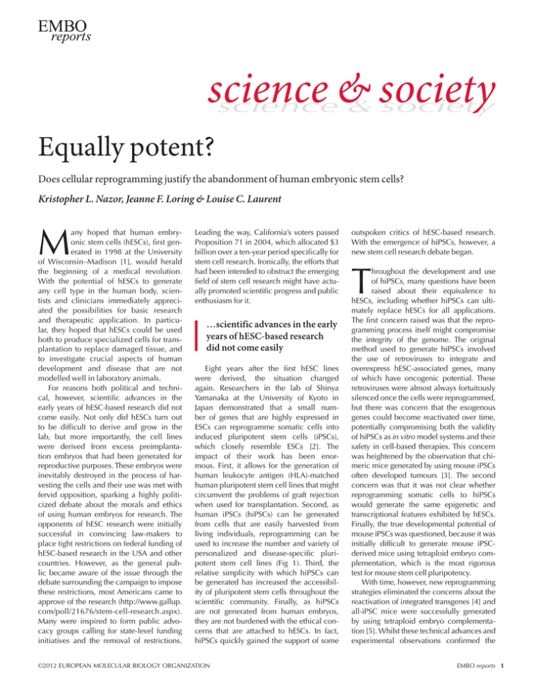
science & society science & society Equally potent? Does cellular reprogramming justify the abandonment of human embryonic stem cells? Kristopher L. Nazor, Jeanne F. Loring & Louise C. Laurent M any hoped that human embryonic stem cells (hESCs), first generated in 1998 at the University of Wisconsin–Madison [1], would herald the beginning of a medical revolution. With the potential of hESCs to generate any cell type in the human body, scientists and clinicians immediately appreciated the possibilities for basic research and therapeutic application. In particular, they hoped that hESCs could be used both to produce specialized cells for transplantation to replace damaged tissue, and to investigate crucial aspects of human develop­ ment and disease that are not modelled­well in laboratory­animals. For reasons both political and technical, however, scientific advances in the early years of hESC-based research did not come easily. Not only did hESCs turn out to be difficult to derive and grow in the lab, but more importantly, the cell lines were derived from excess preimplantation embryos that had been generated for reproductive purposes. These embryos were inevitably destroyed in the process of harvesting the cells and their use was met with fervid opposition, sparking a highly politicized debate about the morals and ethics of using human embryos for research. The opponents of hESC research were initially successful in convincing law-makers to place tight restrictions on federal funding of hESC-based research in the USA and other countries. However, as the general public became aware of the issue through the debate surrounding the campaign to impose these restrictions, most Americans came to approve of the research (http://www.gallup. com/poll/21676/stem-cell-research.aspx). Many were inspired to form public advocacy groups calling for state-level funding initiatives and the removal of restrictions. Leading the way, California’s voters passed Proposition 71 in 2004, which allocated $3 billion over a ten-year period specifically for stem cell research. Ironically, the efforts that had been intended to obstruct the emerging field of stem cell research might have actually promoted scientific progress and public enthusiasm for it. …scientific advances in the early years of hESC-based research did not come easily Eight years after the first hESC lines were derived, the situation changed again. Researchers in the lab of Shinya Yamanaka at the University of Kyoto in Japan demonstrated that a small number of genes that are highly expressed in ESCs can reprogramme somatic cells into induced pluripotent stem cells (iPSCs), which closely resemble ESCs [2]. The impact of their work has been enormous. First, it allows for the generation of human leukocyte antigen (HLA)-matched human pluripotent stem cell lines that might circumvent the problems of graft rejection when used for transplantation. Second, as human iPSCs (hiPSCs) can be generated from cells that are easily harvested from living individuals, reprogramming can be used to increase the number and variety of person­ alized and disease-specific pluri­ potent stem cell lines (Fig 1). Third, the relative simplicity with which hiPSCs can be generated has increased the accessibility of pluripotent stem cells throughout the scientific community. Finally, as hiPSCs are not generated from human embryos, they are not burdened with the ethical concerns that are attached to hESCs. In fact, hiPSCs quickly gained the support of some ©2012 EUROPEAN MOLECULAR BIOLOGY ORGANIZATION out­spoken critics of hESC-based research. With the emergence of hiPSCs, however, a new stem cell research debate began. T hroughout the development and use of hiPSCs, many questions have been raised about their equivalence to hESCs, including whether hiPSCs can ultimately replace hESCs for all applications. The first concern raised was that the reprogramming process itself might compromise the integrity of the genome. The original method used to generate hiPSCs involved the use of retroviruses to integrate and overexpress hESC-associated­genes, many of which have oncogenic potential. These retro­viruses were almost always fortuitously silenced once the cells were reprogrammed, but there was concern that the exogenous genes could become reactivated over time, potentially compromising both the validity of hiPSCs­as in vitro model systems and their safety in cell-based therapies. This concern was heightened by the observation that chimeric mice generated by using mouse iPSCs often developed tumours [3]. The second concern was that it was not clear whether reprogramming somatic cells to hiPSCs would generate the same epigenetic and transcriptional features exhibited by hESCs. Finally, the true develop­mental potential of mouse iPSCs was questioned, because it was initially difficult to generate mouse iPSCderived mice using tetraploid embryo complementation, which is the most rigorous test for mouse stem cell pluripotency. With time, however, new reprogramm­ing strategies eliminated the concerns about the reactivation of integrated transgenes [4] and all-iPSC mice were successfully generated by using tetraploid embryo complementation [5]. Whilst these technical advances and experimental observations confirmed the EMBO reports 1 science & society Isolate The future of stem cell research Cell culture Differentiation Blastocyst Therapeutic use —in clinical trials Drug/toxicity testing, research Inner cell mass Embryonic stem cell Multiple somatic cell types Therapeutic use Adult patient/ donor Drug/toxicity testing, research Somatic cell Somatic cell Multiple somatic cell types Therapeutic use Transfect stem cell genes Somatic cell Drug/toxicity testing, research Pluripotent stem cell Multiple somatic cell types Fig 1 | Simple schematic of the isolation and use of various cell types. In the final part of the scheme, a solid arrow indicates that the cells have been successfully used. A non-solid arrow indicates that clinical trials are ongoing (where indicated) or have not yet begun. pluripotency of iPSCs, questions remained regarding the genomic and epigenetic stability of hESCs and hiPSCs that could only be addressed by further experimentation. B oth hESCs and hiPSCs in culture are prone to genomic instability: recurrent gross chromosomal abnormalities occur, including gains of chromosomes 12, 17 and X. These are probably caused by selection pressures associated with extensive culturing of the cells [6]. Irrespective of their origin, all pluripotent stem cells experience similar selective pressures during long-term culture, so it is not surprising that there is significant overlap in the type and frequency of genetic aberrations between hESCs and hiPSCs. However, these two cell types are derived under markedly different conditions that might impose different selection pressures and could result in systematic­ differences. The derivation of hESCs is a meticulous and painstaking process because human embryos are fragile in culture and require great care to impose as little stress as possible during hESC derivation. Commonly modified variables include the developmental stage of the embryo, embryo quality, oxygen tension, embryo dissection, growth factors, small molecules and the use of extracellular matrix or feeder cells as a growth substrate. Whilst the inner cell mass (ICM) of a blasto­ cyst contains 60–100 cells, as few as 1–6 cells might give rise to an hESC line [7,8]. It is therefore possible that heterogeneity among the ICM cells confers differential fitness during the derivation process. In fact, genetic mosaicism has been shown among cells at all stages of human preimplantation development. As such, the variations in hESC derivation techniques, the mosaicism among cells within a given ICM and the outbred nature of the human population are probably the major sources of the observed variability in behaviour, transcriptional profile, and genomic and epigenetic stability among hESC lines. Whilst the derivation of a hESC line can be interpreted as the stabilization of a transient, pluripotent state, the generation of hiPSCs is more akin to wrestling a differentiated somatic cell into the pluripotent state through forced expression of a few potent regulatory factors. As with hESCs, not all hiPSC lines are generated under the same conditions. In fact, the number of modifiable parameters greatly exceeds those in the hESC derivation process. These include donor age, donor cell type, combinations of reprogramming factors, oxygen tension, growth factors, small molecules and reprogramming factor delivery method, including integrating and non-integrating viral vectors, plasmids, proteins­, mRNAs and miRNAs. G iven the profoundly different ­methods used to generate the cell lines and the different sources of the original cells, there has been interest in determining what genomic and epi­genetic differences might exist between hESCs and hiPSCs. Several studies of the genomic instability and variability among hiPSC and hESC cultures [9,10] show that low-passage hiPSCs contain many copy number variations (CNVs) compared with hESCs, most of which can be traced back to rare cells within the mosaic donor cell population [9,10]. The small number of CNVs that do not seem to originate in the donor populations are probably a direct result of the reprogramming process [11]. However, even shortly after 2 EMBO reports©2012 EUROPEAN MOLECULAR BIOLOGY ORGANIZATION science & society The future of stem cell research reprogramming, clonally derived hiPSC populations are sometimes mosaic for CNVs, suggesting that the mutational events that generated these new CNVs occurred after the initial commitment to pluripotency during the reprogramming process. Intriguingly, most of these reprogramming-associated CNVs seem to place the cells at a selective disadvantage during long-term culture, because they are depleted from the hiPSC cultures during subsequent­passaging [11]. Throughout the development and use of hiPSCs, many questions have been raised about their equivalence to hESCs… Although the overall mutational load of hiPSCs is similar to that of hESCs, the data available so far do not show a perfect overlap between the CNVs observed in each cell type [9–13]. Of course, this could be the result of sampling bias; if this were the case, the examination of a larger number of hiPSC and hESC lines by using high-resolution methods would find an increase in overlap. Alternatively, differences in CNVs between the two cell types could be the result of their origins and methods of derivation. Importantly, there is no direct empirical evidence that the CNVs seen in either hESC or hiPSC lines have an effect on the utility of either cell type. Even so, it is important to identify the range of ‘normal’ CNVs that are observed in hiPSCs derived from healthy individuals, to identify disease-associated CNVs more easily and more accurately in hiPSC-based disease models. In addition, several studies have tried to assess whether differences in genome-wide DNA methylation, chromatin modification and gene expression can distinguish hESCs from hiPSCs, but these have come to conflicting conclusions. The problem is that these experiments are costly and researchers must often decide between breadth—large sample size—and depth—high resolution. Most studies have examined only a few cell lines, and this small sample size has resulted in poor concordance among studies reporting differences between hESCs and hiPSCs; as analysis methods improve and become less costly, more large-scale studies will be done and should resolve these issues. Evidence indicates that cell-type-specific­ genes that have low DNA methylation (hypomethylated) in differentiated somatic cells are consistently highly methylated (hypermethylated) in pluripotent stem cells, suggesting that selective DNA demethyl­ ation takes place as cells differentiate [14]. Another report indicates that regions that are differentially methylated between hiPSCs­and hESCs are hypomethylated in the hiPSCs [15], suggesting that the lower amount of methylation at some loci might be a carry-over from the differentiated cells from which the hiPSCs were generated. Another study has found that whilst some parental somatic-cell-associated gene expression is detectable in early-passage hiPSCs, these differences do not result in an enhanced propensity to differentiate back into the parental cell type [16]. The observation that some hiPSCs and hESCs might have different methyl­ation patterns make it tempting to consider that the differences are due to the failure of genomic loci that are demethylated in a tissuespecific­ manner during normal development to undergo remethylation during reprogramming. If this subset of loci can only be properly methylated through the process of de novo DNA methylation that occurs during gametogenesis, they would be more likely to maintain an appropriate pattern of DNA methylation in hESCs than in hiPSCs­. Such differences could represent bona fide epigenetic differences between hESCs and certain hiPSCs, but it is crucial to keep in mind that large-scale, high-resolution­ studies must be performed to determine whether reproducible DNA methylation differences exist between hESCs and hiPSCs, and whether they truly represent residual DNA methylation patterns characteristic of the original somatic cell type. In addition, there has not yet been any functional significance that has been shown to be attributable to differences in DNA methylation between hiPSCs and hESCs. Whilst there is a perception that hiPSCs can eliminate the need for hESCs, there is also the possibility that direct conversion of cellular identity, or transdifferentiation, between somatic cell types might obviate the need for pluripotent stem cells a­ ltogether in the clinical and preclinical arenas.­Several cell types have been generated in this way, including pancreatic beta islet cells, neurons, neural progenitors, blood and cardio­ myocytes. The primary advantage of direct conversion is that it is typically much faster than sequential generation of hiPSCs and subsequent directed differentiation. However, the process of transdifferentiation is inefficient­ , and in most cases produces ©2012 EUROPEAN MOLECULAR BIOLOGY ORGANIZATION small non-proliferative populations of specific cell types, which limits the utility of this approach for producing cells for replacement therapy or high-throughput applications. Also, human somatic cells such as fibroblasts have finite proliferation ability in vitro, which limits the number of target cells, whilst hiPSCs­can be indefinitely­expanded. Thus, there are several ways to generate specialized cell types for in vitro modelling and cell therapy, including transdifferentiation of somatic cells and directed differentiation of hESCs and hiPSCs. Theoretically, each of these approaches has unique features with particular advantages or disadvantages, depending on the application. It is not yet known whether these techniques produce exactly the same differentiated cell types, but the evidence available so far suggests that hiPSCs and hESCs are similar in their differentiation abilities. …direct conversion of cellular identity, or transdifferentiation, between somatic cell types might obviate the need for pluripotent stem cells altogether in the clinical and preclinical arenas However, some of the most important questions about human pluripotent stem cell differentiated cells derived from either hESCs or hiPSCs have yet to be addressed. How well do the specialized cells generated by differentiation or transdifferentiation mimic the corresponding cells in the body? Do hESCs and hiPSCs differ in their ability to generate new cell types? For example, how well does directed differentiation of hESCs and hiPSCs into neuronal cells recapitulate the epigenetic processes that occur during development of the brain? Do transdifferentiated cells maintain an epi­genetic memory from their parental cell type? Future studies will need to address these questions to determine­the relative value of these approaches. D evelopments in pluripotent stem cell applications will probably have an impact on chronic and untreatable diseases, by providing better models to understand the diseases, improving the efficiency of drug development and, in some cases, providing an alternative therapy consisting of replacement of damaged or destroyed cells. There is enormous pressure EMBO reports 3 science & society A The future of stem cell research Age distribution 1980–2010 (United States) Millions 60 1980 55 1990 50 2000 2010 45 40 35 30 25 20 0–9 10–19 B 20–29 30–39 Years 40–49 50–59 60–69 5.5 5 78 77.5 77 4.5 Alzheimer’s prevalence 76.5 4 76 Life expectancy 75.5 3.5 04 20 05 20 06 20 07 20 08 20 09 20 10 03 20 02 20 01 20 00 20 99 20 98 19 97 19 96 19 95 Disease prevalence (United States) 500 22 450 20 18 400 Diabetes 350 16 300 14 Autism 250 12 10 10 20 09 20 20 08 07 20 06 20 05 20 04 20 03 20 02 20 01 20 00 200 20 Children with autism (0–9 years old; thousands) C 19 94 19 93 19 92 19 91 19 19 19 90 75 Diagnosed diabetics (all ages; millions) Life expectancy (years) 78.5 Alzheimer prevalence (all ages; millions) Life expectancy and Alzheimer prevalence (United States) 79 Fig 2 | Statistics on population growth and disease prevalence in the United States. (A) Distribution of the population by age in 1980, 1990, 2000 and 2010 according to the US Census Bureau. (B) Life expectancy according to the US Census Bureau (http://www.alz.org/alzheimers_disease_21590.asp) and Alzheimer disease prevalence estimations according to the Alzheimer’s Association’s annual ‘Facts & Figures’ reports (http://www.alz.org/alzheimers_disease_21590.asp). (C) Disease prevalence estimations for autism and diabetes according to the Center for Disease Control (http://www.cdc.gov/DataStatistics) and the US Census Bureau. to develop new therapies or preventive measures to ease the social and economic burden of chronic diseases in all developed countries, particularly diseases that are associated with ageing and the steady increase in life expectancy (Fig 2A,B; data from the US Census Bureau, http://www. alz.org/alzheimers_disease_21590.asp). Owing in part to the rising prevalence of Alzheimer disease (Fig 2B; data from the US Census Bureau, http://www.alz.org/ alzheimers_disease_21590.asp), the cost of caring for the growing US population of elderly patients with dementia is estimated at $200 billion per year in the USA alone. At the other end of the age spectrum, the prevalence of autism spectrum disorders increased by nearly 80% from 2000 to 2010 (Fig 2C; data from the Centers for Disease Control and Prevention, http://www.cdc.gov/ DataStatistics). With 1 in 88 children in the USA affected, the care and education of autistic children is challenging the infrastructure of schools and governmental social service programmes, at an estimated annual cost of $126 billion.­We also see a marked increase in the prevalence of diabetes in the USA, such that 11.3% of adults over 20 years of age have diabetes, at a direct cost of $120 billion annually (data from the 2011 National Diabetes Fact Sheet from the Centers for Disease Control and Prevention, http:// www.cdc.gov/diabetes/pubs/estimates11. htm#1). If we consider all chronic diseases, approximately 23% of all Americans are affected by at least one chronic condition, at an annual cost of approximately $1.7 trillion or roughly 12% of the 2012 US GDP (data from the Almanac of Chronic Disease 2009, http://www.fightchronicdisease.org/sites/ fightchronicdisease­.org/files/docs/2009Alm anacofChronicDisease_updated81009.pdf). In The Creative Destruction of Medicine, Eric Topol used a quote from eighteenth century philosopher Voltaire to convey how certain aspects of medicine have remained largely unchanged over the past 250 years: “Doctors are men who prescribe medicines of which they know little, to cure diseases of which they know less, in human beings of whom they know nothing.” However acerbic and rhetorical this assertion might seem, it does make the point that to treat a disease effectively, we must first understand it. It is here where the truly revolutionary potential of stem cell research comes in. By using hiPSC technology, we can begin to explore the fundamental mechanisms that underlie human disease by generating pluripotent cell lines from people suffering with Parkinson disease, cardiomyopathies, Alzheimer disease, diabetes and other disorders. These disease-specific hiPSCs can then be differentiated into disease-relevant cell types such as neurons, cardiac myocytes and beta islet cells, and compared with cells differentiated from hiPSCs from 4 EMBO reports©2012 EUROPEAN MOLECULAR BIOLOGY ORGANIZATION science & society The future of stem cell research healthy individuals. This knowledge will eventually be used to develop new strategies for drug development. Stem cell research is not only bridging the gap between the lab bench and the clinic, but also promoting collaboration between academia and industry. Major pharmaceutical companies have invested in the development of pluripotent stem-cell-based platforms for drug discovery, often in close collaboration with academic scientists. Stem cell research is not only bridging the gap between the lab bench and the clinic, but also promoting collaboration between academia and industry For several degenerative diseases that affect one specific cell type, such as diabetes­and Parkinson disease, there is also great interest in using pluripotent stem-cell-derived cells directly for cell replacement. It will be a few years before we know how well this approach works, although phase I clinical trials for spinal cord injury and macular degeneration are providing some experience of the safety of these therapies. Whilst it might be tempting to assume that hESCs are superior to hiPSCs because they are cultured from a naturally occurring cell type, or that hiPSC-derived or transdifferentiated cells are better suited for transplantation than those from hESCs because they can be generated from specific patients, we must remind ourselves that we do not have enough evidence either for or against these assumptions. The challenges that we face to understand and ultimately conquer human disease are monumental and humbling. To eliminate any ethically acceptable means to reach such an important goal would be a meritless barrier to the advancement of this new era in medicine. ACKNOWLEDGEMENTS K.L.N. is supported by an Autism Speaks Dennis Weatherstone fellowship. K.L.N. and J.F.L. are supported by the California Institute for Regenerative Medicine (CL1‑00502, RT1‑01108, TR1‑01250, RN2‑00931‑1), the National Institutes of Health (NIH; R33MH87925), the Millipore Foundation and the Esther O’Keefe Foundation. L.C.L. is supported by a NIH/National Institute of Child Health & Development K12 Career Development Award and the Hartwell Foundation. CONFLICT OF INTEREST The authors declare that they have no conflict of interest. REFERENCES 1. Thomson JA, Itskovitz-Eldor J, Shapiro SS, Waknitz MA, Swiergiel JJ, Marshall VS, Jones JM (1998) Embryonic stem cell lines derived from human blastocysts. Science 282: 1145–1147 2. Takahashi K, Yamanaka S (2006) Induction of pluripotent stem cells from mouse embryonic and adult fibroblast cultures by defined factors. Cell 126: 663–676 3. Okita K, Ichisaka T, Yamanaka S (2007) Generation of germline-competent induced pluripotent stem cells. Nature 448: 313–317 4. Ban H et al (2011) Efficient generation of transgene-free human induced pluripotent stem cells (iPSCs) by temperature-sensitive Sendai virus vectors. Proc Natl Acad Sci USA 108: 14234–14239 5. Boland MJ, Hazen JL, Nazor KL, Rodriguez AR, Gifford W, Martin G, Kupriyanov S, Baldwin KK (2009) Adult mice generated from induced pluripotent stem cells. Nature 461: 91–94 6. Mitalipova MM, Rao RR, Hoyer DM, Johnson JA, Meisner LF, Jones KL, Dalton S, Stice SL (2005) Preserving the genetic integrity of human embryonic stem cells. Nat Biotechnol 23: 19–20 7. Klimanskaya I, Chung Y, Becker S, Lu SJ, Lanza R (2006) Human embryonic stem cell lines derived from single blastomeres. Nature 444: 481–485 8. Yamagata K, Ueda J, Mizutani E, Saitou M, Wakayama T (2010) Survival and death of epiblast cells during embryonic stem cell derivation revealed by long-term live-cell imaging with an Oct4 reporter system. Dev Biol 346: 90–101 9. Gore A et al (2011) Somatic coding mutations in human induced pluripotent stem cells. Nature 471: 63–67 ©2012 EUROPEAN MOLECULAR BIOLOGY ORGANIZATION 10. Hussein SM et al (2011) Copy number variation and selection during reprogramming to pluripotency. Nature 471: 58–62 11. Laurent LC et al (2011) Dynamic changes in the copy number of pluripotency and cell proliferation genes in human ESCs and iPSCs during reprogramming and time in culture. Cell Stem Cell 8: 106–118 12. Mayshar Y, Ben-David U, Lavon N, Biancotti JC, Yakir B, Clark AT, Plath K, Lowry WE, Benvenisty N (2010) Identification and classification of chromosomal aberrations in human induced pluripotent stem cells. Cell Stem Cell 7: 521–531 13. Amps K et al (2011) Screening ethnically diverse human embryonic stem cells identifies a chromosome 20 minimal amplicon conferring growth advantage. Nat Biotechnol 29: 1132–1144 14. Nazor KL et al (2012) Recurrent variations in DNA methylation in human pluripotent stem cells and their differentiated derivatives. Cell Stem Cell 10: 620–634 15. Lister R et al (2011) Hotspots of aberrant epigenomic reprogramming in human induced pluripotent stem cells. Nature 471: 68–73 16. Ohi Y et al (2011) Incomplete DNA methylation underlies a transcriptional memory of somatic cells in human iPS cells. Nat Cell Biol 13: 541–549 Kristopher L. Nazor and Jeanne F. Loring [middle] are at the Center for Regenerative Medicine, Department of Chemical Physiology, The Scripps Research Institute, La Jolla, California, USA. Jeanne F. Loring also holds an adjunct position at the University of California, San Diego, California, USA. Louise C. Laurent is at the University of California, San Diego, Department of Reproductive Medicine, La Jolla, California, USA. E‑mail: llaurent@ucsd.edu EMBO reports advance online publication 18 September 2012; doi:10.1038/embor.2012.134 EMBO reports 5


