Biceps Dynamometer Elbow Strength and Endurance Testing After Distal Reconstruction with Allograft
advertisement
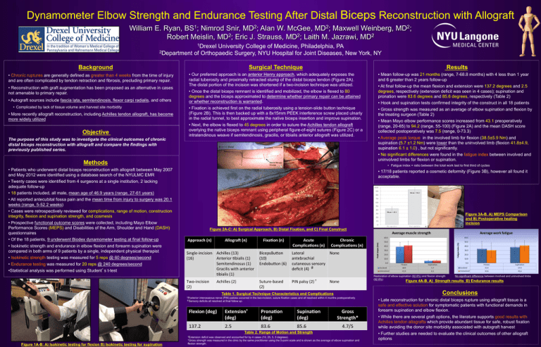
Dynamometer Elbow Strength and Endurance Testing After Distal Biceps Reconstruction with Allograft 1 BS ; 2 MD ; 2 MD ; William E. Ryan, Nimrod Snir, Alan W. McGee, Maxwell Weinberg, 2 2 2 Robert Meislin, MD ; Eric J. Strauss, MD ; Laith M. Jazrawi, MD 2 MD ; 1Drexel University College of Medicine, Philadelphia, PA 2Department of Orthopaedic Surgery, NYU Hospital for Joint Diseases, New York, NY Surgical Technique Background • Chronic ruptures are generally defined as greater than 4 weeks from the time of injury and are often complicated by tendon retraction and fibrosis, precluding primary repair. • Reconstruction with graft augmentation has been proposed as an alternative in cases not amenable to primary repair. • Autograft sources include fascia lata, semitendinosis, flexor carpi radialis, and others • Complicated by lack of tissue volume and harvest site morbidity • More recently allograft reconstruction, including Achilles tendon allograft, has become more widely utilized Objective The purpose of this study was to investigate the clinical outcomes of chronic distal biceps reconstruction with allograft and compare the findings with previously published series. Results • Our preferred approach is an anterior Henry approach, which adequately exposes the radial tuberosity and proximally retracted stump of the distal biceps tendon (Figure 2A). The distal portion of the incision was shortened if a two-incision technique was utilized. • Once the distal biceps remnant is identified and mobilized, the elbow is flexed to 80 degrees and the biceps approximated to determine whether primary repair can be attained or whether reconstruction is warranted. • Fixation is achieved first on the radial tuberosity using a tension-slide button technique (Figure 2B). This is then backed up with a 8x15mm PEEK interference screw placed ulnarly in the radial tunnel, to best approximate the native biceps insertion and improve supination. • Next, the elbow is flexed to 45 degrees in order to suture the Achilles tendon allograft overlying the native biceps remnant using peripheral figure-of-eight sutures (Figure 2C) or a intratendinous weave if semitendinosis, gracilis, or tibialis anterior allograft was utilized. Methods • Isokinetic strength and endurance in elbow flexion and forearm supination were compared in both arms of 9 patients by a single, independent physical therapist • Isokinetic strength testing was measured for 5 reps @ 60 degrees/second • Endurance testing was measured for 20 reps @ 240 degrees/second • Statistical analysis was performed using Student’s t-test • Average peak torque in the involved limb for flexion (38.5±5.9 Nm) and supination (5.7 ±1.2 Nm) were lower than the uninvolved limb (flexion 41.8±4.9, supination 6.1 ± 1.0) , but not significantly. • No significant differences were found in the fatigue index between involved and uninvolved limbs for flexion or supination. • 17/18 patients reported a cosmetic deformity (Figure 3B), however all found it acceptable. Figure 3A-B. A) MEPS Comparison and B) Postoperative healing incision Figure 2A-C: A) Surgical Approach, B) Distal Fixation, and C) Final Construct Approach (n) Allogra. (n) Fixa2on (n) Acute Complica2ons (n) Chronic Complica2ons (n) Single-­‐incision Achilles (13) BicepsBu?on (16) Anterior 5bialis (1) (10) Semitendinosus (1) Endobu?on (6) Gracilis with anterior 5bialis (1) Lateral None antebrachial cutaneous sensory deficit (4) ♯ Two-­‐incision (2) PIN palsy (2) † Achilles (2) Suture-­‐based (2) Restoration of elbow supination (92.6%) and flexion strength (92.8%) None Conclusions †Posterior interosseous nerve (PIN) palsies occurred in the two-incision, suture fixation cases and all resolved within 4 months postoperatively. ♯Sensory deficits all resolved at final follow up. Flexion (deg) Extension† (deg) Prona2on (deg) Supina2on (deg) 137.2 2.5 83.6 85.6 Gross Strength* 4.7/5 Table 2. Range of Motion and Strength †Extension No significant difference between involved and uninvolved limbs Figure 4A-B. A) Strength results B) Endurance results Table 1. Surgical Technique Characteristics and Complications Figure 1A-B: A) Isokinetic testing for flexion B) Isokinetic testing for supination • Gross strength was measured as an average of elbow supination and flexion by the treating surgeon (Table 2) • Mean Mayo elbow performance scores increased from 43.1 preoperatively (range, 20-65) to 94.2 (range, 55-100) (Figure 2A) and the mean DASH score collected postoperatively was 7.5 (range, 0-73.3) • Fatigue index = ratio between the total work last to first third of cyctes • Patients who underwent distal biceps reconstruction with allograft between May 2007 and May 2012 were identified using a database search of the NYULMC EMR • Twenty cases were identified from 4 surgeons at a single institution, 2 lacking adequate follow-up • 18 patients included, all male, mean age of 46.9 years (range, 27-61 years) • All reported antecubital fossa pain and the mean time from injury to surgery was 20.1 weeks (range, 5-52.2 weeks) • Cases were retrospectively reviewed for complications, range of motion, construction integrity, flexion and supination strength, and cosmesis • Prospective functional outcome scores were collected, including Mayo Elbow Performance Scores (MEPS) and Disabilities of the Arm, Shoulder and Hand (DASH) questionnaires • Of the 18 patients, 9 underwent Biodex dynamometer testing at final follow-up • Mean follow-up was 21 months (range, 7-68.8 months) with 4 less than 1 year and 6 greater than 2 years follow-up • At final follow-up the mean flexion and extension were 137.2 degrees and 2.5 degrees, respectively (extension deficit was seen in 4 cases); supination and pronation were 83.6 degrees and 85.6 degrees, respectively (Table 2) • Hook and supination tests confirmed integrity of the construct in all 18 patients deficit was observed and accounted for by 4 cases (10, 25, 5, 5 degrees). *Gross strength was measured in the clinic by the same practitioner using the 5-point scale and is shown as the average of elbow supination and flexion strength. • Late reconstruction for chronic distal biceps rupture using allograft tissue is a safe and effective solution for symptomatic patients with functional demands in forearm supination and elbow flexion. • While there are several graft options, the literature supports good results with Achilles tendon allografts which provide abundant tissue for safe, robust fixation while avoiding the donor site morbidity associated with autograft harvest • Further studies are needed to evaluate the clinical outcomes of other allograft options

