Document 11117925
advertisement
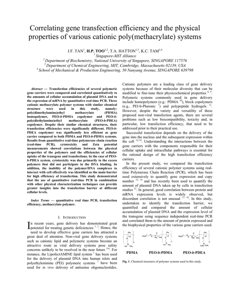
Correlating gene transfection efficiency and the physical properties of various cationic poly(methacrylate) systems J.F. TAN1, H.P. TOO1,2, T.A. HATTON1,3, K.C. TAM1,4 1 Singapore-MIT Alliance 2 Department of Biochemistry, National University of Singapore, SINGAPORE 117576 3 Department of Chemical Engineering, MIT, Cambridge, Massachusetts 02139, USA 4 School of Mechanical & Production Engineering, 50 Nanyang Avenue, SINGAPORE 639798 Abstract — Transfection efficiencies of several polymeric gene carriers were compared and correlated quantitatively to the amounts of cellular accumulation of plasmid DNA and to the expression of mRNA by quantitative real time PCR. Three cationic methacrylate polymer systems with similar chemical structure were used in this study, namely: poly(dimethylamino)ethyl methacrylate (PDMA) homopolymer, PEO-b-PDMA copolymer and PEO-bpoly(diethylamino)ethyl methacrylate (PEO-b-PDEA) copolymer. Despite their similar chemical structures, their transfection efficiencies were significantly different. PEO-bPDEA copolymer was significantly less efficient as gene carrier compared to both PDMA and PEO-b-PDMA systems. Results from quantitative real-time polymerase chain reaction (real-time PCR), cytotoxicity and Zeta potential measurements showed correlations between the physical properties of the polymers and the efficiencies of cellular uptake of the transgene and transfections. In the case of PEOb-PDEA system, cytotoxicity was due primarily to the excess polymers that did not participate in the DNA binding. In addition, the inability of the polymer/DNA complexes to interact with cell effectively was identified as the main barrier for high efficiency of transfection. This study demonstrated that the use of quantitative real-time PCR in combination with other physical characterization techniques can provide greater insights into the transfection barrier at different cellular levels. Index Terms — quantitative real time PCR, transfection efficiency, methacrylate polymer. I. INTRODUCTION I n recent years, gene delivery has demonstrated great potential for treating genetic deficiencies 1, 2. Hence, the need to develop effective gene carriers has attracted a great deal of attention. Non-viral gene delivery systems such as cationic lipid and polymeric systems become an attractive route as viral delivery systems pose safety concerns unlikely to be resolved in the near future 3-6. For instance, the LipofectAMINE lipid system 1 has been used for the delivery of plasmid DNA into human islets and polyethylenimine (PEI) polymeric systems 7 have been used for in vivo delivery of antisense oligonucleotides. Cationic polymers are a leading class of gene delivery systems because of their molecular diversity that can be modified to fine-tune their physicochemical properties 8, 9. Polymeric systems commonly used in gene delivery include homopolymers (e.g.: PDMA 10), block copolymers (e.g., PEI-b-Pluronic 7) and polypeptide hydrogels 11. However, despite the variety and versatility of these proposed non-viral transfection agents, there are several problems such as low biocompatibility, toxicity and, in particular, low transfection efficiency, that need to be addressed prior to their practical use. Successful transfection depends on the delivery of the gene into the nucleus and the subsequent expression within the cell 12-14. Understanding the interactions between the gene carriers with the components responsible for their cellular uptake and intracellular pathways is essential for the rational design of the high transfection efficiency carriers. In the present study, we compared the transfection efficiency of several cationic polymer systems using realtime Polymerase Chain Reaction (PCR), which has been used extensively to quantify gene expression and copy number 15, 16 and has recently been used to quantify the amount of plasmid DNA taken up by cells in transfection studies 12. In general, good correlation between protein and mRNA expression levels is widely observed, but discordant correlation is not unusual 17, 18. In this study, undertaken to identify the transfection barrier, we quantified and compared the amount of cellular accumulation of plasmid DNA and the expression level of the transgene using sequence independent real-time PCR and correlated them to the amount of protein expressed and the biophysical properties of the various gene carriers used. CH3 CH3O O O O m O CH3 O H3C O CH3 O CH3 PDMA O m O H3C O CH3 O n n N H3C CH3 O CH3O N H3C CH3 PEO-b-PDMA n N C2H5 C2H5 PEO-b-PDEA Fig. 1: Chemical structures of polymer systems used in this study. Three cationic polymer systems with similar chemical structures were used in this study (Fig. 1). PDMA is a cationic hydrophilic polymer which has attracted considerable attention for gene therapy applications 10, 19, 20, both as a homopolymer and as block copolymers with polyethers, such as PEO-b-PDMA, which we consider here. PEO-b-PDEA is an amiphiphilic block copolymer capable of forming pH-dependent micelles. At physiological pH (pH 7.4), PDEA chains are somewhat hydrophobic and drive the formation of micelles which are stabilized by the hydrophilic PEO segments. The cationic PDEA segments of the copolymer are able to bind to negatively charged DNA to form polymer/DNA complexes 21, 22 . However, despite the similarities in the chemical structures of PDMA and PDEA, the number of cells transfected by their respective copolymers with PEO was found to be distinctively different. From the quantitative real time PCR results, cytotoxicity data and Zeta potential measurements, a correlation between the biophysical properties of the polymer/DNA polyplexes, cellular accumulation of plasmid DNA and the transfection efficiencies were observed. This study showed that quantifying the accumulation and expression of cellular transgenes, complementing with cytotoxicity studies and in combination with biophysical studies will enable a better understanding of the factors responsible for transfection efficiency. II. EXPERIMENTAL SECTION Preparation of Plasmid DNA. Plasmid GFP (pIREShrGFP-2a), an expression vector containing the fully humanized green fluorescence protein (GFP) driven by CMV promoter was obtained from Stratagene Inc. The plasmid was amplified using Escherichia coli (DH5α) and purified using the Quantum Plasmid Miniprep kit (Biorad). Polymer Synthesis. A full description of the synthesis and characterization of the polymers has recently been reported 21 . The degree of polymerization (DP) for the three polymer systems is about 65 -75 repeat units of methacrylate monomer which was determined using polystyrene GPC standard. The molecular weight of the PEO polymer (Dow Chemical) was Mn ~ 5000 Da. Zeta Potential Measurement. The measurements were carried out using a Brookhaven Zeta PALS (phase analyzer light scattering) as described previously 22. Cell culture and Cytotoxicity (MTS assay) Study. Neuro2A cells (CCL-131, ATCC) were cultured in Dulbecco's Modification of Eagle's Medium (DMEM) supplemented with 10% fetal calf serum (FCS) at 37 oC, 5% CO2 and 95% relative humidity. For cytotoxicity studies, cells (40,000 cells/well) were seeded into each of the 96 wells in a microtiter plate (Nunc, Wiesbaden, Germany) using MTS (3-(4,5-dimethylthiazol-2-yl)-5-(3carboxy-methoxy-phenyl)-2-(4-sulfo-phenyl)-2Htetrazolium) assay; details are provided in an earlier publication 21. Lactate Dehydrogenase (LDH) Assay. The amount of LDH was assayed with the Cytotoxicity Detection Kit (LDH), according to the manufacturer’s instructions (Roche Applied Science). Controls were performed with 0.1% (w/v) Triton X-100 and set as total LDH released. The relative LDH release is defined by the ratio of LDH released over total LDH in the intact cells. Less than 10% LDH release was regarded as a measure of non-toxicity. In vitro transfection. Various cationic polymers at different polycation/DNA ratios were mixed with 4 µg of purified plasmid per well of transfected cells. The polycation/DNA mixtures were diluted to a final volume of 1 ml with serum-free medium (SFM) and allowed to stand at 25 oC for 20 min. Two set of six-well plates (one set was used for plasmid DNA accumulation and the other RNA level measurements), seeded 24 h before transfection with 0.5 x 10⁶ cells per well were prepared. The solutions of polycation/DNA complexes were then added into the wells and incubated at 37 oC, 5% CO2 and 95% relative humidity for six hours. For RNA analysis and expression of transgene, the culture medium was replaced with DMEM supplemented with 10% FCS, and the cells were cultured for 96 h. Thereafter, the number of fluorescent positive cells and total RNA was extracted for real-time PCR. Real time PCR using SYBR Green I. Real-time PCR was carried out on the iCycler iQ (Bio-Rad, Hercules, CA, USA), following an initial denaturation for 3 min at 95 oC,, by 40 cycles of 60 s denaturation at 95 oC, 30 s annealing at 60 oC and 60 s extension at 72 oC. Fluorescence detection was used during the annealing phase. The reaction was carried out in a total volume of 40 µl in 1x XtensaMix-SG™ (BioWORKS), containing 2.5 mM MgCl , 10 pmol of primers and 0.5 U DNA polymerase (DyNAzyme II, Finnzymes Oy, Espoo, Finland). All realtime PCR reactions were carried out simultaneously with linearized plasmid standards and a negative control (water). Forward primer (ATCCGCAGCGACATCAACCT) and reverse primer (ACGCCCTTGCTCTTCATCAG) were designed to amplify a fragment of 238 base pairs within the hrGFP open reading frame based on the vector sequence. III. RESULTS Cytotoxicity. The toxicity of various polymer/DNA complexes in Neuro2A cells was investigated using MTS assay, with the results shown in Fig. 2. A general trend of increasing toxicity with increasing polymer concentration was observed for all polymer systems. However, despite the similarity of the chemical structures of the polymers, the effects on cell viability were different for each of the three polymers. The toxicity varied linearly with concentration for both PDMAEMA and PEO-b-PDMA; as many as 80% of the cells were viable below polymer concentrations of 50 µg/ml. For PEO-b-PDEA system, however, a sigmoidal variation in cell viability with polymer concentration was observed with a sharp increase in the cytotoxicity (< 50% cell viability) for polymer 120% overall surface charge of the system. Fig 3 shows the zeta potentials of the various polymer/DNA complexes as a function of the total polymer concentration. Clearly, the zeta potential of the PEO-b-PDEA polymer is significantly lower than that of the other two systems; at saturation, the zeta potential of the PEO-b-PDEA polyplex is only slightly above zero, while for the PDMA complex it has a value close to +30 mV. 40 PDMA 30 ζ Potential (mV) concentrations above 50 µg/ml. Interestingly, our previous work 22 on the aggregation behavior of the PEO-b-PDEA copolymer with plasmid DNA showed that at concentration of approximately 50 µg/ml, PEO-b-PDEA/DNA complexes undergo structural transformation. Below that concentration, both PDMA homopolymer and PEO-bPDEA copolymer polyplexes have similar morphologies and particle sizes. Above this critical concentration, PEOb-PDEA/DNA complexes undergo significant structural rearrangement to form compact micelle-like structures, driven by the amiphiphilic nature of this copolymer. This observation suggests that the amiphiphilic micelle-like structures induce higher cytotoxicity as compared to homopolymer PDMA/DNA complexes that do not undergo structural transformation. 20 PEO-b -PDMA 10 0 PEO-b -PDEA -10 100% % cell valibility -20 80% PDMA -30 60% 0 PEO-b -PDMA 20 40 60 80 100 120 Polymer Concentration (µg/ml) 40% Fig. 3: Zeta potential of various polyplexes formed with 10 µg of plasmid DNA. 20% PEO-b -PDEA 0% 0 50 100 150 200 250 polymer concentration (µg/ml) Fig. 2: Cytotoxicity of various polymer/DNA complexes systems in Neuro2A cell lines. The cell viability was determined by MTS assay and was shown as mean and the error bars is the standard deviation of five samples. Guided by these toxicity results, we carried out all subsequent transfection studies at polymer concentrations 50 µg/ml and below, where cytotoxicity was minimal for all three polymer systems. In Vitro Transfection of Neuro2A cells. Fluorescence microscopy was used to evaluate the transfection efficiencies at the protein level for the various polymer systems. Significant higher number of GFP-transfected cells was observed with either PDMA or PEO-b-PDMA polyplexes compared to the result in the absence of the polymeric transfection agents (i.e. naked DNA only). These results were consistent with other reports where PDMA and/or its copolymers was found to enhance transfection 10, 23. However, the transfection efficiency of the PEO-b-PDEA copolymer (< 0.1 % cells transfected) was significantly lower than PDMA (12 ± 0.9 % cells transfected), or PEO-b-PDMA (11 ± 0.7 % cells transfected). Despite the similarities in the chemical structures of the polymers there were significant differences in the transfection efficiencies. In an attempt to better understand the structural-functional relationships of the polymers, the zeta potentials were measured and the cellular uptake and expression of the transgene quantified by real-time PCR. Zeta Potential Results. Zeta potential represents the Real-time PCR results. The cellular uptake and expression of the transgene of the various optimized transfection systems were compared using real time RTPCR (Fig. 4). Quantification at the mRNA expression level (amount of transgene inside the nucleus) and accumulation of the plasmid DNA at the cellular level are shown in Figs. 4b and 4c, respectively. Amplification of plasmid standards that was also carried out simultaneously is shown in Fig. 6a. As a control in quantifying mRNA levels, no significant amplification of the transgene was observed when reverse transcription was omitted (data not shown). At the exponential phase of PCR, the amplification process can be described by the equation 24, Tn = To(1+ε)n (1) where Tn is the number of target molecules at cycle n, To is the initial number of target molecules, ε is the efficiency of amplification (a value between 0 to 1) and n is the number of cycles. Taking logarithm, rearranging equation (1) and setting n = Ct (the threshold cycle) we obtain the equation Ct = log(TC ) t log(1+ ε ) − log(T0 ) log(1+ ε ) (2) The efficiency of amplification (ε) of a target molecule can be calculated from the slope of the curve of Ct versus log(Tc). For maximum efficiency of ε = 1, the slope will have a value of 3.32. The amplification of the standards in Fig. 6a yielded a slope of the curve of 3.47, corresponding to a high amplification efficiency of about 0.90. The dynamic range of amplification was greater than 5 log dilutions (10-16 – 10-21 moles). Amplification signals detected in the no-template control (W) case were due to the formation of primer-dimer, as verified by gel electrophoresis (data not shown). 1000 RFU y = -3.47x + 6.89 2 R = 0.996 100 10 0 5 10 15 20 25 30 35 40 Cycles 1000 PEO-b -PDEA RFU PEO-b -PDMA PDMA 100 DNA only Cells only 10 5 10 15 20 25 30 Cycles 1000 PEO-b -PDEA RFU PEO-b -PDMA PDMA 100 Cells only DNA only accumulation at the cellular level are shown in Fig. 4c. Six hours after the transfection, DNA (both genomic and plasmid DNA) were extracted and the plasmid DNA was then quantified by real-time PCR. The overall trends for the various systems at the DNA cellular level were similar to those observed at the mRNA level. The results for PDMA and PEO-b-PDMA were very much comparable at different concentrations for both mRNA and plasmid DNA accumulation at cellular levels, implying that the presence of PEO does not have a significant effect on the transfection pathway, which is of no surprise. For the PEO-b-PDEA polyplexes system, the cellular plasmid DNA content was significantly lower than the PDMA and PEO-b-PDMA systems. This observation suggests that the possible barrier to the use of the PEO-bPDEA system may reside in the inability of the polyplexes to be associated and/or transported across the cellular membrane as compared to other two polymer systems. Lactate Dehydrogenase (LDH) Assay. LDH is a cytoplasmic enzyme that is normally not secreted outside the cells and is often used as an indicator of cell membrane disruption 25, 26. In this experiment, the LDH assay was used to compare the extent of cell membrane disruption in the presence of various polymer systems. Fig. 5 shows that neither PDMA nor PEO-b-PDMA polymer systems induce significant damage to the cell membrane, (< 10% of LDH was released). However, PEO-b-PDEA caused a significant disruption of the cell membrane. More importantly, it was observed that the % LDH released in the presence of free PEO-b-PDEA polymer (59% LDH released) was significantly higher than the PEO-b-PDEA + DNA (32% LDH released), under which both polyplexes and excess polymer chains were present. 10 5 10 15 20 25 100% 30 Polymer only Cycles Fig 4: Real-time PCR results. (a) Amplification of five log10 dilution of GFP standards. Negative controls with no templates (W) were carried out simultaneously. (b) Amplification of various optimized transfection agents at mRNA level. (c) Amplification of various optimized transfection agents at DNA cellular uptake/binding level. The real-time PCR results at the mRNA expression level shown in Fig. 4b indicate high expression levels of GFP transcripts for both PDMA and PEO-b-PDMA polyplexes (Ct value ~ 12) when compared to cells transfected with naked GFP (Ct value ~ 22 (high Ct values indicate low levels of gene expression and vice versa). The difference in the Ct value of naked DNA and PDMA polyplexes was about 10 cycles, an approximately 1000 fold difference of the amounts of transgene uptaken. On the other hand, the amount of GFP transgene uptaken in cells exposed to PEOb-PDEA polyplexes (Ct value ~ 18) was significantly lower than the DMA/polyplexes systems. Since similar trend was observed between the protein and mRNA expression levels were observed for the various polymer systems, the translation of mRNA to protein is unlikely to be a major transfection barrier for these polymer systems as gene carrier. The real-time PCR results for plasmid DNA % LDH released 80% Polymer + DNA 60% 40% 20% 0% PDMA PEO-b-PDMA PEO-b-PDEA Fig. 5: The effect of polymer (50 µg/ml) on membrane integrity as quantified by the release of the cytoplasmic enzyme LDH. Differences in the % LDH released between PEO-b-PDEA polymer and polyplexes were statistically significant based on the paired two-tailed t-test. A value of P < 0.05 was considered significant (P=0.0083). Each value represents the mean + standard deviation of five determinations. IV. DISCUSSION We have evaluated three cationic polymers with similar chemical structures for their DNA transfection efficiency. The number of cells expressing green fluorescence protein, the expression levels of the transgene, and the cellular content of the plasmid DNA all clearly showed that, despite their similarity in structures, these different polymers exhibit significant differences in transfection capability. A closer examination of the real-time PCR results (Fig. 4) provides insights into the disparities between the transfection efficiencies of the various polymer systems. In the case of PEO-b-PDEA, both the mRNA level and the plasmid DNA accumulation level were significantly lower than their corresponding levels of the two PDMA systems. If the main transfection barrier occurred at the intracellular level before the transgene reached the nucleus, the real-time PCR results should show high plasmid DNA accumulation levels with low mRNA expression levels. However, in the case of PEO-b-PDEA system, both plasmid DNA accumulation level and mRNA expression levels were low, indicating that the transfection barrier probably occurred at the extra-cellular membrane level. The bottleneck is probably due to the inability of the polyplexes to be transported across the cellular membrane, which is generally the first hurdle along the transfection pathway. However, why were the PEO-b-PDEA polyplexes not able to broach the cellular membrane (as the RT-PCR results suggest) and yet they exhibited high toxicity at high polymer concentration (as the cytotoxicity results indicate)? The results of the LDH assays shed some light on this apparent contradiction. Fig. 5 reveals that the membrane disruption effect was much smaller with PEO-b-PDEA polyplexes than the free PEO-b-PDEA polymers only. In other words, membrane disruption appears to be due primarily to the free PEO-b-PDEA polymers (i.e., those that are not involved in the DNA binding) rather than due to the PEO-b-PDEA/DNA polyplexes themselves. Similar observations showing that free polymers cause higher cytotoxicity than do their polyplexes have also been reported for other systems 27, 28. On the other hand, the sigmoid cytotoxicity of PEO-b-PDEA polymer is probably due to the excessive membrane disruption due to its amiphiphilic nature of the PEO-b-PDEA polymers chains. In recent years, a large number of novel cationic polymer and surfactant systems have been reported and characterized for gene transfection. But in some instances, the transfection systems may have good biophysical characteristics such as ‘enhanced endosomal escape 29’ or ‘resistance to enzymatic degradation 30’ or even ‘with targeted moiety 31’ but also low protein expression level. The reasons for the poor correlation between their biophysical properties and transfection results are not well investigated. The strategies and approaches outlined in this study could probably enable future non-viral gene delivery researches to gain greater insights into the transfection barriers at different cellular levels. V. CONCLUSIONS We have evaluated the transfection efficiency of three cationic poly(methacrylate) systems at various levels: protein, mRNA and plasmid DNA accumulation. Although the three polymers are similar structurally, their transfection efficiencies were not always comparable. Cytotoxicity, real-time PCR and zeta potential measurements showed that, for PEO-b-PDEA, transport across the cellular membrane is probably the major barrier to transfection, because interactions between the polyplex and the membrane wall are weak. Furthermore, the somewhat amiphiphilic nature of the free polymer allows it to penetrate and disrupt the cell wall, leading to significant cytotoxicity at high concentrations. For the PDMA and PEO-b-PDMA polymers, on the other hand, relatively higher transfection efficiencies were observed, attributed to the electrostatic adsorption of the polyplexes to the cell surface that facilitates its uptake by endocytosis, with significantly smaller cytotoxicity. This study has demonstrated that transfection barriers can be investigated well by correlating the protein expression levels, mRNA expression levels and cellular plasmid DNA levels with the physical properties of the gene carrier,. These insights into the structure-transfection relationship could prove to be helpful for the optimization of polymeric gene delivery system. VI. ACKNOWLEDGEMENT J.F. acknowledges the financial support provided by the Singapore-MIT (SMA) Alliance, and thanks Yong Li Foong and Peng Zhong Ni from NUS for several fruitful discussions and suggestions. REFERENCES 1. Mahato, R. I.; Henry, J.; Narang, A. S.; Sabek, O.; Fraga, D.; Kotb, M.; Gaber, A. O., Cationic lipid and polymer-based gene delivery to human pancreatic islets. Mol. Ther. 2003, 7, (1), 89-100. 2. Pinthus, J. H.; Waks, T.; Kaufman-Francis, K.; Schindler, D. G.; Harmelin, A.; Kanety, H.; Ramon, J.; Eshhar, Z., Immuno-gene therapy of established prostate tumors using chimeric receptor-redirected human lymphocytes. Cancer Res. 2003, 63, (10), 2470-2476. 3. Roth, C. M.; Sundaram, S., Engineering synthetic vectors for improved DNA delivery: Insights from intracellular pathways. Annu. Rev. Biomed. Eng. 2004, 6, 397-426. 4. Pannier, A. K.; Shea, L. D., Controlled release systems for DNA delivery. Mol. Ther. 2004, 10, (1), 19-26. 5. Niidome, T.; Huang, L., Gene therapy progress and prospects: Nonviral vectors. Gene Ther. 2002, 9, (24), 1647-1652. 6. Pack, D. W.; Hoffman, A. S.; Pun, S.; Stayton, P. S., Design and Development of polymers for gene delivery. Nature Reviews 2005, 4, 581593. 7. Belenkov, A. I.; Alakhov, V. Y.; Kabanov, A. V.; Vinogradov, S. V.; Panasci, L. C.; Monia, B. P.; Chow, T. Y. K., Polyethyleneimine grafted with pluronic P85 enhances Ku86 antisense delivery and the ionizing radiation treatment efficacy in vivo. Gene Ther. 2004, 11, (22), 16651672. 8. Putnam, D.; Gentry, C. A.; Pack, D. W.; Langer, R., Polymer-based gene delivery with low cytotoxicity by a unique balance of side-chain termini. Proc. Natl. Acad. SCI. U.S.A. 2001, 98, (3), 1200-1205. 9. Azzam, T.; Eliyahu, H.; Makovitzki, A.; Linial, M.; Domb, A. J., Hydrophobized dextran-spermine conjugate as potential vector for in vitro gene transfection. J. Controlled Release 2004, 96, (2), 309-323. 10. Mastrobattista, E.; Kapel, R. H. G.; Eggenhuisen, M. H.; Roholl, P. J. M.; Crommelin, D. J. A.; Hennink, W. E.; Storm, G., Lipid-coated polyplexes for targeted gene delivery to ovarian carcinoma cells. Cancer gene ther. 2001, 8, (6), 405-413. 11. Megeed, Z.; Haider, M.; Li, D. Q.; O'Malley, B. W.; Cappello, J.; Ghandehari, H., In vitro and in vivo evaluation of recombinant silkelastinlike hydrogels for cancer gene therapy. J. Controlled Release 2004, 94, (2-3), 433-445. 12. Varga, C.; Tedford, N.; Thomas, M.; Klibanov, A.; Griffith, L.; Lauffenburger, D., Quantitative comparison of polyethylenimine formulations and adenoviral vectors in terms of intracellular gene delivery processes. Gene Ther. 2005, 1-10. 13. Chesnoy, S.; Huang, L., Structure and function of lipid-DNA complexes for gene delivery. Annu. Rev. Biophys. Biomol. Struct. 2000, 29, 27-47. 14. Wang, K. C.; Wu, J. C.; Chung, Y. C.; Ho, Y. C.; Chang, D. T.; Hu, Y. C., Baculovirus as a Highly Efficient Gene Delivery Vector for the Expression of Hepatitis Delta Virus Antigens in Mammalian Cells. Biotech. & Bioeng. 2004, 89, (4), 464-473. 15. Too, H. P., Real time PCR quantification of GFR alpha-2 alternatively spliced isoforms in murine brain and peripheral tissues. Mol. Brain Res. 2003, 114, (2), 146-153. 16. Giulietti, A.; Overbergh, L.; Valckx, D.; Decallonne, B.; Bouillon, R.; Mathieu, C., An overview of real-time quantitative PCR: applications to quantify cytokine gene expression. Methods 2001, 25, 386-401. 17. Gygi, S. P.; Rochon, Y.; Franza, B. R.; Aebersold, R., Correlation between protein and mRNA abundance in yeast. Mol. Cell. Proteomics 1999, 19, (3), 1720-1730. 18. Chen, G. A.; Gharib, T. G.; Huang, C. C.; Taylor, J. M. G.; Misek, D. E.; Kardia, S. L. R.; Giordano, T. J.; Iannettoni, M. D.; Orringer, M. B.; Hanash, S. M.; Beer, D. G., Discordant protein and mRNA expression in lung adenocarcinomas. Mol. Cell. Proteomics 2002, 1, (4), 304-313. 19. Zabner, J.; Fasbender, A.; Moninger, T.; Poellinger, K.; Welsh, M., Cellular and molecular barriers to gene transfer by a cationic lipid. J. Biol. Chem. 1995, 270, 18997-19004. 20. Bromberg, L.; Deshmukh, S.; Temchenko, M.; Iourtchenko, L.; Alakhov, V.; Alvarez-Lorenzo, C.; Barreiro-Iglesias, R.; Concheiro, A.; Hatton, T. A., Polycationic block copolymers of poly(ethylene oxide) and poly(propylene oxide) for cell transfection. Bioconjugate Chem. 2005, 16, (3), 626-633. 21. Tan, J. F.; Ravi, P.; Too, H. P.; Hatton, T. A.; Tam, K. C., Association behavior of biotinylated and non-biotinylated PEO-bPDEAEMA. Biomacromolecules 2005, 6, (1), 498-506. 22. Tan, J. F.; Too, H. P.; Hatton, T. A.; Tam, K. C., Aggregation Behavior and Thermodynamics of binding between PEO-PDEAEMA and plasmid DNA. Langmuir 2005. 23. van Steenis, J. H.; van Maarseveen, E. M.; Verbaan, F. J.; Verrijk, R.; Crommelin, D. J. A.; Storm, G.; Hennink, W. E., Preparation and characterization of folate-targeted pEG-coated pDMAEMA-based polyplexes. J. Controlled Release 2003, 87, (1-3), 167-176. 24. Rasmussen, R., Quantification on the LightCycler. In Rapid Cycle Real-Time PCR, Meuer, S.; Wittwer, C.; Nakagawara, K. I., Eds. Springer: 2001; pp 21-34. 25. Moghimi, S. M.; Symonds, P.; Murray, J. C.; Hunter, A. C.; Debska, G.; Szewczyk, A., A Two-Stage Poly(ethylenimine)-Mediated Cytotoxicity: Implications for Gene Transfer/Therapy. Mol. Ther. 2005, 11, (6), 990-995. 26. Vihola, H.; Laukkanen, A.; Valtola, L.; Tenhu, H.; Hirvonen, J., Cytotoxicity of thermosensitive polymers poly(N-isopropylacrylamide), poly(N-vinylcaprolactam) and amphiphilically modified poly(Nvinylcaprolactam). Biomaterials 2005, 26, (16), 3055-3064. 27. Kunath, K.; von Harpe, A.; Fischer, D.; Kissel, T., Galactose-PEI– DNA complexes for targeted gene delivery: degree of substitution affects complex size and transfection efficiency. J. Controlled Release 2003, 88, 159-172. 28. Fischer, D.; Li, Y. X.; Ahlemeyer, B.; Krieglstein, J.; Kissel, T., In vitro cytotoxicity testing of polycations: influence of polymer structure on cell viability and hemolysis. Biomaterials 2003, 24, (7), 1121-1131. 29. Funhoff, A. M.; van Nostrum, C. F.; Koning, G. A.; Schuurmans, N. M. E.; Crommelin, D. J. A.; Hennink, W. E., Endosomal escape of polymeric gene delivery complexes is not always enhanced by polymers buffering at low pH. Biomacromolecules 2004, 5, (1), 32-39. 30. Fischer, D.; Dautzenberg, H.; Kunath, K.; Kissel, T., Poly(diallyldimethylammonium chlorides) and their N-methyl-Nvinylacetamide copolymer-based DNA-polyplexes: role of molecular weight and charge density in complex formation, stability, and in vitro activity. Int. J. Pharm. 2004, 280, 253-269. 31. Dauty, E.; Remy, J. S.; Zuber, G.; Behr, J. P., Intracellular Delivery of Nanometric DNA Particles via the Folate Receptor. Bioconjugate Chem. 2002, 13, 831-839.
