Scanning transmission electron microscopy investigation of the Si 111
advertisement

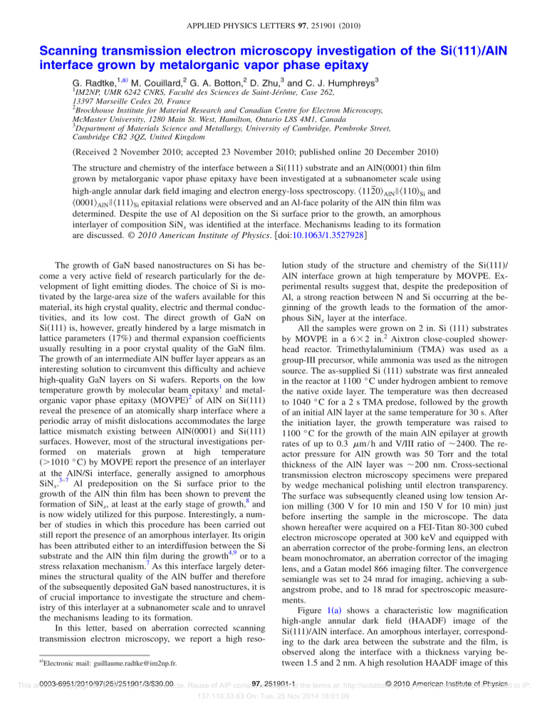
APPLIED PHYSICS LETTERS 97, 251901 共2010兲 Scanning transmission electron microscopy investigation of the Si„111…/AlN interface grown by metalorganic vapor phase epitaxy G. Radtke,1,a兲 M. Couillard,2 G. A. Botton,2 D. Zhu,3 and C. J. Humphreys3 1 IM2NP, UMR 6242 CNRS, Faculté des Sciences de Saint-Jérôme, Case 262, 13397 Marseille Cedex 20, France 2 Brockhouse Institute for Material Research and Canadian Centre for Electron Microscopy, McMaster University, 1280 Main St. West, Hamilton, Ontario L8S 4M1, Canada 3 Department of Materials Science and Metallurgy, University of Cambridge, Pembroke Street, Cambridge CB2 3QZ, United Kingdom 共Received 2 November 2010; accepted 23 November 2010; published online 20 December 2010兲 The structure and chemistry of the interface between a Si共111兲 substrate and an AlN共0001兲 thin film grown by metalorganic vapor phase epitaxy have been investigated at a subnanometer scale using high-angle annular dark field imaging and electron energy-loss spectroscopy. 具112̄0典AlN 储 具110典Si and 具0001典AlN 储 具111典Si epitaxial relations were observed and an Al-face polarity of the AlN thin film was determined. Despite the use of Al deposition on the Si surface prior to the growth, an amorphous interlayer of composition SiNx was identified at the interface. Mechanisms leading to its formation are discussed. © 2010 American Institute of Physics. 关doi:10.1063/1.3527928兴 The growth of GaN based nanostructures on Si has become a very active field of research particularly for the development of light emitting diodes. The choice of Si is motivated by the large-area size of the wafers available for this material, its high crystal quality, electric and thermal conductivities, and its low cost. The direct growth of GaN on Si共111兲 is, however, greatly hindered by a large mismatch in lattice parameters 共17%兲 and thermal expansion coefficients usually resulting in a poor crystal quality of the GaN film. The growth of an intermediate AlN buffer layer appears as an interesting solution to circumvent this difficulty and achieve high-quality GaN layers on Si wafers. Reports on the low temperature growth by molecular beam epitaxy1 and metalorganic vapor phase epitaxy 共MOVPE兲2 of AlN on Si共111兲 reveal the presence of an atomically sharp interface where a periodic array of misfit dislocations accommodates the large lattice mismatch existing between AlN共0001兲 and Si共111兲 surfaces. However, most of the structural investigations performed on materials grown at high temperature 共⬎1010 ° C兲 by MOVPE report the presence of an interlayer at the AlN/Si interface, generally assigned to amorphous SiNx.3–7 Al predeposition on the Si surface prior to the growth of the AlN thin film has been shown to prevent the formation of SiNx, at least at the early stage of growth,8 and is now widely utilized for this purpose. Interestingly, a number of studies in which this procedure has been carried out still report the presence of an amorphous interlayer. Its origin has been attributed either to an interdiffusion between the Si substrate and the AlN thin film during the growth4,9 or to a stress relaxation mechanism.7 As this interface largely determines the structural quality of the AlN buffer and therefore of the subsequently deposited GaN based nanostructures, it is of crucial importance to investigate the structure and chemistry of this interlayer at a subnanometer scale and to unravel the mechanisms leading to its formation. In this letter, based on aberration corrected scanning transmission electron microscopy, we report a high resoa兲 Electronic mail: guillaume.radtke@im2np.fr. lution study of the structure and chemistry of the Si共111兲/ AlN interface grown at high temperature by MOVPE. Experimental results suggest that, despite the predeposition of Al, a strong reaction between N and Si occurring at the beginning of the growth leads to the formation of the amorphous SiNx layer at the interface. All the samples were grown on 2 in. Si 共111兲 substrates by MOVPE in a 6 ⫻ 2 in.2 Aixtron close-coupled showerhead reactor. Trimethylaluminium 共TMA兲 was used as a group-III precursor, while ammonia was used as the nitrogen source. The as-supplied Si 共111兲 substrate was first annealed in the reactor at 1100 ° C under hydrogen ambient to remove the native oxide layer. The temperature was then decreased to 1040 ° C for a 2 s TMA predose, followed by the growth of an initial AlN layer at the same temperature for 30 s. After the initiation layer, the growth temperature was raised to 1100 ° C for the growth of the main AlN epilayer at growth rates of up to 0.3 m / h and V/III ratio of ⬃2400. The reactor pressure for AlN growth was 50 Torr and the total thickness of the AlN layer was ⬃200 nm. Cross-sectional transmission electron microscopy specimens were prepared by wedge mechanical polishing until electron transparency. The surface was subsequently cleaned using low tension Arion milling 共300 V for 10 min and 150 V for 10 min兲 just before inserting the sample in the microscope. The data shown hereafter were acquired on a FEI-Titan 80-300 cubed electron microscope operated at 300 keV and equipped with an aberration corrector of the probe-forming lens, an electron beam monochromator, an aberration corrector of the imaging lens, and a Gatan model 866 imaging filter. The convergence semiangle was set to 24 mrad for imaging, achieving a subangstrom probe, and to 18 mrad for spectroscopic measurements. Figure 1共a兲 shows a characteristic low magnification high-angle annular dark field 共HAADF兲 image of the Si共111兲/AlN interface. An amorphous interlayer, corresponding to the dark area between the substrate and the film, is observed along the interface with a thickness varying between 1.5 and 2 nm. A high resolution HAADF image of this 0003-6951/2010/97共25兲/251901/3/$30.00 97,is251901-1 © 2010 American InstituteDownloaded of Physics to IP: This article is copyrighted as indicated in the article. Reuse of AIP content subject to the terms at: http://scitation.aip.org/termsconditions. 137.110.33.63 On: Tue, 25 Nov 2014 18:01:09 251901-2 Appl. Phys. Lett. 97, 251901 共2010兲 Radtke et al. FIG. 1. 共Color online兲 共a兲 Low-magnification HAADF view of the Si共111兲/ AlN interface. 共b兲 High-resolution HAADF image of the interface acquired in the 具110典 zone axis of the Si wafer. 共c兲 Detail of the atomic structure of the film showing the Al-polarity of AlN observed in HAADF and 共d兲 in ABF. FIG. 2. 共Color online兲 EELS spectrum-image of the Si共111兲/AlN interface: 共a兲 HAADF image, 共b兲 Si map extracted from the Si-L23 edge 共99 eV兲, 共c兲 N map extracted from the N-K edge 共401 eV兲, 共d兲 Al map extracted from the Al-L23 edge 共77 eV兲, and 共e兲 EELS profiles obtained by integrating the signal perpendicularly to the interface. The dotted lines represent the approximate position of the interfacial layer. dicularly to the interface and normalized to their maximum interface, acquired with a 75 mrad ADF inner angle in the Si height. Note that no detectable O signal was recorded in this 具110典 zone axis is displayed in Fig. 1共b兲. Interestingly, dearea. As these experiments were performed with a probe size spite the presence of this continuous layer, the epitaxial reof about 1 Å, the broad distribution of the chemical profiles lations 具112̄0典AlN 储 具110典Si and 具0001典AlN 储 具111典Si are fairly presented here is intrinsic to the sample and does not arise from an experimental broadening. This chemical analysis well respected. A detailed investigation of the atomic strucconfirms that the amorphous zone observed in the HAADF at ture of AlN, shown in Fig. 1共c兲, reveals the Al-face polarity the interface corresponds to a SiNx layer. The chemical proof the film. The short Al–N interatomic distance in this profiles shown in Fig. 2共e兲 bring, however, further information. jection 共1.1 Å兲 and the presence of the neighboring strongly First, the distribution of Si 共3 nm wide兲 extends beyond the scattering Al make the N appear as an elongation of the Al SiNx interlayer into the AlN film. This signal can be attribalternatively pointing diagonally left and right. For compariuted either to overlapping AlN and SiNx signals over the son, an image of the same area acquired in annular bright thickness of the specimen or to a heavily doped AlN interfield 共ABF兲 with a 12–26 mrad detector, allowing a better mediate phase. Second, the profiles of N and Al, respectively, visibility of the light N atomic columns,10 is displayed in Fig. 2 and 2.5 nm wide, are similar but shifted with respect to 1共d兲 and confirms the Al-face polarity of the thin film. each other by 1.8 nm. A non-negligible Al signal is therefore In a second step, the chemical analysis of this interface only observed above the SiNx interlayer, in the AlN film. was carried out using electron energy-loss spectroscopy In order to confirm our analysis of the interlayer, energy共EELS兲 spectrum imaging. Figure 2 shows the twoloss near edge structures 共ELNESs兲 of the Si-L23, Al-L23, and dimensional maps obtained from a 175⫻ 36 pixels N-K edges, displayed in Fig. 3, were recorded for the three spectrum-image acquired at 300 keV without drift correction, phases with a high-energy resolution 共⬃0.3 eV兲 in monoa dwell time of 30 ms/pixel, and a collection semiangle of 80 chromated mode. Whereas the signatures recorded in the Si mrad. Figure 2共a兲 shows the HAADF image acquired simulsubstrate and the AlN thin film exhibit very fine structures taneously with the EELS spectra. Figures 2共b兲–2共d兲 show, characteristic of the bulk references,11,12 the ELNESs acrespectively, the Si, N, and Al maps obtained by multiple quired in the interlayer are much broader, as expected for a linear least squares fit of the spectrum-image with reference substoichiometric amorphous material. The chemical shift spectra acquired in the same conditions. This procedure was observed on the Si-L23 edge between the interlayer and the Si required for a proper extraction of the Al and Si signals from substrate 共2.5⫾ 0.5 eV兲, attributed in first approximation to their overlapping L23 edges. Finally, Fig. 2共e兲 shows the elemental profiles obtained by integrating the signal perpena charge transfer from Si to N atoms, also appears as a direct to IP: This article is copyrighted as indicated in the article. Reuse of AIP content is subject to the terms at: http://scitation.aip.org/termsconditions. Downloaded 137.110.33.63 On: Tue, 25 Nov 2014 18:01:09 251901-3 Appl. Phys. Lett. 97, 251901 共2010兲 Radtke et al. FIG. 3. 共Color online兲 High-energy resolution ELNES obtained from bulk Si 共solid line兲, bulk AlN 共dashed line兲, and from the interfacial layer 共dotted line兲. The spectra displayed here are the sum of spectra acquired by scanning the probe parallel to the interface as indicated by the dots in the inset. evidence for the formation of Si–N chemical bonds in the amorphous phase. Our chemical analysis of the interface shows that Al predeposition is unable to prevent the strong reaction occurring between N and the Si substrate, at least at high temperatures. Elemental profiles across the interface favor the hypothesis of the formation of the SiNx layer underneath the surface at the very beginning of the growth. An Al and Si rich surfactant layer could indeed form simultaneously, owing to their relatively low surface energies 共⬃1200 mJ/ m2兲. This is supported by the large proportion of Si found above the amorphous layer and by the absence of detectable Al in the Si wafer and the SiNx region. After reaching a critical thickness, the SiNx layer acts as a barrier, considerably slowing down the diffusion of Si from the wafer toward the surface. The increasing concentration of Al at the surface would then trigger the growth of the AlN thin film. The same analysis has been carried out on a 20 nm thick AlN epilayer sample grown under the same conditions for a shorter time 共⬃4 min兲. The observation of an amorphous interlayer with the same characteristics further supports the hypothesis of a rapid formation of SiNx as a first step of the growth process. Within this framework, the crystallographic relationship between the AlN layer and the Si substrate, as well as the Al-face polarity of the film, also reported previously in the presence of a sharp Si共111兲/AlN interface,2 remains unexplained. Different hypotheses have been formulated to clarify this point, particularly in the case of growths preceded by a nitridation step.13 A plausible explanation resides in the presence of contact points between the Si substrate and the AlN film due to discontinuities in the SiNx interlayer. Electron microscopy was carried out at the Canadian Centre for Electron Microscopy, a facility supported by the NSERC 共Canada兲 and McMaster University. C.J.H. and G.A.B. are grateful for funding from the Ministry of Research and Innovation of Ontario 共ISOP program兲. G.R. and G.A.B. acknowledge the Fonds France-Canada pour la Recherche for partial support of this collaboration. G.R. thanks A. Saúl 共CINaM-CNRS, Marseille兲 for helpful discussions. 1 A. Bourret, A. Barski, J. L. Rouvière, G. Renaud, and A. Barbier, J. Appl. Phys. 83, 2003 共1998兲. 2 R. Liu, F. A. Ponce, A. Dadgar, and A. Krost, Appl. Phys. Lett. 83, 860 共2003兲. 3 K. Y. Zang, S. J. Chua, L. S. Wang, and C. V. Thompson, Phys. Status Solidi C 0, 2067 共2003兲. 4 S.-J. Lee, S.-H. Jang, S.-S. Lee, and C.-R. Lee, J. Cryst. Growth 249, 65 共2003兲. 5 S. Tanaka, Y. Kawaguchi, N. Sawaki, M. Hibino, and K. Hiramatsu, Appl. Phys. Lett. 76, 2701 共2000兲. 6 S. Kaiser, M. Jakob, J. Zweck, W. Gebhardt, O. Ambacher, R. Dimitrov, A. T. Schremer, J. A. Smart, and J. R. Shealy, J. Vac. Sci. Technol. B 18, 733 共2000兲. 7 G. Q. Hu, X. Kong, L. Wan, Y. Q. Wang, X. F. Duan, Y. Lu, and X. L. Liu, J. Cryst. Growth 256, 416 共2003兲. 8 K. Y. Zang, L. S. Wang, S. J. Chua, and C. V. Thompson, J. Cryst. Growth 268, 515 共2004兲. 9 D. Xi, Y. Zheng, P. Chen, Z. Zhao, P. Chen, S. Xie, B. Shen, S. Gu, and R. Zhang, Phys. Status Solidi A 191, 137 共2002兲. 10 S. D. Findlay, N. Shibata, H. Sawada, E. Okunishi, Y. Kondo, T. Yamamoto, and Y. Ikuhara, Appl. Phys. Lett. 95, 191913 共2009兲. 11 V. Serin, C. Colliex, R. Brydson, S. Matar, and F. Boucher, Phys. Rev. B 58, 5106 共1998兲. 12 P. E. Batson, Phys. Rev. B 47, 6898 共1993兲. 13 E. Arslan, M. K. Ozturk, Ö. Duygulu, A. Arslan Kaya, S. Ozcelik, and E. Ozbay, Appl. Phys. A: Mater. Sci. Process. 94, 73 共2009兲. This article is copyrighted as indicated in the article. Reuse of AIP content is subject to the terms at: http://scitation.aip.org/termsconditions. Downloaded to IP: 137.110.33.63 On: Tue, 25 Nov 2014 18:01:09


