Current Biology
Dispatches
Cytoskeletal Chirality: Swirling Cells Tell Left
from Right
Alex Mogilner1,2,* and Ben Fogelson1
1Courant
Institute of Mathematical Sciences, New York University, 251 Mercer Street, New York, NY 10012, USA
of Biology, New York University, 100 Washington Square East, 1009 Silver Center, New York, NY 10003, USA
*Correspondence: mogilner@cims.nyu.edu
http://dx.doi.org/10.1016/j.cub.2015.04.039
2Department
A new study reports that dynamic actin fibers in cells on circular islands self-organize into a swirling counterclockwise pattern and describes a basic cytoskeletal mechanism for the establishment of left–right
asymmetry that is based on myosin contraction and twisting of the formin–actin filament.
How developing embryos acquire
left–right asymmetry is a tantalizing
question of great physiological
importance [1,2]. Asymmetric organ
morphogenesis follows asymmetric
signaling cascades, which in turn follow
asymmetric events on the cellular
scale — for example, rotating cilia that
generate a right-to-left fluid flow in mouse
embryos [3]. Underlying such cell-scale
asymmetries must be some molecular
event that generates chirality — the
asymmetric property of a structure
whereby it is distinguishable from its
mirror image. A prime candidate for the
molecular system governing chirality is
the cytoskeleton, which contains innately
handed molecules, such as the
right-handed actin helix. Indeed,
cytoskeleton-dependent chiralities have
been identified in many organisms, from
Drosophila to Caenorhabditis elegans to
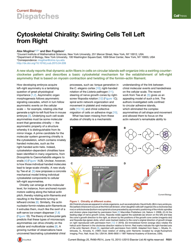
snails [4] (Figure 1A,B). Unclear, however,
is how those individual handed molecules
lead to large-scale chirality. A new study
by Tee et al. [5] now proposes a concrete
mechanical model linking individual
cytoskeletal components to cellular
left–right asymmetries.
Chirality can emerge at the molecular
level, for instance, from anchored myosin
motors walking along the helical actin
pitch, thereby rotating actin filaments and
resulting in the filaments turning in
leftward circles [6]. Similarly, the actin
nucleator formin rotates actin filaments as
it extrudes them, much like the swirl of a
soft-serve ice-cream dispenser [7,8]
(Figure 1D). The theory of active polar gels
predicts that these types of microscopic
asymmetries can drive chirality on the
cellular and multicellular scales [9]. A
growing number of observations have
uncovered fascinating cytoskeletal chiral
processes, such as: torque generation in
the C. elegans cortex [10]; right-handed
rotation of the Listeria pathogen [11];
steering of nerve growth cones by rightscrew filopodia rotation [12] (Figure 1C);
spiral actin network organization and
movement in platelet and melanophore
cells [13,14]; and chiral collective
migration of cells on adhesive rings [15].
What has been missing from these
studies of chirality is a mechanistic
understanding of the link between
chiral molecular events and handedness
on the cellular scale. The recent
work from Tee et al. [5] gives us an
appealing model of such a link. The
authors investigated cells confined
to circular adhesive islands,
which eliminated the complex
asymmetries of cell shape and movement
and allowed them to focus on the
actin network’s remarkable ability to
Figure 1. Chirality at different scales.
(A) Chiral structures are apparent in whole organisms, such as snails (photo: David Huth). (B) In many embryos,
the earliest chiral event occurs at the third cell division, when daughter cells self-organize into a clockwise (top
row) or anticlockwise (bottom row) spiral structure preceded by asymmetric positioning of the mitotic spindles
and division plane (reprinted by permission from [2] Macmillan Publishers Ltd, Nature ª 2008). (C) At the
leading edge of nerve growth cones, filopodia rotate against the substrate (as shown on the left) and bias
the cone’s growth direction to the right, as shown by the positions of the growth cone center (magenta dot)
and filopodia tips (green dots), which were tracked relative to the cone’s original direction of growth (image
on right reproduced with permission from [12] ª 2010). (D) Immobilized formin (orange disc) rotates the
helical actin filament in the clockwise direction relative to formin. Red marks indicate the right-handed axis
of the actin filament. (From [8], reprinted with permission from AAAS. Adapted from Sase, I., Miyata, H.,
Ishiwata, S., Kinosita, K. (1997) Axial rotation of sliding actin filaments revealed by single-fluorophore
imaging. Proc. Natl. Acad. Sci. USA 94, 5646–5650 ª 1997 National Academy of Sciences, USA).
Current Biology 25, R490–R514, June 15, 2015 ª2015 Elsevier Ltd All rights reserved R501
Current Biology
Dispatches
Current Biology
Figure 2. Mechanical chiral instability.
Anticlockwise rotation of radial actin filaments (green arrow) develops via a ‘rack and pinion’ mechanism
(center inset) from an initially radial structure (top left), when formin-nucleated actin filaments (red)
are rotated by formins immobilized at focal adhesions (blue circles). A small leftwards tilt (top right)
of radial actin filaments, pivoting at the focal adhesions, causes contractile transverse fibers (yellow) on
either side of the tilting radial fiber to slide relative to one another (bottom left; yellow arrows illustrate
contractile force). This sliding generates a torque imbalance which amplifies the tilt (bottom right).
self-organize into a beautiful array of
patterns [16].
Tee et al. [5] observed a familiar
cobweb-like system of radial and
transverse fibers (RFs and TFs,
respectively) [17]. The RFs grew
centripetally from the cell periphery, like
spokes on a wheel. These fibers — which
resemble the ‘dorsal’ stress fibers of
migrating cells — extended inward from
focal adhesions, were rich in the
crosslinking protein a-actinin-1 and did
not contain myosin II. TFs — which
resemble previously described ‘transverse
arcs’ — ran parallel to the cell edge, linking
the RFs together. An elegant experiment
showed that nanoparticles conjugated
with myosin V molecules travelled
bidirectionally along TFs, indicating that
there was no left–right asymmetry in these
fibers. The nanoparticles did not bind to
the RFs, so the organization of actin in
RFs remains unclear. Electron microscopy
showed that the two systems of fibers
passed through each other, hinting at a
likely physical interaction. Photobleaching
and pharmacological perturbations
demonstrated that RFs grew inward,
likely from formins at the focal adhesions,
and that the formin-independent TFs
moved inward more quickly than the
RFs, meaning that TFs were sliding relative
to the RFs. Inhibition of myosin revealed
that this TF sliding was generated by
myosin-powered contraction.
Unexpectedly, a couple of hours after
the cobweb radial pattern emerged, the
RFs broke radial symmetry and started to
swirl. This swirling lasted for a few hours
before giving way to a linear pattern of
fibers spanning the whole cell with both
ends associated with focal adhesions at
the cell edge. Swirling almost always
happened in an anticlockwise direction.
Serendipitously, Tee et al. [5] noticed that
when they overexpressed the crosslinker
a-actinin-1, swirling was less frequent
but could also go in the opposite,
i.e. clockwise, direction.
These results raise two burning
questions: why do the RFs tilt, and why
do they overwhelmingly tilt in the
anticlockwise direction? Since these
questions could not be answered by cell
biological or biophysical methods, the
authors turned to a simple computational
model of TFs as contractile springs that
can slide along RFs. The simulations
predicted an instability: when RFs
undergo a slight tilt relative to the radius
of the cell, the tilt tends to increase
(Figure 2). Understanding this instability is
highly nontrivial, which is why modelling
was so crucial. If the TFs were not sliding
relative to RFs, then the non-chiral
symmetric position of the RFs would have
been stable. However, if TF sliding is fast
and a RF pivots to the left with respect
to the focal adhesion (as shown in
Figure 2), then the TFs pulling it to the left
R502 Current Biology 25, R490–R514, June 15, 2015 ª2015 Elsevier Ltd All rights reserved
slide down to the turning tip of the RF,
while the TFs to the right slide closer up
towards the focal adhesion. This sliding
changes the lever arms for the left- and
right-pulling TFs, generating a net torque
that tilts the RF further from the radial
direction.
This model explains the emergence of
swirling, but not its predominantly
anticlockwise direction. The authors
hypothesized that, as RF filaments grow
inwards from formins embedded in focal
adhesions, the filaments would rotate in a
clockwise direction (if one looks at the
filament from its barbed end at the focal
adhesion; Figures 1D and 2). If these
rotating radial filaments act like gears
driving the TFs (Figure 2), then the RFs will
propel the TFs in an anticlockwise
direction in the cell. Note that in order for
this ‘rack and pinion’ explanation to work,
TFs have to be located higher than the
RFs relative to the substrate, which is not
explicitly stated in the new study [5]. What
about the clockwise swirling when the
a-actinin crosslinkers are
overexpressed? With high levels of
crosslinking, formin-generated twist is
stored elastically in the filament tips rather
than rotating filaments like a gear, and
from time to time this twist is relieved
abruptly by quick anticlockwise rotations
of the filament, pushing the RFs in the
clockwise direction in the cell. Note that
the formin-generated twist does not have
to be significant — even a slight bias can
tweak the tilting instability into a preferred
direction. One of the lessons from the
model is that torque, a mechanical factor
that remains underappreciated but is
gathering attention [8,9,18], plays an
important role in cells.
Many open questions remain. Are the
TFs really located higher than the RFs
relative to the substrate? Electron
microscopy images are inconclusive. Are
actin filaments in RFs really oriented with
their barbed ends outward? Is the TF
sliding fast enough to explain the tilting
instability? These gaps in our
understanding will no doubt be filled via
follow-up studies. There are larger
questions: when the swirling actin pattern
evolves into the linear pattern, does some
part of the chirality survive? What
happens to chirality in cells that are
geometrically unconstrained? Does this
self-organization work in a more
physiologically relevant setting, for
Current Biology
Dispatches
example, with cells of highly irregular
shape embedded into a 3D extracellular
matrix?
Most importantly, is this selforganization an epiphenomenon or does it
have a function? Alignment of linear actin
fibers, similar to that observed when the
swirling ends, is an important stage of
spontaneous polarization in some cell
types [19]. A few years ago it was reported
that neutrophils polarize to the left of an
arrow drawn from the center of the
nucleus of an unpolarized cell to
its centrosome [20]. Thus, although the
mechanism reported in the new study [5]
depends on actin dynamics and is
independent of microtubules, it could be
that the transient handedness of the actin
network is only able to sense left and
right in a polarized cell, which could be
useful for motile cells navigating complex
chemical and physical gradients. It is
also possible that the tilting and swirling
enable the cell or its organelles to rotate
[12–14], or mechanically ease the
transition from the ‘spokes-on-a-wheel’
pattern into the linear fiber arrangement,
or create skewed transportation tracks or
stresses in the cell.
Even with all these questions
unanswered, for the first time we have a
concrete physical understanding of how
actin self-organization could use chiral
molecular motors to give the cell a sense
of right and left.
REFERENCES
1. Coutelis, J.-B., González-Morales, N.,
Géminard, C., and Noselli, S. (2014). Diversity
and convergence in the mechanisms
establishing L/R asymmetry in metazoa.
EMBO Rep. 15, 926–937.
2. Grande, C., and Patel, N.H. (2008). Nodal
signalling is involved in left-right asymmetry in
snails. Nature 457, 1007–1011.
3. Nonaka, S., Tanaka, Y., Okada, Y., Takeda, S.,
Harada, A., Kanai, Y., Kido, M., and Hirokawa,
N. (1998). Randomization of left-right
asymmetry due to loss of nodal cilia generating
leftward flow of extraembryonic fluid in mice
lacking KIF3B motor protein. Cell 95, 829–837.
4. Rose, L., and Gönczy, P. (2014). Polarity
establishment, asymmetric division and
segregation of fate determinants in early C.
elegans embryos. WormBook, 1–43.
5. Tee, Y.H., Shemesh, T., Thiagarajan, V.,
Hariadi, R.F., Anderson, K.L., Page, C.,
Volkmann, N., Hanein, D., Sivaramakrishnan,
S., Kozlov, M.M., et al. (2015). Cellular chirality
arising from the self-organization of the actin
cytoskeleton. Nat. Cell Biol. 17, 445–457.
6. Pyrpassopoulos, S., Feeser, E.A., Mazerik,
J.N., Tyska, M.J., and Ostap, E.M. (2012).
Membrane-bound myo1c powers asymmetric
motility of actin filaments. Curr. Biol. 22,
1688–1692.
7. Shemesh, T., Otomo, T., Rosen, M.K.,
Bershadsky, A.D., and Kozlov, M.M. (2005). A
novel mechanism of actin filament processive
capping by formin: solution of the rotation
paradox. J. Cell Biol. 170, 889–893.
8. Mizuno, H., Higashida, C., Yuan, Y., Ishizaki,
T., Narumiya, S., and Watanabe, N. (2011).
Rotational movement of the formin mDia1
along the double helical strand of an actin
filament. Science 331, 80–83.
9. Fürthauer, S., Strempel, M., Grill, S.W., and
Jülicher, F. (2013). Active chiral processes in
thin films. Phys. Rev. Lett. 110, 048103.
10. Naganathan, S.R., Fürthauer, S., Nishikawa,
M., Jülicher, F., and Grill, S.W. (2014). Active
torque generation by the actomyosin cell
cortex drives left-right symmetry breaking.
eLife 3, e04165.
11. Robbins, J.R., and Theriot, J.A. (2003). Listeria
monocytogenes rotates around its long axis
during actin-based motility. Curr. Biol. 13,
R754–R756.
12. Tamada, A., Kawase, S., Murakami, F., and
Kamiguchi, H. (2010). Autonomous rightscrew rotation of growth cone filopodia drives
neurite turning. J. Cell Biol. 188, 429–441.
13. Hagmann, J. (1993). Pattern formation and
handedness in the cytoskeleton of human
platelets. Proc. Natl. Acad. Sci. USA 90,
3280–3283.
14. Yamanaka, H., and Kondo, S. (2015). Rotating
pigment cells exhibit an intrinsic chirality.
Genes Cells 20, 29–35.
15. Wan, L.Q., Ronaldson, K., Park, M., Taylor, G.,
Zhang, Y., Gimble, J.M., and VunjakNovakovic, G. (2011). Micropatterned
mammalian cells exhibit phenotype-specific
left-right asymmetry. Proc. Natl. Acad. Sci.
USA 108, 12295–12300.
16. Vignaud, T., Blanchoin, L., and Théry, M.
(2012). Directed cytoskeleton selforganization. Trends Cell Biol. 22, 671–682.
17. Burnette, D.T., Shao, L., Ott, C., Pasapera,
A.M., Fischer, R.S., Baird, M.A., Der Loughian,
C., Delanoe-Ayari, H., Paszek, M.J., Davidson,
M.W., et al. (2014). A contractile and
counterbalancing adhesion system controls
the 3D shape of crawling cells. J. Cell Biol. 205,
83–96.
18. Lacayo, C.I., Soneral, P.A.G., Zhu, J.,
Tsuchida, M.A., Footer, M.J., Soo, F.S., Lu, Y.,
Xia, Y., Mogilner, A., and Theriot, J.A. (2012).
Choosing orientation: influence of cargo
geometry and ActA polarization on actin comet
tails. Mol. Biol. Cell 23, 614–629.
19. Prager-Khoutorsky, M., Lichtenstein, A.,
Krishnan, R., Rajendran, K., Mayo, A., Kam, Z.,
Geiger, B., and Bershadsky, A.D. (2011).
Fibroblast polarization is a matrix-rigiditydependent process controlled by focal
adhesion mechanosensing. Nat. Cell Biol. 13,
1457–1465.
20. Xu, J., Van Keymeulen, A., Wakida, N.M.,
Carlton, P., Berns, M.W., and Bourne, H.R.
(2007). Polarity reveals intrinsic cell chirality.
Proc. Natl. Acad. Sci. USA 104, 9296–9300.
Palaeontology: In a Flap About Flaps
Gregory D. Edgecombe
Department of Earth Sciences, The Natural History Museum, Cromwell Road, London SW7 5BD, UK
Correspondence: g.edgecombe@nhm.ac.uk
http://dx.doi.org/10.1016/j.cub.2015.04.029
An anomalocaridid from the Ordovician exposes a second set of body flaps and reopens the question of how
the two branches of arthropod legs evolved.
Sorting out the evolutionary
transformations of the legs of arthropods
is a vexing problem. 1.2 million known
living species and a vast diversity of
fossils that span 520 million years
demonstrate that legs on different
segments have been modified for
walking, swimming, feeding, breeding
and breathing in ways that make
Current Biology 25, R490–R514, June 15, 2015 ª2015 Elsevier Ltd All rights reserved R503


