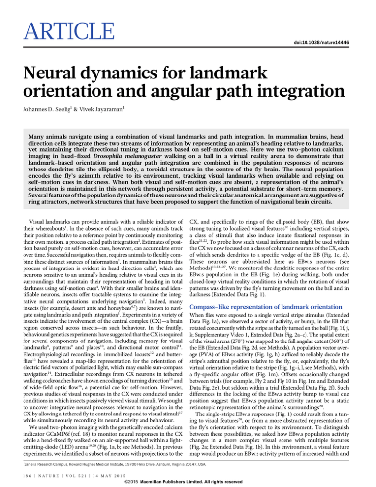
ARTICLE
doi:10.1038/nature14446
Neural dynamics for landmark
orientation and angular path integration
Johannes D. Seelig1 & Vivek Jayaraman1
Many animals navigate using a combination of visual landmarks and path integration. In mammalian brains, head
direction cells integrate these two streams of information by representing an animal’s heading relative to landmarks,
yet maintaining their directional tuning in darkness based on self-motion cues. Here we use two-photon calcium
imaging in head-fixed Drosophila melanogaster walking on a ball in a virtual reality arena to demonstrate that
landmark-based orientation and angular path integration are combined in the population responses of neurons
whose dendrites tile the ellipsoid body, a toroidal structure in the centre of the fly brain. The neural population
encodes the fly’s azimuth relative to its environment, tracking visual landmarks when available and relying on
self-motion cues in darkness. When both visual and self-motion cues are absent, a representation of the animal’s
orientation is maintained in this network through persistent activity, a potential substrate for short-term memory.
Several features of the population dynamics of these neurons and their circular anatomical arrangement are suggestive of
ring attractors, network structures that have been proposed to support the function of navigational brain circuits.
Visual landmarks can provide animals with a reliable indicator of
their whereabouts1. In the absence of such cues, many animals track
their position relative to a reference point by continuously monitoring
their own motion, a process called path integration2. Estimates of position based purely on self-motion cues, however, can accumulate error
over time. Successful navigation then, requires animals to flexibly combine these distinct sources of information1. In mammalian brains this
process of integration is evident in head direction cells3, which are
neurons sensitive to an animal’s heading relative to visual cues in its
surroundings that maintain their representation of heading in total
darkness using self-motion cues4. With their smaller brains and identifiable neurons, insects offer tractable systems to examine the integrative neural computations underlying navigation5. Indeed, many
insects (for example, desert ants and honeybees6,7) are known to navigate using landmarks and path integration1. Experiments in a variety of
insects indicate the involvement of the central complex (CX)—a brain
region conserved across insects—in such behaviour. In the fruitfly,
behavioural genetics experiments have suggested that the CX is required
for several components of navigation, including memory for visual
landmarks8, patterns9 and places10, and directional motor control11.
Electrophysiological recordings in immobilized locusts12 and butterflies13 have revealed a map-like representation for the orientation of
electric field vectors of polarized light, which may enable sun-compass
navigation14. Extracellular recordings from CX neurons in tethered
walking cockroaches have shown encodings of turning direction15 and
of wide-field optic flow16, a potential cue for self-motion. However,
previous studies of visual responses in the CX were conducted under
conditions in which insects passively viewed visual stimuli. We sought
to uncover integrative neural processes relevant to navigation in the
CX by allowing a tethered fly to control and respond to visual stimuli17
while simultaneously recording its neural activity and behaviour.
We used two-photon imaging with the genetically encoded calcium
indicator GCaMP6f (ref. 18) to monitor neural responses in the CX
while a head-fixed fly walked on an air-supported ball within a lightemitting-diode (LED) arena19,20 (Fig. 1a, b; see Methods). In previous
experiments, we identified a subset of neurons with projections to the
1
CX, and specifically to rings of the ellipsoid body (EB), that show
strong tuning to localized visual features20 including vertical stripes,
a class of stimuli that also induce innate fixational responses in
flies21,22. To probe how such visual information might be used within
the CX we now focused on a class of columnar neurons of the CX, each
of which sends dendrites to a specific wedge of the EB (Fig. 1c, d).
These neurons are abbreviated here as EBw.s neurons (see
Methods)13,23–27. We monitored the dendritic responses of the entire
EBw.s population in the EB (Fig. 1e) during walking, both under
closed-loop virtual reality conditions in which the rotation of visual
patterns was driven by the fly’s turning movement on the ball and in
darkness (Extended Data Fig. 1).
Compass-like representation of landmark orientation
When flies were exposed to a single vertical stripe stimulus (Extended
Data Fig. 1a), we observed a sector of activity, or bump, in the EB that
rotated concurrently with the stripe as the fly turned on the ball (Fig. 1f, j,
k; Supplementary Video 1, Extended Data Fig. 2a–c). The spatial extent
of the visual arena (270u) was mapped to the full angular extent (360u) of
the EB (Extended Data Fig. 2d, see Methods). A population vector average (PVA) of EBw.s activity (Fig. 1g, h) sufficed to reliably decode the
stripe’s azimuthal position relative to the fly, or, equivalently, the fly’s
virtual orientation relative to the stripe (Fig. 1g–i, l, see Methods), with
a fly-specific angular offset (Fig. 1m). Offsets occasionally changed
between trials (for example, Fly 2 and Fly 10 in Fig. 1m and Extended
Data Fig. 2e), but seldom within a trial (Extended Data Fig. 2f). Such
differences in the locking of the EBw.s activity bump to visual cue
position suggest that EBw.s population activity cannot be a static
retinotopic representation of the animal’s surroundings20.
The single-stripe EBw.s responses (Fig. 1) could result from a tuning to visual features20, or from a more abstracted representation of
the fly’s orientation with respect to its environment. To distinguish
between these possibilities, we asked how EBw.s population activity
changes in a more complex visual scene with multiple features
(Fig. 2a; Extended Data Fig. 1b). In this environment, a visual feature
map would produce an EBw.s activity pattern of increased width and
Janelia Research Campus, Howard Hughes Medical Institute, 19700 Helix Drive, Ashburn, Virginia 20147, USA.
1 8 6 | N AT U R E | VO L 5 2 1 | 1 4 M AY 2 0 1 5
G2015
Macmillan Publishers Limited. All rights reserved
ARTICLE RESEARCH
f
b
MB
120°
Optic lobe
Gall
FB
LAL
t = 1.8 s
EB
LAL
−3π/4
Fly holder
Protocerebral bridge
5 4 3 2 1 1 2 3 4 5
6
7
8
7
8
L
g
R
6R
6L
3L
3R
2R
8L
OddL
Gall
7R
7L
EvenR
1R 1L
2L
8R
EvenL
h %ΔF/F
900
EB
1 16
Wedge 1
PVA
estimate
100% ΔF/F
Gall
300
300
0
200
Wedge 9
100
250
200
150
100
50
0
1 3 5 7 9 11 13 15
π/2
π/3
π/6
0
Fly
1 3 5 7 9 11 13 15
Fly
40
60
80
100
π
i
120
PVA estimate
Visual cue position
π/2
0
−π/2
0
l
2π/3
20
−π
Correlation of
PVA estimate vs
cue position
k
FWHM of bump (rad)
Number of bumps
1.4
1.2
1.0
0.8
0.6
0.4
0.2
0
0
0
Wedge vector
Population vector average
j
%ΔF/F
600 Wedge 16
Wedge 1
Wedge 16
e
70 max
60
50
40
30
20
10
0 min
PVA
amplitude
%ΔF/F
Wedge 9
PB
t = 113.4 s
F
t = 1.8 s
8 9
Ellipsoid body
d
t = 60.0 s
3π/4
7 8 9 10
6
11
12
5
4
13
3
14
2
15
1 16
4R 5L 5R 4L
Gall OddR
t = 43.4 s
7 8 9 10
6
11
12
5
4
13
3
14
2
15
1 16
0°
6
t = 38.1 s
Optic lobe
No
270°
LED
arena
c
t = 33.3 s
MB
PB
Gall
20
40
m
1.0
0.5
0
–0.5
–1.0
60
80
Time (s)
1 3 5 7 9 11 13 15
Fly
EB to cue position
offset (rad)
Two-photon laser
scanning microscope
Azimuth (rad)
a
100
120
140
π
π/2
0
−π/2
−π
9 7 14 11 12 3 2 5
8 4 1 6 10 15 13
Fly
Figure 1 | Ellipsoid body activity tracks azimuth of visual cue. a, Schematic
of setup. Inset, close-up of fly on air-supported ball (modified from ref. 20).
b, Schematic of fly central brain and CX: ellipsoid body (EB), fan-shaped body
(FB), protocerebral bridge (PB), paired noduli (NO), lateral accessory lobe
(LAL) and gall (Gall). MB: mushroom body. c, Each EBw.s neuron receives
inputs from an EB wedge and sends outputs to a corresponding PB column, and
to the gall24,26. The PB has 18 columns24, but EBw.s neurons only innervate the
central 16. OddR, EvenR, OddL and EvenL, odd and even PB columns, right
and left side of brain, respectively. d, GFP-labelled EBw.s neurons in a brain
counterstained with nc82 (maximum intensity projection (MIP), reproduced
with permission from Janelia FlyLight Image Database23). e, MIP of twophoton imaging stack (5 frames, 5 mm apart, see Methods) showing EB
processes of GCaMP6f-labelled EBw.s neurons. f, Top, closed-loop walking
with a vertical stripe. Bottom, EBw.s activity is measured in 16 regions of
interest (ROIs). Sample frames from calcium imaging time series (Fly 15)
showing MIP of EB activity bump (see Methods) as fly rotates visual cue around
arena (top). F, fluorescence intensity (arbitrary units). Arrows near top of
h indicate frame times. g, Steps to compute PVA based on EBw.s population
activity. EB is unwrapped from Wedge 1 to Wedge 16 to display population
time series in h. Superimposed is PVA estimate that incorporates trial-specific
offset (m; see Methods). h, EBw.s fluorescence transients during a single trial
(same trial as f). Colour scale at right. Superimposed brown line indicates PVA
estimate of angular orientation of visual cue. Top, horizontal greyscale stripe
shows PVA amplitude; intensity scale at left. i, PVA estimate of angular
orientation plotted against actual orientation of visual cue (see Methods).
j, Mean and standard deviation (s.d.) of number of activity bumps in EBw.s
population activity across trials for each of 15 flies (see Methods). k, Mean and
s.d. of full width at half maximum (FWHM) of activity bump across trials and
flies (see Methods). l, Mean and s.d. of correlation between PVA estimate and
actual orientation (pattern position) (see Methods). m, Mean and s.d. of
angular offsets between PVA position and pattern position (see Methods,
Extended Data Fig. 2e, f). All scale bars, 20 mm.
complexity. Instead, consistent with EBw.s activity representing the
fly’s orientation, we observed a single bump of similar width (Fig. 2a–c,
Supplementary Video 2, Extended Data Fig. 3a–c), the spatial extent
of the arena was once again mapped onto the EB (Extended Data
Fig. 3d), and the PVA estimate of the fly’s azimuth remained accurate
(Fig. 2d–g, Extended Data Fig. 3e, f).
All the cues used thus far provided the fly with landmarks that
uniquely define its orientation in the environment. An activity bump
could thus represent either the fly’s angular position within the visual
scene or its orientation relative to a specific landmark within it. We next
placed the fly in a visual scene with two identical vertical stripes positioned to map exactly opposite each other on the EB ring (Extended
Data Fig. 1c, Extended Data Fig. 4a). Consistent with EBw.s activity
representing orientation by flexibly locking to a single landmark, the
resulting EBw.s representation again involved a single bump (Extended
Data Fig. 4b–f; Supplementary Video 3), with the same mapping of the
visual scene onto the EB as before (Extended Data Fig. 4g). PVA estimates were well correlated with the orientation of the fly in the scene
(Extended Data Fig. 4h–m), regardless of which stripe was directly in
front of the fly (Supplementary Video 3). In a few cases, however, we
observed that EBw.s activity transitioned from one offset to another
relative to the visual cues (Extended Data Fig. 5a–c, Supplementary
Video 4), potentially reflecting the ambiguity inherent in determining
landmark-guided orientation in an environment with multiple indistinguishable visual landmarks. Taken together, these data are consistent
with a function for the EBw.s population as an internal compass that
adaptively represents the fly’s orientation relative to visual landmarks.
Visual landmarks dominate over self-motion cues
Our closed-loop virtual reality experiments directly couple the fly’s
turning movements to the rotation of the visual scene. To disambiguate the contributions of visual landmark position and self-motion cues
to the EBw.s representation of the fly’s orientation, we performed two
sets of manipulations with a single stripe pattern. First, having
observed hints of landmark-tethered activity in bump transitions in
the two-stripe environment (Extended Data Fig. 5), we instantaneously shifted cue position during a period of closed-loop walking
1 4 M AY 2 0 1 5 | VO L 5 2 1 | N AT U R E | 1 8 7
G2015
Macmillan Publishers Limited. All rights reserved
RESEARCH ARTICLE
t = 15.8 s
t = 18.7 s
t = 57.1 s
t = 60.1 s
b
t = 66.9 s
1.4
Number
of bumps
a
F
7 8 9 10
11
6
5
12
13
4
14
3
2
15
1 16
%ΔF/F
400
300
200
100
0
20
40
60
80
100
Azimuth (rad)
π
120
PVA estimate
Panorama
position
π/2
−π/2
−π
0
20
40
60
80
Time (s)
100
120
π/2
π/3
π/6
140
1 23 45 6 7 89
π
π/2
0
−π/2
−π
9135 46872
g
0
1 23 45 6 7 89
2π/3
0
EB to panorama
position offset (rad)
0
FWHM of
bump (rad)
f
PVA
amplitude
1,000
Wedge 16
500
Wedge 9
100
Wedge 1
e
0
c
60
40
20
0 min
%ΔF/F
0.6
0.2
80 max
Correlation of
PVA estimate vs
panorama position
d
1.0
1.0
Figure 2 | Ellipsoid body is not a retinotopic
map of visual scene, but represents the fly’s
orientation relative to visual landmarks.
a, Closed-loop experiment in visual
environment with multiple features (Fly 1 in
b). b, Mean and s.d. of number of bumps across
trials for each of 9 flies. c, Mean and s.d. of
FWHM of bump. Distribution of bump widths
is not significantly different from that for
single-stripe stimulus (Fig. 1k); P 5 0.14 (see
Methods), mean width 5 84.9u 6 12.6u for
multiple feature trials versus 82.3u 6 11.5u for
single stripe trials. d, EBw.s fluorescence
transients (same trial as a). e, PVA estimate of
the fly’s angular orientation compared to actual
orientation. f, Mean and s.d. of angular offsets
(see Methods, Extended Data Fig. 3e, f).
g, Correlation between PVA estimate and
actual orientation. Scale bar, 20 mm.
0.5
0
–0.5
–1.0
123456789
Fly
(Fig. 3a; also see Methods). If EBw.s responses arise from self-motion
cues rather than landmark orientation, the abrupt shift in landmark
azimuthal position should not by itself induce matching shifts in
EBw.s activity. Instead, we observed that EBw.s. population activity
moved to match the cue shift, preserving the initial offset between the
EBw.s bump and visual cue azimuth (Extended Data Fig. 6a, b, Fig. 3b;
Supplementary Video 5). Thus, local landmarks rather than selfmotion cues appear to determine the position of the EBw.s bump.
Interestingly, the bump did not always move instantaneously. In response to the first landmark shift in Extended Data Fig. 6b (see also
Supplementary Video 5), for example, the bump rapidly tracks the
shifted visual landmark, but the second abrupt displacement of the
landmark elicits a much slower response.
As a second manipulation, we varied the closed-loop gain that
coupled the fly’s rotational movements on the ball to the movement
of the visual cue (Fig. 3c). If EBw.s activity is determined by the fly’s
orientation relative to the landmark, the bump should move in lockstep
with landmark rotation rather than with the fly’s turning movements.
Indeed, in almost all cases, activity in the EBw.s population faithfully
followed the visual cue (Fig. 3d–g, Extended Data Fig. 6c). Consistent
with this, the relationship between walking rotation and EBw.s bump
movement was strongly dependent on closed-loop gain (Fig. 3h),
whereas the relationship between visual cue movement and rotation
of the EBw.s bump scaled only slightly with gain (Fig. 3i). Nevertheless,
we occasionally observed examples of EBw.s activity being more influenced by the animal’s rotation than cue movement, particularly in
situations of low gain (Extended Data Fig. 6d–f). Overall, as has been
observed in the mammalian head direction system4, the EBw.s compass
predominantly relies on visual landmarks, but its computation of the
fly’s orientation is also influenced by the animal’s angular movements.
Angular path integration with no visual cues
The influence of self-motion cues on EBw.s population activity
(Extended Data Fig. 6d–f) suggests that visual information can be
overridden in determining compass heading. We next sought to
uncover the contribution of self-motion cues in an environment that
provided no visual information. A key feature of mammalian head
direction cells is their ability to retain their compass-like function in
the absence of visual information using path integration4. We
searched for evidence of angular path integration, that is, tracking
of orientation by self-motion cues, by imaging EBw.s population
responses of flies walking in complete darkness without prior exposure to a closed-loop visual scene on the ball (Fig. 4a). As in all visually
stimulated conditions, EBw.s activity in the dark settled into a single
bump, and then tracked the fly’s turning movements on the ball
(Fig. 4b–d; Supplementary Videos 6–8, Extended Data Fig. 7a–e, h).
However, the PVA estimate of orientation based on the activity in the
EBw.s population often degraded over time (Fig. 4d, Extended Data
Fig. 7b, f, h, i), with EBw.s activity not tracking very small or slow
angular movements (Extended Data Fig. 7g, j, and Extended Data
Fig. 8). Although the fly’s potentially impaired ability to track its rotation when tethered on a ball may contribute to the measured drift, it
likely also reflects a common limitation of path integrators in the
absence of corrective feedback4,28. We also noted some fly-to-fly variability in the effective (measured) gain between ball rotation and EBw.s
bump movement in these experiments (Extended Data Fig. 7f, i). The
observation that the EBw.s system can operate at different gains raises
the possibility that the compass can not only adjust to various visual
closed-loop gains (as seen in Fig. 3), but also perhaps tune the gain
between ball rotation and EBw.s activity through experience. However,
prior exposure to specific closed-loop gains in visual surroundings only
had a negligible influence on the effective gain between ball rotation and
EBw.s activity in darkness (Extended Data Fig. 9). Overall, these results
show that the EBw.s population performs angular path integration in
darkness by relying exclusively on self-motion cues, albeit with a gradual accumulation of error in its orientation estimate.
Orientation represented by persistent activity
Having established that both visual landmark information and selfmotion cues contribute to EBw.s population activity, we next asked
how the system responds to the absence of both sources of orientation
information. Specifically, we examined EBw.s activity during epochs
when the fly stopped walking while in the dark. In almost all such cases,
the EBw.s population maintained a representation of the fly’s orientation (Fig. 4e–i; Extended Data Fig. 10a–f; Supplementary Videos 7 and
8). This representation persisted even when EBw.s calcium activity was
low, as evident in the fact that renewed bouts of movement caused a
bump to reappear in exactly the wedges expected based on the last
orientation of the fly (Fig. 4h and Extended Data Fig. 10a, c, e,
Supplementary Videos 7 and 8). This representation of orientation
sometimes persisted for more than 30 s (Fig. 4i, Extended Data
Fig. 10b, d, f). Such persistence was also a feature of EBw.s dynamics
1 8 8 | N AT U R E | VO L 5 2 1 | 1 4 M AY 2 0 1 5
G2015
Macmillan Publishers Limited. All rights reserved
ARTICLE RESEARCH
a
Frame t
Frame t + 1
Discussion
b
c
Offset before cue jump
(rad)
Actual offset
Offset if bump were to not follow cue
Closed loop protocols
High gain
Normal gain
Low gain
π
0
−π
−π
0
π
Offset after cue jump (rad)
Visual stimulus rotation (arena)
Walking rotation (ball)
d
%ΔF/F
300
200
100
0
Low gain
Wedge 16
Wedge 9
Wedge 1
0
Accumulated rotation
(rad)
e
15
20
40
60
80
100
120
PVA estimate
Visual cue
Walking
10
5
0
0
20
40
60
f
80
Time (s)
100
120
High gain
Wedge 16
%ΔF/F
300
200
100
0
Wedge 9
Wedge 1
Accumulated rotation
(rad)
20
40
60
80
100
120
60
80
Time (s)
100
120
PVA estimate
Visual cue
Walking
40
30
20
10
0
0
20
40
i
1.5
1.0
0.5
0
0
0.5 1.0 1.5
Closed loop gain
Gain of visual cue rotation
vs PVA estimate
h
0
Gain of walking rotation
vs PVA estimate
g
140
1.5
1.0
0.5
0
0
0.5 1.0 1.5
Closed loop gain
Figure 3 | EBw.s activity tracks landmark orientation cues over angular
rotation when these cues are in conflict. a, In cue-shift experiments, fly is in
closed-loop control of stripe position until cue is abruptly shifted to new
position (see Methods). b, Offset between PVA estimate and actual orientation
relative to visual cue before and after cue shift. Plot compares actual offsets with
those expected if EBw.s activity did not follow cue position (see Methods).
N 5 50 shifts (n 5 6 flies), r 5 0.85, pr 5 0. Fit: slope 5 0.78 6 0.07, pslope 5 0,
R2 5 0.72. c, In closed-loop gain-change experiments, ball rotation drives
movement of visual stimulus with different closed-loop gains. d, Fluorescence
transients during trial with low gain of 0.6 (Fly 15 from Fig. 1j). e, Comparison
of PVA estimate versus accumulated rotation of visual cue and walking rotation
on ball (trial in d). Walking rotation exceeds visual cue angular rotation in this
low-gain trial. f, Similar to d, but with high closed-loop gain of 1.3 (Fly 3 from
Fig. 1j). g, Similar to e, but with high gain (trial in f). h, Effective gain between
walking rotation and PVA estimate for different closed-loop gains (r 5 0.69,
pr 5 0, Fit: slope 5 0.85 6 0.07, pslope 5 0, n 5 172, R2 5 0.48, see Methods).
i, Effective gain between visual cue rotation and PVA estimate for different
closed-loop gains (r 5 0, pr 5 15.1 3 1023, Fit: slope 5 20.17 6 0.07,
pslope 5 0.02, n 5 172, R2 5 0.03, see Methods).
when the fly remained standing in a visual environment (Extended
Data Fig. 10g–r), extending beyond durations that elicit adaptation in
early visual circuits29. Thus, even in the absence self-motion cues, EBw.s
population activity maintains a stable representation of the fly’s orientation in its environment with or without visual landmarks.
The ability of animals to combine continuous path integration with
potentially intermittent landmark-based orienting enables navigation
in a wide diversity of environmental conditions1,6. We studied the activity
dynamics of a complete population of identified CX neurons in tethered
walking flies and found that this network uses information from both
landmark-based and angular path integration systems to create a compass-like representation of the animal’s orientation in the environment.
Previous studies have described static visual maps in the CX12,13,20.
Such maps may allow navigating insects to maintain a sun-compassbased heading direction12,13,27,30. Here we found that EBw.s neurons
track the fly’s orientation relative to visual landmarks in a variety of
different visual environments (Figs 1 and 2), suggesting that the CX
dynamically adapts to estimate the fly’s orientation within its visual
surroundings (Extended Data Figs 2d, 3d and 4g). Subsets of ring
neurons are likely to bring information about spatially localized visual
features20 to specific rings of the ellipsoid body31. It is not yet clear how
this information is converted into an abstract and flexible representation of the animal’s orientation relative to landmarks32, but EBw.s
responses in a symmetric environment with two indistinguishable
cues (Extended Data Figs 4 and 5) hint at an underlying winnertake-all process for landmark selection33. Combining landmark orientation with information about the animal’s movement effectively
creates an internal reference frame for the animal in its surroundings.
Many of the proposed functions of the CX in directed locomotion11,15,
visual place learning10, and action-selection34, may rely on this
internal reference. Although the EBw.s population tracks the fly’s
rotational movements in darkness, we do not yet know where and
how translational motion, an important component of a complete
navigational system, is incorporated. Additionally, although the calcium sensor we chose for our imaging experiments has the temporal
resolution necessary to capture EBw.s representations of the fly’s
angular rotation (see Methods), it lacks the precision necessary for
us to determine whether EBw.s activity represents the fly’s predicted
future orientation or its estimate of current orientation.
Our observation that EBw.s activity was maintained in the absence
of self-motion suggests that internal dynamics play a significant role in
shaping neural activity in the fly brain, much as they do in the brains of
larger animals. Persistent activity in the CX can maintain compass
information when the fly is standing in darkness for 30 s—two orders
of magnitude longer than might be explained by calcium sensor decay
kinetics18. Persistent activity has been shown to support maintenance
of eye position in the goldfish35 and has been proposed to underlie
working memory in mammals36. In the CX, this activity may allow the
fly to retain a short-term orientation memory even when landmarks
are temporarily out of sight8. Consistent with this notion, the EBw.s
activity bump largely remained tethered to the position of one landmark even in the presence of another identical landmark in front of the
fly (Extended Data Fig. 4i). The bump also did not always shift instantaneously following an abrupt displacement of visual landmarks, as if
temporarily retaining the original orientation reference before locking
on to its new position (Extended Data Fig. 6b).
Several models have been proposed to explain how visual landmark and self-motion cues are integrated at the level of head direction cell activity in mammals37. Most rely on circuits organized as
ring attractors: neurons are schematized as being arranged in a
circle based on their preferred directions38, with connection
strengths that depend on their angular separation37. With initial
sensory input and an appropriate balance of recurrent excitation
and inhibition, such a circuit can generate and sustain a localized
activity bump. The bump’s position on the circle corresponds to
the animal’s heading which is then updated by directional drive
from self-motion signalling neurons. Direct experimental evidence
in support of these models has been difficult to obtain in mammals
owing to the distributed nature of the underlying circuits.
Although the functional connectivity between EBw.s neurons is
1 4 M AY 2 0 1 5 | VO L 5 2 1 | N AT U R E | 1 8 9
G2015
Macmillan Publishers Limited. All rights reserved
RESEARCH ARTICLE
a
c
In dark
%ΔF/F
1,100
800
500
200
PVA
amplitude
Wedge 16
%ΔF/F
400
300
200
100
0
Wedge 9
Wedge 1
0
1.6
1.4
1.2
1.0
0.8
0.6
0.4
0.2
0
Number of bumps
b
e
Accumulated
rotation (rad)
d
40
60
80
100
PVA estimate
Walking
4
2
0
0
20
40
1 3 5 7 9 11
Fly
t = 62.9 s
t = 72.8 s
t = 77.1 s
60
80
Time (s)
t = 81.7 s
100
t = 88.1 s
80
70
60
50
40
30
20
10
0
h
PVA
amplitude
%ΔF/F
Wedge 16
300
200
Wedge 9
100
Wedge 1
0
20
40
60
80
100
10
5
0
0
20
40
60
80
Time (s)
100
i
120
Online Content Methods, along with any additional Extended Data display items
and Source Data, are available in the online version of the paper; references unique
to these sections appear only in the online paper.
8.
9.
10.
11.
12.
13.
14.
15.
16.
17.
18.
Received 1 December 2014; accepted 9 April 2015.
19.
3.
4.
5.
6.
7.
min
0
π
π
0
PVA estimate
before stop (rad)
0
−π
10
20
30
Time (s)
not yet known, we have observed several of the expected features of
ring attractor models37,39,40 in the dynamics of this population of
CX neurons: organization of activity into a localized bump, movement of the bump to neighbouring wedges based on self-motion,
drift in bump location in darkness, persistent activity, and both
abrupt jumps and gradual transitions of the activity bump when
triggered by strong visual input. Cell-intrinsic mechanisms could
also underlie some of these features, including, for example, persistent activity35,41,42. The genetic tools available in Drosophila to
target and manipulate the activity of identified cell types should
allow different models for visually guided orientation and angular
path integration to be discriminated at the level of synaptic, cellular
and network mechanism.
2.
max
−π
−π
Difference in PVA
estimate (before stop
- at restart) (rad)
15
Accumulated
rotation (rad)
0
PVA estimate
Walking
Standing
g
120
120
π
PVA estimate
at restart (rad)
800
600
400
200
0
1.
F
8 9 10
11
12
13
14
2
15
1 16
7
f
%ΔF/F
120
–2
t = 57.8 s
6
5
4
3
20
8
6
Figure 4 | Path integration, drift and
persistence in EBw.s activity in total
darkness. a, Experiments with flies walking in
total darkness. b, Mean and s.d. of number of
bumps across trials for each of 11 flies.
c, Fluorescence transients during trial in
darkness (Fly 9 in b). d, Accumulated ball
rotation plotted against accumulated PVA
estimate of fly’s rotation. e, Sample frames from
time series showing that EBw.s activity is
maintained in absence of both visual cues and
rotation (Fly 3 in b). Scale bar, 20 mm.
f, Fluorescence transients during trial in
e. g, Representation of fly’s angular orientation
is maintained in the absence of rotation and
resumes from previous wedge after long delay
(grey rectangles indicate epochs of fly standing).
h, Comparison of PVA estimate of orientation
before stop and at restart for different standing
bouts across n 5 11 flies (r 5 0.7, pr 5 0, Fit:
slope 5 0.96 6 0.17, pslope 5 0, n 5 499, R2 5
0.879). i, Durations of standing bouts in h (tmean
5 6.7 6 5.1 s, DPVAmean 5 0.017 6 0.76 rad).
Collett, T. S. & Graham, P. Animal navigation: Path integration, visual landmarks
and cognitive maps. Curr. Biol. 14, R475–R477 (2004).
Mittelstaedt, M. L. & Mittelstaedt, H. Homing by path integration in a mammal.
Naturwissenschaften 67, 566–567 (1980).
Taube, J. S., Muller, R. U. & Ranck, J. B. Head-direction cells recorded from the
postsubiculum in freely moving rats. 1. Description and quantitative analysis.
J. Neurosci. 10, 420–435 (1990).
Taube, J. S. The head direction signal: origins and sensory-motor integration.
Annu. Rev. Neurosci. 30, 181–207 (2007).
Huston, S. J. & Jayaraman, V. Studying sensorimotor integration in insects. Curr.
Opin. Neurobiol. 21, 527–534 (2011).
Wehner, R. Desert ant navigation: how miniature brains solve complex tasks.
J. Comp. Physiol. A 189, 579–588 (2003).
Collett, T. S. & Collett, M. Path integration in insects. Curr. Opin. Neurobiol. 10,
757–762 (2000).
20.
21.
22.
23.
24.
25.
Neuser, K., Triphan, T., Mronz, M., Poeck, B. & Strauss, R. Analysis of a spatial
orientation memory in Drosophila. Nature 453, 1244–1247 (2008).
Liu, G. et al. Distinct memory traces for two visual features in the Drosophila brain.
Nature 439, 551–556 (2006).
Ofstad, T. A., Zuker, C. S. & Reiser, M. B. Visual place learning in Drosophila
melanogaster. Nature 474, 204–207 (2011).
Strauss, R. The central complex and the genetic dissection of locomotor behaviour.
Curr. Opin. Neurobiol. 12, 633–638 (2002).
Heinze, S. & Homberg, U. Maplike representation of celestial E-vector orientations
in the brain of an insect. Science 315, 995–997 (2007).
Heinze, S. & Reppert, S. M. Sun compass integration of skylight cues in migratory
monarch butterflies. Neuron 69, 345–358 (2011).
Pfeiffer, K. & Homberg, U. Organization and functional roles of the central complex
in the insect brain. Annu. Rev. Entomol. 59, 165–184 (2014).
Guo, P. & Ritzmann, R. E. Neural activity in the central complex of the cockroach
brain is linked to turning behaviors. J. Exp. Biol. 216, 992–1002 (2013).
Kathman, N. D., Kesavan, M. & Ritzmann, R. E. Encoding wide-field motion and
direction in the central complex of the cockroach Blaberus discoidalis. J. Exp. Biol.
217, 4079–4090 (2014).
Dombeck, D. A. & Reiser, M. B. Real neuroscience in virtual worlds. Curr. Opin.
Neurobiol. 22, 3–10 (2012).
Chen, T. W. et al. Ultrasensitive fluorescent proteins for imaging neuronal activity.
Nature 499, 295–300 (2013).
Seelig, J. D. et al. Two-photon calcium imaging from head-fixed Drosophila during
optomotor walking behavior. Nature Methods 7, 535–540 (2010).
Seelig, J. D. & Jayaraman, V. Feature detection and orientation tuning in the
Drosophila central complex. Nature 503, 262–266 (2013).
Bausenwein, B., Muller, N. R. & Heisenberg, M. Behavior-dependent activity
labeling in the central complex of Drosophila during controlled visual stimulation.
J. Comp. Neurol. 340, 255–268 (1994).
Strauss, R. & Pichler, J. Persistence of orientation toward a temporarily invisible
landmark in Drosophila melanogaster. J. Comp. Physiol. A 182, 411–423 (1998).
Jenett, A. et al. A GAL4-driver line resource for Drosophila neurobiology. Cell Rep. 2,
991–1001 (2012).
Wolff, T., Iyer, N. A. & Rubin, G. M. Neuroarchitecture and neuroanatomy of the
Drosophila central complex: a GAL4-based dissection of protocerebral bridge
neurons and circuits. J. Comp. Neurol. 523, 997–1037 (2015).
Hanesch, U., Fischbach, K. F. & Heisenberg, M. Neuronal architecture of the central
complex in Drosophila melanogaster. Cell Tissue Res. 257, 343–366 (1989).
1 9 0 | N AT U R E | VO L 5 2 1 | 1 4 M AY 2 0 1 5
G2015
Macmillan Publishers Limited. All rights reserved
ARTICLE RESEARCH
26. Lin, C. Y. et al. A comprehensive wiring diagram of the protocerebral bridge for visual
information processing in the Drosophila brain. Cell Rep. 3, 1739–1753 (2013).
27. Heinze, S., Gotthardt, S. & Homberg, U. Transformation of polarized light information in the central complex of the locust. J. Neurosci. 29, 11783–11793 (2009).
28. Mizumori, S. J. Y. & Williams, J. D. Directionally selective mnemonic properties of
neurons in the lateral dorsal nucleus of the thalamus of rats. J. Neurosci. 13,
4015–4028 (1993).
29. Laughlin, S. B. The role of sensory adaptation in the retina. J. Exp. Biol. 146, 39–62
(1989).
30. Bockhorst, T. & Homberg, U. Amplitude and dynamics of polarization-plane
signaling in the central complex of the locust brain. J. Neurophysiol. http://
dx.doi.org/10.1152/jn.00742.2014 (2015).
31. Young, J. M. & Armstrong, J. D. Structure of the adult central complex in Drosophila:
organization of distinct neuronal subsets. J. Comp. Neurol. 518, 1500–1524 (2010).
32. Zeil, J. Visual homing: an insect perspective. Curr. Opin. Neurobiol. 22, 285–293
(2012).
33. Koch, C. & Ullman, S. Shifts in selective visual attention: towards the underlying
neural circuitry. Hum. Neurobiol. 4, 219–227 (1985).
34. Strausfeld, N. J. & Hirth, F. Deep homology of arthropod central complex and
vertebrate basal ganglia. Science 340, 157–161 (2013).
35. Aksay, E. et al. Functional dissection of circuitry in a neural integrator. Nature
Neurosci. 10, 494–504 (2007).
36. Durstewitz, D., Seamans, J. K. & Sejnowski, T. J. Neurocomputational models of
working memory. Nature Neurosci. 3, 1184–1191 (2000).
37. Knierim, J. J. & Zhang, K. C. Attractor dynamics of spatially correlated neural
activity in the limbic system. Annu. Rev. Neurosci. 35, 267–285 (2012).
38. Peyrache, A., Lacroix, M. M., Petersen, P. C. & Buzsaki, G. Internally organized
mechanisms of the head direction sense. Nature Neurosci. 18, 569–575 (2015).
39. Arena, P., Maceo, S., Patané, L. & Strauss, R. A spiking network for spatial memory
formation: towards a fly-inspired ellipsoid body model. Intl. Joint Conf. Neural
Networks http://dx.doi.org/10.1109/IJCNN.2013.6706882 (2013).
40. Haferlach, T., Wessnitzer, J., Mangan, M. & Webb, B. Evolving a neural model of
insect path integration. Adapt. Behav. 15, 273–287 (2007).
41. Yoshida, M. & Hasselmo, M. E. Persistent firing supported by an intrinsic cellular
mechanism in a component of the head direction system. J. Neurosci. 29,
4945–4952 (2009).
42. Major, G. & Tank, D. Persistent neural activity: prevalence and mechanisms. Curr.
Opin. Neurobiol. 14, 675–684 (2004).
Supplementary Information is available in the online version of the paper.
Acknowledgements We thank T. Wolff and G. Rubin for sharing information about CX
neuron morphology. We thank Janelia Fly Core, and in particular K. Hibbard and
S. Coffman, for support, J. Liu for technical support, and V. Iyer for ScanImage support.
We are grateful to A. Karpova, A. Leonardo, S. S. Kim, H. Haberkern, D. Turner-Evans,
C. Dan, S. Wegener and R. Franconville for discussions and comments on the
manuscript. We thank W. Denk, S. Druckmann, J. Dudman, A. Lee, K. Longden, M. Reiser,
S. Romani, G. Rubin, Y. Sun, and T. Wolff for feedback on the manuscript. This work was
supported by the Howard Hughes Medical Institute.
Author Contributions Both authors designed the study, performed data analysis and
wrote the manuscript. J.D.S. carried out all experiments.
Author Information Reprints and permissions information is available at
www.nature.com/reprints. The authors declare no competing financial interests.
Readers are welcome to comment on the online version of the paper. Correspondence
and requests for materials should be addressed to V.J. (vivek@janelia.hhmi.org).
1 4 M AY 2 0 1 5 | VO L 5 2 1 | N AT U R E | 1 9 1
G2015
Macmillan Publishers Limited. All rights reserved
RESEARCH ARTICLE
METHODS
Fly stocks. All calcium imaging experiments were performed with 8–11 days old
female UAS-GCaMP6f;R60D05-GAL4 flies. Flies were randomly picked from
their housing vials for all experiments.
Nomenclature. EBw.s neurons are referred to variously as eb-pb-vbo25, EIP26 and
EBw.s-PBg.b-gall.b24 neurons in the fly literature. They may be homologous to
CL1a neurons in the locust27 and butterfly13.
Fly preparation for imaging during walking. The fly was anaesthetized on ice
and transferred to a cold plate at 4 uC. The fly’s proboscis was pressed onto its
head and immobilized with wax. To maximize the fly’s field of view we used a
two-piece pyramidal stainless steel shim holder19 similar to those previously used
for tethered flying fly experiments20,43. The fly was glued to a pin and positioned in
the holder using a micromanipulator and fixed in the holder with UV gel as
previously described19,20. The fly body axis was angled at 31u 6 5u (measured
for 5 flies) with respect to the tracking system to orient the EB optimally with
respect to the microscope’s focal plane. To stop brain movement due to the
pulsation of muscle M16, we cut the muscle—or the nerves innervating the
muscle—with dissection needles if necessary. The fly holder (including the micromanipulator) was then transferred to the two-photon microscope and secured
using magnetic mounts. As previously described19,20, the fly was positioned on an
air-supported ball with a three-axis micromanipulator and the walking velocity of
the fly was monitored using a camera system. For all experiments described in the
main figures we used a 6-mm diameter, 40-mg ball19. For the experiments in the
dark described in Extended Data Fig. 7h–j, we used a 10-mm diameter, 175-mg
ball. All balls were made of polyurethane foam.
For experiments with visual cues, we removed parts of the antennae (funiculus
and arista) to reduce the fly’s tendency to touch the holder.
For experiments in which flies walked in the dark we additionally coated the
eyes of a subset of flies with black paint (Premiere Acrylic Colours, Mars Black).
For the 6-mm-ball experiments, Flies 4–11 had coated eyes, while Flies 2–13 had
coated eyes for the 10-mm-ball experiments. The number of trials per fly was as
follows (Fly (number of trials)). 6-mm ball: 1(15), 2(8), 3(8), 4(8), 5(11), 6(10),
7(8), 8(10), 9(17), 10(10), 11(11). 10-mm ball: 1(7), 2(3), 3(8), 4(12), 5(10), 6(8),
7(6), 8(5), 9(3), 10(14), 11(8), 12(6), 13(11). All trials across all conditions
lasted 140 s.
Two-photon calcium imaging. Calcium imaging was performed using a custombuilt two-photon microscope controlled with ScanImage 4.244. We used an
Olympus 340 objective (LUMPlanFl/IR, NA 0.8) and a GaAsP photomultiplier
tube (H7422PA-40, Hamamatsu). A Chameleon Ultra II laser (Coherent, Santa
Clara, CA) tuned to 920 nm was used as the excitation source with the power
adjusted to below 20 mW at the sample. We used the same saline as in previous
studies20 but adjusted the calcium concentration to 2.5 mM. Focal planes were
selected to optimize coverage of the part of the EB innervated by EBw.s neurons.
We imaged from 5-plane volumes at a rate of 8.507 Hz with an equal spacing of
between 4 mm to 6 mm between individual focal planes.
The calcium signals we measured may reflect synaptic input, action potential
output or some combination of both. We chose GCaMP6f18 for our experiments
because it offers the temporal resolution necessary to capture EBw.s representation of the fly’s angular rotation. Based on in vivo measurements of responses to
20 Hz spiking at the Drosophila larval neuromuscular junction, GCaMP6f has a
time-to-peak of ,141 ms (close to the 8.507 Hz frame rate of our imaging system)
and a decay time of ,380 ms (ref. 18). Assuming that one complete rotation of the
fly is represented by activity moving through the 16 wedges of the EB, each wedge
represents 22.5u of rotation. The maximum average rotational velocity reached by
a fly in our experiments was , 35u s21 (Extended Data Fig. 1), which would result
in a bump of activity moving across a wedge no faster than ,640 ms on average.
Thus, possible lags in the calcium signals introduced by the rise and decay times of
GCaMP6f would not compromise the detection of these activity changes.
Although we do not know the actual change in electrical activity associated with
the calcium transients we see, the kinetics of GCaMP6f provide a considerable
margin of error.
Visual stimulation. Visual arena. Visual stimuli were presented on a cylindrical
LED display45 spanning 270u in azimuth and 120u in elevation, and tilted by 10u
towards the fly. The display was covered with a colour filter and a diffuser as
previously described19,20.
Visual stimuli for closed-loop walking experiments. We used three different visual
stimuli: condition 1, a bright vertical stripe spanning 120u in elevation and 15u in
azimuth; condition 2, two bright stripes of the same dimensions separated by 135u
(resulting in a pattern that was invariant to rotations by 135u); and condition 3,
a pattern containing several vertical and horizontal stripes (Extended Data
Fig. 1a–c). The number of trials (in brackets) for each fly for each of these conditions was: trial 1, 1(7), 2(6), 3(9), 4(11), 5(15), 6(13), 7(3), 8(7), 9(7), 10(6), 11(5),
12(9), 13(9), 14(10), 15(6); trial 2, 1(12), 2(8), 3(10), 4(3), 5(6), 6(8), 7(16); trial 3,
G2015
1(6) (same as fly 1 in trial 2), 2(2) (same as fly 2 in trial 2), 3(5) (same as fly 4 in
trial 2), 4(3), 5(7) (same as fly 5 in trial 2), 6(11), 7(4), 8(11), 9(12).
In cue-shift experiments each trial consisted of two cue shifts by the same
angular distance within each trial (either by 60u or 120u) after at least 50 s of
closed-loop behaviour. The first cue shift was counter clockwise from the current
position and the next, 50 s later, clockwise by the same angular amount from the
current position. Cue shift experiments were performed with a subset of the flies
in condition 1. A 60u cue shift was used for flies 7(2), 11(3), 13(3), 15(2) and
120ucue shift for flies 7(2), 8(3), 9(1), 11(3), 13(3), 15(2).
In experiments that tested the influence of prior exposure to visual stimuli in
closed loop on the gain between walking rotation and PVA estimate in darkness
(Extended Data Fig. 9), we exposed the flies to 65 s of closed-loop walking with
either low gain (mean closed-loop gain 5 0.47 6 0.04, close to the fly’s default
gain on the ball without prior closed-loop walking experience) or higher gain
(mean set gain 5 0.9 6 0.16) with a single stripe, after which the stripe disappeared and the trial continued for another 60 s in darkness. For these experiments, we only used flies that showed strong rotational movement with low
drift—as assessed at the onset of the experiment—to increase the accuracy of
the gain calculation. We only recorded a small number of trials per fly, because the
fly usually rotated more at the onset of experiments and walked forward more
towards the end, leading to increased drift. For a subset of flies, we also tested the
intrinsic gain of the flies walking in darkness without prior exposure to the closedloop stimulus. The number of trials for experiments in which we combined
closed-loop walking and walking in darkness were (fly number (trials with disappearing stripe/trials in darkness before exposure to closed-loop condition)):
1(5/0), 2(4/1), 3(3/1), 4(5/0), 5(7/1), 6(3/2), 7(4/1), 8(6/2), 9(5/1), 10(4/1), 11(4/
1), 12(3/2), 13(5/2), 14(5/1), 15(1/2), 16(5/2), 17(3/2), 18(6/2), 19(4/2), 20(4/2),
21(5/2), 22(4/2), 23(4/1), 24(2/2), 25(4/2), 26(3/2).
Closed-loop gains to convert rotation on the ball to displacement of the stripe
around the arena were close to 1 (‘normal gain’), but were varied from 0.4 to 1.6 in
experiments to explore the effect of gain change on EBw.s representation. Actual
values of the gain were verified by fitting changes in ball displacement to changes
in pattern displacement on the arena. All patterns were displaced directly from
one edge of the 270u arena to the other behind the fly rather than having them
progress virtually through the 90u of visual field not represented by the arena.
This was done to prevent abrupt changes in light intensity and to keep the number
of features in the fly’s visual field constant.
All experiments with visual stimulation were performed in closed loop46. The
voltage position signal of the tracking system was read with a DAQ board and
discretized in 20-ms intervals using custom LabVIEW software which was also
used to update the position of the visual stimulus45.
Data analysis. We used MATLAB (MathWorks, Inc., Natick, MA) and the
Circular Statistics Toolbox47 for data analysis. All errors and error bars shown
are standard deviation (s.d.). No statistical methods were used to predetermine
sample size.
Calculation of fluorescence changes. Each imaged volume (stack of five frames)
was averaged for analysis—we refer to this average as a ‘frame’. Each frame was
spatially filtered with a 2-pixel-wide Gaussian filter after which background
fluorescence was subtracted. Calcium transients recorded from behaving flies
were smoothed with a 3rd order Savitzky–Golay filter over 7 frames (822 ms)
for comparisons with behavioural data. The baseline for calculating DF/F was
computed by averaging over the 10% of lowest-intensity frames in each trial. For
display only, MIP fluorescence intensity images shown in Figs 1e, f, 2a, 4e, and
Extended Data Fig. 5a were filtered with a 20-pixel-wide, 10-pixel-s.d. Gaussian
filter (the size of each image is 216 pixels by 216 pixels).
ROI selection. ROIs corresponding to 16 wedges of the EB were selected manually in videos of DF/F by drawing an ellipse (with a central hole, as depicted in
Fig. 1f, g) that surrounded the EB, and then equally subdividing the ellipse into 16
wedges each spanning 22.5u. The number of wedges was selected based on the
well-characterized EB wedge and PB glomerular innervation patterns of EBw.s
neurons labelled by the R60D05-Gal4 line24. Some EBw.s neurons are known to
arborize only in half- or demi-wedges24. Thus, our ROI selection and population
analysis strategy may underestimate the actual resolution of the EBw.s system.
Population vector average (PVA). The PVA was computed as the weighted
average of EB wedge angles ranging from 0 to 360u, with average DF/F values
for each wedge used as a weight. This PVA estimate was smoothed with a box-car
filter over 3 frames (352 ms). We used brewermap (S. Cobeldick, MathWorks file
exchange) with colour schemes from http://colorbrewer2.org/ to generate colour
maps for all PVA plots except for PVA amplitude, which we display in greyscale.
For display of PVA estimates of orientation or walking rotation, raw PVA was
offset by the median difference (circular distance) between PVA and either the
visual cue position (for closed-loop trials in the arena) or the walking rotation
signal (for trials in the dark). We computed the offset using epochs of walking in
Macmillan Publishers Limited. All rights reserved
ARTICLE RESEARCH
the final 80% of a trial, a period during which PVA estimates were typically more
stable. The offset adjustment was necessitated by the fact that there was no
stereotyped relationship between cue positions and EBw.s signal across flies
(Fig. 1m, Extended Data Fig. 2e, f, Fig. 2f, Extended Data Figs 3e, f, 4j, l, m).
The magnitude of the offset in many animals (Fig. 1m, Extended Data Figs 2e, f,
3e, f, 4j, l) greatly exceeded the slight variance in the angle at which the tethered
fly’s head was fixed relative to the LED arena. The offset also occasionally changed
between trials for the same fly. We did not monitor the fly’s walking between
trials, leaving open the possibility that these differences in offset arose purely from
rotational movements of the fly (in the absence of closed-loop visual feedback)
before initiation of the next closed-loop trial.
Analysis of number and width of activity bumps. All ROIs with calcium transients above a set threshold were included in an activity bump. Each contiguous
set of ROIs above threshold defined an individual bump. We used two separate
methods to set the threshold. In Method 1 (used for all the main figures), the
threshold was defined as 1-s.d. above the mean of calcium transients across ROIs
for each imaging frame. In Method 2 (see Extended Data Figs 2, 3, 4 and 7), the
threshold was defined as the mean of calcium transients across ROIs over the
entire trial. The width of a bump in each frame was, in all cases, defined as the full
width at half maximum (with minimum in each frame subtracted). We used the
Kolmogorov–Smirnov two-sample test for tests of the null hypothesis that bump
widths for two different stimulus conditions are drawn from the same distribution. P values for this test are shown in the relevant figure legends.
Offset between pattern position and PVA estimate. The offset between the
pattern position and the PVA estimate was calculated as the circular distance
between the PVA and the leftmost pixel of the pattern in Extended Data Fig. 1a–c
across the entire trial for Extended Data Figs 2e, 3e and 4l, and averaged over all
trials for each fly in Figs 1m and 2f and Extended Data Fig. 4j. The s.d. of the offset
was calculated as the circular s.d.47 of the offset signal in each trial, and averaged
across all trials. The pattern position from 0u to 270u was mapped to 0u to 360u, as
explained below.
Mapping of 2706 visual arena onto 3606 EB. To compute the mapping of the
visual pattern onto the EB we calculated the gain—slope of a linear fit—between
the unwrapped (see below) pattern position and the unwrapped PVA estimate for
those trials in which the pattern moved over at least half the display. Since EBw.s
activity in response to cues on the 270u LED arena was uniformly mapped to 360u
of the EB (Extended Data Figs 2d, 3d, 4g), visual cue positions on the 270u arena
were mapped linearly to an arena spanning 360u in all plots and analyses to
facilitate comparisons of cue position with PVA estimates and walking rotation.
Walking behaviour analysis. Ball movement was recorded at a sampling rate of 4
kHz19. Ball displacement and stimulus position were down-sampled to match the
corresponding two-photon scan rate (8.507 Hz). Velocities in Extended Data Fig.
1 were calculated over 20 frames (2.35 s) and averaged over the entire trial.
Walking traces were subdivided into walking and standing epochs—only epochs
that lasted at least 20 frames were considered for such classification. We only
labelled epochs as ‘standing’ if the fly was standing for at least 20 successive
imaging frames (2.35 s).
Correlation analysis. Pearson’s correlation coefficients were computed between
two entire ‘unwrapped’ time series, which is the cumulative sum of all angular
displacements. For display only, we ‘wrapped’ the angular data into the –p to p
range. Only trials in which the fly walked for more than 30% of the time were used
in summary plots of correlations and gains. For Fig. 1, we can reject the null
hypothesis that true single-trial correlations are 0 with P , 0.05 for all trials
except for 1/12 trials of Fly 3, 1/15 trials of Fly 5 and 1/9 trials of Fly 13. For
G2015
these trials, the correlations are 0.028, 0.053 and 0.024 respectively. For Fig. 2g
(multiple features) and Extended Data Fig. 4k (two stripes), P values for correlations for all trials of all flies are ,0.05. For Extended Data Fig. 7b (walking in
darkness), P . 0.05 for the correlations for only the following trials: 1/8 trials of
Fly 3, 1/8 trials of Fly 4, 1/9 trials of Fly 6, 1/10 for Fly 8, 1/7 for Fly 9, 1/10 for Fly
10. For these trials, correlations are 0.056, 0.037, 20.023, 20.054, 0.039, and
20.026 respectively.
Computation of gains. Closed-loop gains that translated ball rotation into movements of visual patterns on the LED arena were set to fixed values for each trial.
However, differences in infrared lighting conditions affected the optical mouse
sensor chip system’s computation of ball rotation slightly19, resulting in small
variations in effective closed-loop gains. To compute true gain values, the ball’s
rotation about the vertical axis was linearly fit against the pattern position. The
slope of this line was considered the actual gain for the trial.
The relationship between rotation of activity around the EB and either walking
rotation or visual cue rotation on the arena was captured by a linear fit. The gain
between EBw.s activity rotation and either behaviour or stimulus was computed
as the slope of this line.
In all cases above, gains were computed across 200 frames (23.51 s) or over the
entire walking epoch for data in Fig. 3h, i. Two-dimensional distributions of
correlation values for flies walking in darkness were computed using a window
of 200 frames sliding along the time series in steps of 25 frames (2.94 s). For
Fig. 3h, i we only included walking epochs for which the visual cue moved over at
least half of the display, and calculated gains over the entire walking bout.
Comparison of angular velocity with PVA-estimated velocity. For Extended
Data Fig. 8, we computed angular velocity and PVA-estimated angular velocity
using a 20-frame window (2.35 s) and plotted the values against each other for all
trials in darkness for each fly. Points were then coloured based on the mean PVA
amplitude during the 20-frame epoch.
Analysis of persistent activity. To compare changes in PVA estimates during
periods when the fly was not walking, we selected epochs of alternating walkingstanding-walking bouts, with walking bouts each lasting at least 5 frames (588 ms)
and non-walking bouts lasting at least 20 frames (2.35 s), well beyond the persistence of calcium signals attributable to the decay kinetics of the indicator. All
values were averaged over 5 frames (588 ms) before or after the stop and restart of
walking, respectively.
Analysis of responses to cue shifts. Changes in the offset between the visual cue
position (the leftmost pixels of the cue seen from the fly’s perspective) and the
PVA estimate were computed as the mean circular distance over 100 frames
(11.76 s). We compared the 100-frame mean offset before the first cue jump to
the offset before the second cue jump, and the offset before the second cue jump to
the offset at the end of the trial. For comparison, we also computed the expected
change in PVA-cue offset if the PVA were not to follow visual cue position, in
which case the PVA-cue offset would change by the magnitude of the cue jump.
43.
44.
45.
46.
47.
Maimon, G., Straw, A. D. & Dickinson, M. H. Active flight increases the gain of visual
motion processing in Drosophila. Nature Neurosci. 13, 393–399 (2010).
Pologruto, T. A., Sabatini, B. L. & Svoboda, K. ScanImage: flexible software for
operating laser scanning microscopes. Biomed. Eng. Online 2, 13 (2003).
Reiser, M. B. & Dickinson, M. H. A modular display system for insect behavioral
neuroscience. J. Neurosci. Methods 167, 127–139 (2008).
Bahl, A., Ammer, G., Schilling, T. & Borst, A. Object tracking in motion-blind flies.
Nature Neurosci. 16, 730–738 (2013).
Berens, P. CircStat: A MATLAB toolbox for circular statistics. J. Stat. Softw. 31,
1–21 (2009).
Macmillan Publishers Limited. All rights reserved
RESEARCH ARTICLE
Extended Data Figure 1 | Visual stimuli, walking velocities and fraction of
time walking across flies and conditions. a, Single-stripe pattern. b, Pattern
with multiple features. c, Pattern with two identical stripes positioned
symmetrically on the 270u visual display. In all closed-loop experiments, visual
stimuli wrapped around the 270u arena, going directly from 0u to 270u and vice
versa. d–g, Walking performance during closed-loop walking with a single
stripe: d, forward velocity; e, magnitude of sideslip velocity; f, magnitude of
G2015
rotational velocity; g, fraction of time walking across all trials. h–k, Same as
d–g for the pattern with multiple features. l–o, Same as d–g for pattern with two
stripes. p–s, Same as d–g for walking in the dark on a 6-mm diameter ball.
t–w, Same as d–g for walking in the dark on a 10-mm diameter ball. x–aa, same
as d–g for experiments with trials that combined epochs of closed-loop walking
with epochs of walking in darkness (Extended Data Fig. 9).
Macmillan Publishers Limited. All rights reserved
ARTICLE RESEARCH
Extended Data Figure 2 | Closed-loop walking in visual environment with
single stripe pattern. a, Mean and s.d. of the number of activity bumps as
measured by Method 2 (see Methods) during all trials of all flies shown in Fig. 1.
b, Mean and s.d. of the number of successive calcium imaging frames (recorded
at 8.507 Hz) with more than one bump, measured using Method 1 (see
Methods), for all flies shown in Fig. 1. c, Same as b, but computed using Method
2. d, Histogram of slopes of the linear fit between PVA estimate and pattern
position during walking epochs, that is, the gain between unwrapped PVA
estimate and unwrapped pattern position. The pattern was mapped from 0u-to-
G2015
270u to 0u-to-360u for PVA calculations (see Methods). Thus, a slope of 1
corresponds to a visual pattern on the 270u arena that maps to the entire ring of
the ellipsoid body. Only those walking epochs during which the pattern moved
over at least half of the visual display were included so as to obtain an accurate
estimate of the slope (mean slope 5 0.92 6 0.32, n 5 172 walking epochs, see
Methods). e, Mean and s.d. of angular offsets between PVA position and
pattern position for each trial (140 s, see Methods) for all flies. f, Mean and s.d.
of s.d. of angular offset between PVA position and pattern position.
Macmillan Publishers Limited. All rights reserved
RESEARCH ARTICLE
Extended Data Figure 3 | Closed-loop walking in visual environment with
multiple features. a, Mean and s.d. of the number of activity bumps as
measured by Method 2 (see Methods) during all trials of all flies shown in Fig. 2.
b, Mean and s.d. of the number of successive calcium imaging frames with more
than one bump, measured using Method 1 (see Methods), for all flies shown in
G2015
Fig. 2. c, Same as b, but computed using Method 2. d, Same as Extended Data
Fig. 2d for the pattern with multiple features (mean slope 5 0.97 6 0.43, n 5 74
walking epochs). e, Mean and s.d. of angular offsets between PVA position and
pattern position for each trial (140 s) for all flies. f, Mean and s.d. of s.d. of
angular offset between PVA position and pattern position.
Macmillan Publishers Limited. All rights reserved
ARTICLE RESEARCH
Extended Data Figure 4 | Single activity bump during closed-loop walking
in visual environment with two stripes. a, Closed-loop experiment in visual
environment with two identical and symmetrically placed stripes. b, Mean and
s.d. of number of bumps in EBw.s population activity across trials for each of 7
flies. c, Mean and s.d. of FWHM of bump. Distribution of bump widths is
significantly different from that for single-stripe stimulus (Fig. 1k); P 5 4.5 3
1026 (see Methods), mean width 5 78.7u 6 15.6u for two-stripe trials versus
82.3u 6 11.5u for single-stripe trials. d, Mean and s.d. of the number of activity
bumps as measured by Method 2 (see Methods) during all trials for all flies.
e, Mean and s.d. of the number of successive calcium imaging frames with more
G2015
than one bump, measured using Method 1 (see Methods). f, Same as e, but
computed using Method 2. g, Same as Extended Data Fig. 2d for the pattern
with two stripes (mean slope 5 1.08 6 0.41, n 5 96 walking epochs). h, EBw.s
fluorescence transients during trial with two-stripe pattern (Fly 2 in b). i, PVA
estimate of the fly’s angular orientation compared to actual orientation. j, Mean
and s.d. of angular offsets between PVA position and pattern position in all flies.
k, Correlation between PVA estimate and actual orientation of original left
stripe for all flies. l, Mean and s.d. of angular offsets between PVA position and
pattern position for each trial for all flies. m, Mean and s.d. of s.d. of angular
offset between PVA position and pattern position.
Macmillan Publishers Limited. All rights reserved
RESEARCH ARTICLE
Extended Data Figure 5 | Example of EBw.s activity bump transitioning
between locking to one of two identical visual cues placed symmetrically on
LED arena. a, Sample frames from a calcium imaging time series showing
single bump of EBw.s activity as the two-stripe pattern moved around the arena
in a trial in which correlation between EBw.s activity and PVA estimate changes
over a 4-s period (Fly 6 in Extended Data Fig. 4b). Frames during jump
G2015
indicated by red time stamps. Scale bar, 20 mm. b, EBw.s fluorescence transients
during trial displayed in a. c, Decoding of fly’s angular orientation using
unwrapped PVA of EBw.s activity plotted against the fly’s unwrapped
orientation with respect to stripe 1 and stripe 2 in the visual scene with two
stripes. Red box corresponds to period when the EB activity bump switches
from locking to one stripe to locking to the other (identical) stripe.
Macmillan Publishers Limited. All rights reserved
ARTICLE RESEARCH
Extended Data Figure 6 | Competing influences of visual cue and selfmotion on EBw.s activity. a, Fluorescence transients during cue shift trial (Fly
9 from Fig. 1j). Red box highlights epochs during which cue abruptly shifted to
new position. b, Comparison of PVA estimate versus actual orientation.
c, Correlations between PVA estimates and actual orientation relative to visual
cue across trials and flies for different closed-loop gain values. d, Fluorescence
transients in the EB during closed-loop trial with a low gain of 0.58 (Fly 6 in
Fig. 1j-m). Superimposed brown line indicates PVA estimate of orientation.
e, Decoding of fly’s angular orientation using PVA of EBw.s activity plotted
along with the pattern position and the fly’s walking rotation. PVA closely
matches walking rotation rather than visual cue rotation. Note that walking
rotation exceeds visual cue angular rotation in this low gain trial. f, Comparison
of PVA estimate versus accumulated rotation of visual cue and accumulated
walking rotation on the ball shows PVA estimate more closely matches walking
rotation than visual cue rotation.
G2015
Macmillan Publishers Limited. All rights reserved
RESEARCH ARTICLE
Extended Data Figure 7 | EBw.s activity when flies walk in darkness on balls
of two different diameters. a, Mean and s.d. of FWHM of bump when walking
in darkness on 6-mm ball. Distribution of bump widths is significantly different
from that for single-stripe stimulus (Fig. 1k); P 5 8 3 1029 (see Methods),
mean width 5 90.9u 6 11.2u for walking in darkness versus 82.3u 6 11.5u for
single stripe. b, Correlations between accumulated PVA and walking rotation
in the dark across flies for walking on 6-mm diameter ball. c, Mean and s.d. of
the number of activity bumps as measured by Method 2 (see Methods) during
all trials (6-mm ball). d, Mean and s.d. of the number of successive calcium
imaging frames with more than one bump, measured using Method 1 (see
G2015
Methods, 6-mm ball). e, Same as d, but computed using Method 2 (6-mm ball).
f, Gain between accumulated PVA estimates of orientation and accumulated
walking rotation across flies for 6-mm ball. g, Sliding window correlations (200
frames with a step size of 25 frames) between accumulated PVA estimate and
accumulated walking rotation for different levels of s.d. of walking rotation for
6-mm ball (s.d. cutoff shown included 97% of epochs). Brown line connects
highest-frequency bins. h, Correlations between accumulated PVA and
walking rotation across flies when walking in the dark on 10-mm diameter ball.
i, Same as f for 10-mm ball. j, Same as g for 10-mm ball.
Macmillan Publishers Limited. All rights reserved
ARTICLE RESEARCH
Extended Data Figure 8 | Low rotational velocities during walking in
darkness are not well captured by EBw.s activity. Comparison of angular
velocity against PVA-estimated angular velocity for all flies walking in darkness
on 6-mm ball (Fig. 4, see Methods). Each point is computed across a 20-frame
window, and coloured based on the strength of the PVA during that epoch.
Three features are prominent: (1) rotational velocity and PVA-estimated
angular velocity are correlated, but with some spread and with different slopes
G2015
for different flies, that is, effective walking-rotation-to-PVA gains can be
different for different flies (see Extended Data Fig. 7f, i). (2) Low rotational
velocities are not always well captured by EB activity which can drift under such
conditions (see points near 0 of the y axis). (3) Most cases of EB activity drift
seem to occur in phases when the PVA strength is low (as marked by dark blue
points arranged in a horizontal line for low velocities).
Macmillan Publishers Limited. All rights reserved
RESEARCH ARTICLE
Extended Data Figure 9 | Gain and correlation coefficients for flies walking
with a bright stripe and after the stripe has disappeared. a, Distribution of
gains between accumulated walking rotation and accumulated PVA estimate
for flies walking in the dark before exposure to visual stimulus in closed-loop
experiment (mean 5 0.47 6 1.2, n 5 397 walking bouts). b, Distribution of
gains between accumulated walking rotation and PVA estimate of flies walking
with a bright stripe with high (light red, mean 5 0.86 6 0.64, n 5 147 walking
bouts) or low (light blue, mean 5 0.54 6 0.5, n 5 132) closed-loop gain. All
gains used were close to the likely ‘natural’ gain. c, Distribution of gains between
accumulated walking rotation and PVA estimate of flies walking in darkness
after walking with a stripe under closed-loop control in high (red, mean 5 0.57
6 0.84, n 5 150) or low (blue, mean 5 0.46 6 0.7, n 5 134) gain conditions.
d, Distribution of correlation coefficients between accumulated walking
rotation and accumulated PVA estimate for flies walking in darkness before
visual experience in the closed-loop setup (mean 5 0.6 6 0.42). e, Distribution
of correlation coefficients between accumulated walking rotation and
accumulated PVA estimate for flies walking with a stripe under closed-loop
control with high (light red, mean 5 0.79 6 0.34) or low (light blue, mean 5
0.85 6 0.18) closed-loop gain. f, Distribution of correlation coefficients
between accumulated walking rotation and accumulated PVA estimate for flies
G2015
walking in darkness after walking with a stripe under closed-loop control with
high (red, mean 5 0.48 6 0.43) or low (blue, mean 5 0.49 6 0.49) gain. P values
(Kolmogorov–Smirnov two-sample test) for tests of the null hypothesis that the
correlations from two different conditions are drawn from the same
distribution are as follows. The null hypothesis can be rejected at P , 0.05 for:
gainDarkAfterHighGain vs gainDarkAfterLowGain: P 5 0.04; gainDarkNaive vs
gainDarkAfterHighGain: P 5 0.01; gainStripeHighGain vs gainStripeLowGain: P 5 4 3
1028; gainStripeHighGain vs gainDarkAfterHighGain: P 5 3 3 1027; gainStripeLowGain vs
gainDarkAfterLowGain: P 5 0.05; gainStripeLowGain vs gainDarkNaive: P 5 0.001;
gainStripeHighGain vs gainDarkNaive: P 5 1 3 10215. It cannot be rejected for:
gainDarkNaive vs gainDarkAfterLowGain: P 5 0.2. Subscripts indicate conditions of
the relevant experiments. DarkNaive: in darkness without previous exposure to
closed-loop visual stimulus; DarkAfterLowGain: walking in darkness after a
period of walking in closed loop with a single-stripe stimulus under low closedloop gain conditions; DarkAfterHighGain: walking in darkness after a period of
walking in closed loop with a single-stripe stimulus under high closed-loop gain
conditions; StripeHighGain: walking with a single stripe under high closedloop gain; StripeLowGain: walking with a single stripe under low closed-loop
gain.
Macmillan Publishers Limited. All rights reserved
ARTICLE RESEARCH
G2015
Macmillan Publishers Limited. All rights reserved
RESEARCH ARTICLE
Extended Data Figure 10 | Maintenance of EB representation of orientation
with persistent activity when the fly is standing. a, PVA estimate before stop
compared to PVA estimate before restart for the 6-mm ball (r 5 0.5, P 5 1 3
10229, n 5 449, linear fit slope 5 0.96 6 0.02, P 5 0, intercept: 0.2 6 0.06, P 5
0.0006, R2 5 0.83). b, Difference in PVA before stop and before restart plotted
against duration over which the fly was standing (mean standing time, tmean 5
6.6 6 5.1 s, mean PVA difference, DPVAmean 5 0.09 6 1). c, Same as a for the
10-mm ball (r 5 0.56, P 5 1 3 10231, n 5 374, intercept 5 0.1 6 0.06, P 5 0.09,
slope 5 0.97 6 0.016, P 5 0, n 5 374, R2 5 0.903). d, Same as b for the 10-mm
ball (tmean 5 6.2 6 4.5 s, DPVAmean 5 0.03 6 0.8). e, PVA estimate before stop
compared to PVA estimate at restart for the 10-mm ball (r 5 0.48, P 5 1 3
10222, n 5 374, slope 5 0.96 6 0.02, P 5 0, intercept 5 0.13 6 0.06, P 5 0.02,
R2 5 0.91). f, Difference in PVA estimate before stop and at restart for the 10mm ball and duration over which the fly was standing (tmean 5 6.1 6 4.47 s,
DPVAmean 5 0.04 6 0.9). g, PVA estimate before stop compared to PVA
estimate before restart during closed-loop behaviour with a single stripe (r 5
0.64, P 5 1.5 3 10246, n 5 388, intercept 5 0.03 6 0.07, P 5 0.6, slope 5 1 6
0.02, P 5 0, R2 5 0.85). h, Difference in PVA before stop and before restart in
G2015
single stripe closed-loop trial plotted against duration for which the fly was not
walking (tmean 5 4.85 6 3.0 s, DPVAmean 5 0.04 6 0.74). i, PVA estimate
before stop compared to PVA estimate at restart during closed-loop behaviour
with a single stripe (r 5 0.67, P 5 5 3 10252, n 5 388, intercept 5 0.1 6 0.06, P
5 0.1, slope 5 0.97 6 0.02, P 5 0, R2 5 0.88). j, Difference in PVA estimate
before stop and at restart during closed-loop behaviour with a single stripe
(tmean 5 4.97 6 3.0 s, DPVAmean 5 0.02 6 0.65). k–n, Same as g–j for closedloop walking with the pattern with multiple features. g, r 5 0.66, P 5 2 3 10219,
n 5 146, intercept 5 0.2 6 0.1, P 5 0.05, slope 5 0.9 6 0.03, P 5 0, R2 5 0.85.
h, r 5 0.6, P 5 1.6 3 10214, n 5 146, intercept 5 0.19 6 0.11, P 5 0.07, slope 5
0.91 6 0.03, P 5 2.1 3 10264, R2 5 0.87. i, tmean 5 6.3 6 7.4 s, DPVAmean 5
20.1 6 0.8. j, tmean 5 6.4 6 7.4 s, DPVAmean 5 20.04 6 0.8. o–r, Same as
g–j for closed-loop walking with two stripes. o, r 5 0.6, P 5 5.1 3 10215, n 5
139, intercept 5 0.19 6 0.11, P 5 0.08, slope 5 0.93 6 0.03, P 5 0, R2 5 0.88.
p, r 5 0.7, P 5 1.4 3 10221, n 5 139, intercept 5 0.2 6 0.1, P 5 0.03, slope 5
0.95 6 0.03, P 5 0, R2 5 0.9. q, tmean 5 5.6 6 5.8 s, DPVAmean 5 0.01 6 0.7.
r, tmean 5 5.8 6 5.8 s, DPVAmean 5 0.1 6 0.66.
Macmillan Publishers Limited. All rights reserved

