Linckia laevigata Genetic population structure of the blue starfish
advertisement
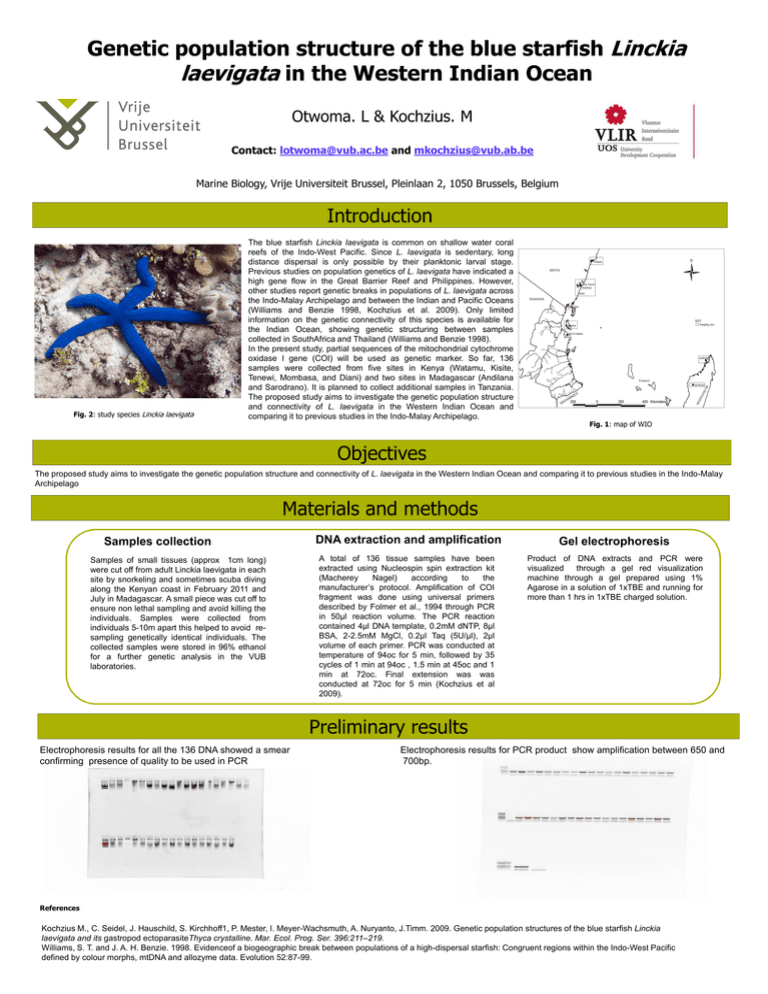
Genetic population structure of the blue starfish Linckia laevigata in the Western Indian Ocean Otwoma. L & Kochzius. M Contact: lotwoma@vub.ac.be and mkochzius@vub.ab.be Marine Biology, Vrije Universiteit Brussel, Pleinlaan 2, 1050 Brussels, Belgium Introduction L. major L. major E. densa Fig. 2: study species Linckia laevigata The blue starfish Linckia laevigata is common on shallow water coral reefs of the Indo-West Pacific. Since L. laevigata is sedentary, long distance dispersal is only possible by their planktonic larval stage. Previous studies on population genetics of L. laevigata have indicated a high gene flow in the Great Barrier Reef and Philippines. However, other studies report genetic breaks in populations of L. laevigata across the Indo-Malay Archipelago and between the Indian and Pacific Oceans (Williams and Benzie 1998, Kochzius et al. 2009). Only limited information on the genetic connectivity of this species is available for the Indian Ocean, showing genetic structuring between samples collected in SouthAfrica and Thailand (Williams and Benzie 1998). In the present study, partial sequences of the mitochondrial cytochrome oxidase I gene (COI) will be used as genetic marker. So far, 136 samples were collected from five sites in Kenya (Watamu, Kisite, Tenewi, Mombasa, and Diani) and two sites in Madagascar (Andilana and Sarodrano). It is planned to collect additional samples in Tanzania. The proposed study aims to investigate the genetic population structure and connectivity of L. laevigata in the Western Indian Ocean and comparing it to previous studies in the Indo-Malay Archipelago. Fig. 1: map of WIO Objectives The proposed study aims to investigate the genetic population structure and connectivity of L. laevigata in the Western Indian Ocean and comparing it to previous studies in the Indo-Malay Archipelago Materials and methods Samples collection Samples of small tissues (approx 1cm long) were cut off from adult Linckia laevigata in each site by snorkeling and sometimes scuba diving along the Kenyan coast in February 2011 and July in Madagascar. A small piece was cut off to ensure non lethal sampling and avoid killing the individuals. Samples were collected from individuals 5-10m apart this helped to avoid resampling genetically identical individuals. The collected samples were stored in 96% ethanol for a further genetic analysis in the VUB laboratories. DNA extraction and amplification Gel electrophoresis A total of 136 tissue samples have been extracted using Nucleospin spin extraction kit (Macherey Nagel) according to the manufacturer’s protocol. Amplification of COI fragment was done using universal primers described by Folmer et al., 1994 through PCR in 50µl reaction volume. The PCR reaction contained 4µl DNA template, 0.2mM dNTP, 8µl BSA, 2-2.5mM MgCl, 0.2µl Taq (5U/µl), 2µl volume of each primer. PCR was conducted at temperature of 94oc for 5 min, followed by 35 cycles of 1 min at 94oc , 1.5 min at 45oc and 1 min at 72oc. Final extension was was conducted at 72oc for 5 min (Kochzius et al 2009). Product of DNA extracts and PCR were visualized through a gel red visualization machine through a gel prepared using 1% Agarose in a solution of 1xTBE and running for more than 1 hrs in 1xTBE charged solution. Preliminary results Electrophoresis results for all the 136 DNA showed a smear confirming presence of quality to be used in PCR Electrophoresis results for PCR product show amplification between 650 and 700bp. References Kochzius M., C. Seidel, J. Hauschild, S. Kirchhoff1, P. Mester, I. Meyer-Wachsmuth, A. Nuryanto, J.Timm. 2009. Genetic population structures of the blue starfish Linckia laevigata and its gastropod ectoparasiteThyca crystalline. Mar. Ecol. Prog. Ser. 396:211–219. Williams, S. T. and J. A. H. Benzie. 1998. Evidenceof a biogeographic break between populations of a high-dispersal starfish: Congruent regions within the Indo-West Pacific defined by colour morphs, mtDNA and allozyme data. Evolution 52:87-99.
