Aim:
advertisement
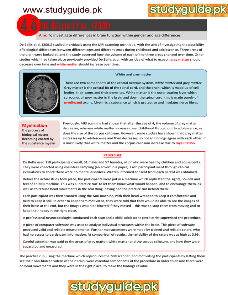
www.studyguide.pk Aim: To investigate differences in brain function within gender and age differences De Bellis et al. (2001) studied individuals using the MRI scanning technique, with the aim of investigating the possibility of biological differences between different ages and different sexes during childhood and adolescence. Three areas of the brain were looked at, and the study observed how the volume of each of the three areas changed over time. Other studies which had taken place previously provided De Bellis et al. with an idea of what to expect: grey matter should decrease over time and white matter should increase over time. White and grey matter There are two components of the central nervous system, white matter and grey matter. Grey matter is the central bit of the spinal cord, and the brain, which is made up of cell bodies, their axons and their dendrites. White matter is the outer coating layer which surrounds all grey matter in the brain and down the spinal cord: this is made purely of myelinated axons. Myelin is a substance which is protective and insulates nerve fibres Myelination the process of biological matter becoming coated by the substance myelin Previously, MRI scanning had shown that after the age of 4, the volume of grey matter decreases, whereas white matter increases over childhood throughout to adolescence, as does the size of the corpus callosum. However, some studies have shown that grey matter increases up to adolescence and then decreases, so not all findings agree with each other. It is most likely that white matter and the corpus callosum increase due to myelination. PROCEDURE De Bellis used 118 participants overall, 61 males and 57 females, all of who were healthy children and adolescents. They were collected using volunteer sampling (an advert in a paper). Each participant went through clinical evaluations to check there were no mental disorders. Written informed consent from each parent was obtained. Before the actual study took place, the participants were put in a machine which replicated the sights, sounds and feel of an MRI machine. This was a ‘practice run’ to let them know what would happen, and to encourage them, as well as to reduce head movements in the real thing, having had the practice run behind them. Each participant was then scanned using the MRI machine, with their head wrapped to keep it comfortable and held to keep it still. In order to keep them motivated, they were told that they would be able to see the images of their brain at the end, but the images would be blurred if they moved – this was to stop them from moving and to keep their heads in the right place. A professional neuroradiologist conducted each scan and a child-adolescent psychiatrist supervised the procedure. A piece of computer software was used to analyse individual structures within the brain. This piece of software produced valid and reliable measurements. Further measurements were made by trained and reliable raters, who had no access to participant information. At comparison of results, the reliability of the raters was as high as 0.99. Careful attention was paid to the areas of grey matter, white matter and the corpus callosum, and how they were separated and measured. The practice run, using the machine which reproduces the MRI scanner, and motivating the participants by letting them see their non-blurred videos of their brain, were essential components of the procedure in order to ensure there were no head movements and they were in the right place, to make the findings reliable. www.aspsychology101.wordpress.com www.studyguide.pk FINDINGS The results showed age-related changes between genders: The grey matter, white matter and corpus callosum all showed significant differences when sex by age interactions were tested Girls showed significant developmental changes with age, but at a slower rate than boys Males had a 19.1% reduction in grey matter between 6 and 17 years of age compared to 4.7% in females Males had a 45.1% increase in white matter compared with a 17.1% increase in females Males had a 58.5% increase in the corpus callosum compared with a 27.4% increase in females DATA ANALYSIS & CONCLUSIONS Overall, the volume of grey matter showed the decrease which was expected, as did the white matter and corpus callosum increase as expected, with age. There were also sex differences, where females increased or decreased the volumes of their respective brain areas at a slower rate and by smaller amounts. This shows further differences between the male and the female brain An evaluation by De Bellis et al. (2001) De Bellis et al. offered their own evaluation of their study. Here are some of their strengths and weaknesses: The participants used made it a cross-sectional study (i.e. different participants were studied all at one time), and needs to be replicated as a longitudinal study (i.e. the same participants are tested over time for a better and more reliable series of results) There were quite large differences between participants of similar ages, which suggests a need for investigation Grey and white matter had to be separated by estimation, as they are not easily-measured areas The sampling and procedure were carefully controlled Giedd et al. (1999) found similar increases and decrease, adding to the reliability of the findings EVALUATION The study had strong controls, including trained raters who did not have access to the participants’ information Each participant was positioned carefully in the same place, providing more strong controls, so reliability can be tested Although the sampling was volunteer sampling, they were matched as best as possible with regard to socioeconomic status, IQ and race, providing a group control It was hard to operationalise the dependent variable which means it was difficult to measure the grey and white matter – estimation of results could mean the findings might not be reliable, or valid The overall sample used high-functioning young people with higher-than-average IQs of an average of 116, which could have affected the results (however, all the participants were like this, so it was still controlled) www.aspsychology101.wordpress.com
