Quantification of Short-TE Metabolite Signals in Human Brain Using QUEST...
advertisement
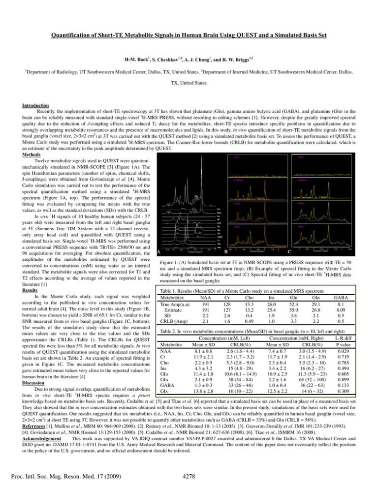
Quantification of Short-TE Metabolite Signals in Human Brain Using QUEST and a Simulated Basis Set H-M. Baek1, S. Cheshkov1,2, A. J. Chang1, and R. W. Briggs1,2 1 Department of Radiology, UT Southwestern Medical Center, Dallas, TX, United States, 2Department of Internal Medicine, UT Southwestern Medical Center, Dallas, TX, United States Introduction Recently, the implementation of short-TE spectroscopy at 3T has shown that glutamate (Glu), gamma amino butyric acid (GABA), and glutamine (Gln) in the brain can be reliably measured with standard single-voxel 1H-MRS PRESS, without resorting to editing schemes [1]. However, despite the greatly improved spectral quality due to the reduction of J-coupling effects and reduced T2 decay for the metabolites, short-TE spectra introduce specific problems in quantification due to strongly overlapping metabolite resonances and the presence of macromolecules and lipids. In this study, in vivo quantification of short-TE metabolite signals from the basal ganglia (voxel size, 2×3×2 cm3) at 3T was carried out with the QUEST method [2] using a simulated metabolite basis set. To assess the performance of QUEST, a Monte Carlo study was performed using a simulated 1H-MRS spectrum. The Cramer-Rao lower bounds (CRLB) for metabolite quantification were calculated, which is an estimate of the uncertainty in the peak amplitude determined by QUEST. Methods Twelve metabolite signals used in QUEST were quantummechanically simulated in NMR-SCOPE [3] (Figure 1A). The spin Hamiltonian parameters (number of spins, chemical shifts, J-couplings) were obtained from Govindaraju et al. [4]. Monte Carlo simulation was carried out to test the performance of the spectral quantification method using a simulated 1H-MRS spectrum (Figure 1A, top). The performance of the spectral fitting was evaluated by comparing the means with the true values, as well as the standard deviations (SDs) with the CRLB. In vivo 1H signals of 10 healthy human subjects (24 - 57 years old) were measured from the left and right basal ganglia at 3T (Siemens Trio TIM System with a 12-channel receiveonly array head coil) and quantified with QUEST using a simulated basis set. Single-voxel 1H-MRS was performed using a conventional PRESS sequence with TR/TE= 2500/30 ms and 96 acquisitions for averaging. For absolute quantification, the amplitudes of the metabolites estimated by QUEST were Figure 1. (A) Simulated basis set at 3T in NMR-SCOPE using a PRESS sequence with TE = 30 converted to concentrations (mM) using water as an internal ms and a simulated MRS spectrum (top), (B) Example of spectral fitting in the Monte Carlo standard. The metabolite signals were also corrected for T1 and study using the simulated basis set, and (C) Spectral fitting of in vivo short-TE 1H-MRS data T2 effects according to the average of values reported in the measured on the basal ganglia literature [1] Results Table 1. Results (Mean/SD) of a Monte Carlo study on a simulated MRS spectrum In the Monte Carlo study, each signal was weighted Metabolites NAA Cr Cho Ins Glu Gln GABA according to the published in vivo concentration values for True Amp(a.u) 191 128 13.3 26.0 52.4 29.1 8.1 normal adult brain [4]. The noise level in this study (Figure 1B, Estimate 191 127 13.2 25.4 55.0 26.8 8.09 bottom) was chosen to yield a SNR of 65:1 for Cr, similar to the SD 2.2 2.6 0.4 1.9 3.8 2.1 0.5 SNR measured from in vivo basal ganglia (Figure 1C, bottom). CRLB (Amp) 2.1 1.6 0.49 1.0 3.3 2.2 0.5 The results of the simulation study show that the estimated Table 2. In vivo metabolite concentrations (Mean/SD) in basal ganglia (n = 10, left and right) mean values are very close to the true values and the SDs Concentration (mM, Left) Concentration (mM, Right) L-R diff approximate the CRLBs (Table 1). The CRLBs for QUEST Metabolite Mean ± SD CRLB(%) Mean ± SD CRLB(%) P value spectral fits were less than 5% for all metabolite signals. In vivo NAA 8.1 ± 0.6 2.6 (1.6 - 4.4) 7.4 ± 0.7 3.0 (1.5 - 4.9) 0.029 results of QUEST quantification using the simulated metabolite Cr 11.9 ± 2.1 2.3 (1.7 – 3.2) 11.7 ± 1.9 2.1 (1.4 - 2.9) 0.719 basis set are shown in Table 2. An example of spectral fitting is Cho 2.2 ± 0.5 5.3 (2.8 – 9.0) 2.3 ± 0.4 5.5 (2.3 – 10) 0.785 given in Figure 1C. The measured metabolite concentrations Ins 4.3 ± 3.2 15 (4.8 - 29) 3.4 ± 2.2 16 (6.2 - 27) 0.494 gave estimated mean values very close to the reported values for Glu 11.4 ± 1.8 10.6 (8.1 – 14.9) 10.9 ± 2.5 11.3 (5.9 – 23) 0.605 human brain in the literature [4]. Gln 2.1 ± 0.9 58 (34 – 84) 2.2 ± 1.6 65 (32 – 100) 0.899 Discussion GABA 1.3 ± 0.3 33 (26 – 46) 1.0 ± 0.4 36 (22 – 63) 0.133 Due to strong signal overlap, quantification of metabolites Glx 13.8 ± 2.8 16 (10 – 22) 12.5 ± 2.2 14 (6 – 32) 0.309 1 from in vivo short-TE H-MRS spectra requires a priori knowledge based on metabolite basis sets. Recently, Cudalbu et al. [5] and Tkac et al. [6] reported that a simulated basis set can be used in place of a measured basis set. They also showed that the in vivo concentration estimates obtained with the two basis sets were similar. In the present study, simulations of the basis sets were used for QUEST quantification. Our results suggested that six metabolites (i.e., NAA, Ins, Cr, Cho, Glu, and Glx) can be reliably quantified in human basal ganglia (voxel size, 2×3×2 cm3) at short TE using 3T. However, it was not possible to quantify other metabolites such as GABA (CRLB = 33%) and Gln (CRLB = 58%). References [1]. Mullins et al., MRM 60: 964-969 (2008). [2]. Ratiney et al., NMR Biomed 18: 1-13 (2005). [3]. Graveron-Demilly et al. JMR 101:233-239 (1993). [4]. Govindaraju et al., NMR Biomed 13:129-153 (2000). [5]. Cudalbu et al., NMR Biomed 21: 627-636 (2008). [6]. Tkac et al., ISMRM 16 (2008). Acknowledgement This work was supported by VA IDIQ contract number VA549-P-0027 awarded and administered b the Dallas, TX VA Medical Center and DOD grant no. DAMD 17-01-1-0741 from the U.S. Army Medical Research and Material Command. The content of this paper does not necessarily reflect the position or the policy of the U.S. government, and no official endorsement should be inferred. Proc. Intl. Soc. Mag. Reson. Med. 17 (2009) 4278
