Burstein lab, GNM 8/2007
advertisement
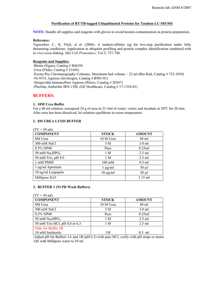
Burstein lab, GNM 8/2007 Purification of BT/TB-tagged Ubiquitinated Proteins for Tandem LC-MS/MS NOTE: Handle all supplies and reagents with gloves to avoid keratin contamination in protein preparation. Reference: Tagwerker, C., K. Flick, et al. (2006). A tandem-affinity tag for two-step purification under fully denaturing conditions: Application in ubiquitin profiling and protein complex identification combined with in vivo cross-linking. Mol Cell Proteomics, Vol 5, 737-748. Reagents and Supplies: -Biotin (Sigma, Catalog # B4639) -Urea (Fluka, Catalog # 51456) -Econo-Pac Chromatography Columns, Maximum bed volume ~ 22 ml (Bio-Rad, Catalog # 732-1010) -Ni-NTA Agarose (Invitrogen, Catalog # R901-01) -Strepavidin ImmunoPure Agarose (Pierce, Catalog # 20347) -PlusOne Amberlite IRN-150L (GE Healthcare, Catalog # 17-1326-01) BUFFERS: 1. 10M Urea Buffer For a 40 ml solution, resuspend 24 g of urea in 21.3ml of water, vortex and incubate at 30oC for 20 min. After urea has been dissolved, let solution equilibrate to room temperature. 2. 8M UREA LYSIS BUFFER (TV = 50 ml) COMPONENT 8M Urea 300 mM NaCl 0.5% NP40 50 mM Na2HPO4 50 mM Tris, pH 8.0 1 mM PMSF 1 ng/ml Aprotinin 10 ng/ml Leupeptin Millipore H2O STOCK 10 M Urea 5M Pure 1M 1M 100 mM 1 µg/ml 10 µg/ml AMOUNT 40 ml 3.0 ml 0.25ml 2.5 ml 2.5 ml 0.5 ml 50 µl 50 µl 1.15 ml 3. BUFFER 1 (Ni PD Wash Buffers) (TV = 50 ml) COMPONENT STOCK AMOUNT 8M Urea 10 M Urea 40 ml 300 mM NaCl 5M 3.0 ml 0.5% NP40 Pure 0.25ml 50 mM Na2HPO4 1M 2.5 ml 50 mM Tris-HCl, pH 8.0 or 6.3 1M 2.5 ml Only for Buffer 1B 10 mM Imidazole 1M 0.5 ml -Adjust pH for Buffers 1A and 1B (pH 6.3) with pure HCl, verify with pH strips or meter. -QS with Millipore water to 50 ml Burstein lab, GNM 8/2007 4. BUFFER 2 (Elution Buffer) (TV = 50 ml) COMPONENT STOCK AMOUNT 8M Urea 10 M Urea 40 ml 200 mM NaCl 5M 2.0 ml 50 mM Na2HPO4 1M 2.5 ml 2% SDS Pure 1g 10 mM EDTA 0.5 M 1 ml 50 mM Tris-HCl, pH 4.3 1M 2.5 ml -Adjust pH with pure HCl to pH 4.3, verify with pH strips or meter. -QS with Millipore water to 50 ml 5. BUFFER 3 (Stepavidin PD wash buffers) (TV = 50 ml) COMPONENT 8M Urea 200 mM NaCl 0.2% SDS 50 mM Tris-HCl, pH 8.0 Millipore H2O -Buffer 3A: 5 ml 20% SDS, 0.5 ml H2O -Buffer 3B: No SDS, 5.5 ml H2O STOCK 10 M Urea 5M 20% SDS 1M AMOUNT 40 ml 2.0 ml 0.5 ml 2.5 ml 5.0 ml 6. BUFFER 4 (Final wash buffer) (TV = 50 ml) COMPONENT 50 mM Tris-HCl, pH 8.0 0.5 mM EDTA 1 mM DTT Millipore H2O STOCK 1M 0.5 M 1M AMOUNT 2.5 ml 50 µl 50 µl 47.4 ml EXPERIMENTAL PROCEDURES 1. Seed 7 x 10⁶ HEK 293 cells in 15 cm dishes ~ 18 hrs prior to transfection. 2. Transfect cells using conventional calcium phosphate method. The amounts of plasmid indicated below were adopted from small scale experiments, in which ployubiquitinated forms were readily observed by immunoblotting after two-step affinity purification. Per 15 cm dish: 30 µg pEBB-BT/TB-fusion protein 3 µg pEBB RGS-His6-Human Ubiquitin 3. Replace media ~8 hrs post-transfection with fresh warm media supplemented with Biotin (4 µm). NOTE: Biotinylation via the endogenous pool of biotin will occur. However, optimal biotinylation requires exogenous biotin for highly expressed fusion proteins. 4. Prepare fresh 10M Urea buffer in Millipore water, and incubate for 1 hr at room temperature with 1% Amberlite ion exchange resin to remove isocyanate impurities. NOTE: The presence of isocyanate impurities can lead to carbamylation of lysine residues and interfere with protein analysis. Burstein lab, GNM 8/2007 5. Two days post-transfection, aspirate media from cells, gently wash with 5 ml room temperature 1x PBS, and lyse cells with 1.5 ml 8M urea lysis buffer per plate. Incubate plates at room temperature for 20 min, scrape, and transfer lysate to an appropriate vessel. 6. Sonicate samples to reduce lysate viscosity. For HEK 293 cells, 25 pulses (output control 2.5, 75% duty cycle) is typically sufficient to reduce the viscosity. Verify by pipetting the lysate. -Sonicate ~10 ml lysate in a single 50 ml conical tube. 7. Remove cell debris by centrifuging lysate at 15,000g for 15 min at 4oC. Transfer supernatant to a fresh conical tube and determine protein concentration by Bradford assay. NOTE: Save small aliquot for Input sample. 8. Pre-equilibrate Ni-NTA bead slurry with Buffer 1: Load column with 30 µl of a 50% slurry for each milligram of total protein lysate. Apply 5 bead volumes of Buffer 1 (see below) and allow to drain by gravity flow. 9. Precipitate ubiquitinated proteins with Ni-NTA beads: Apply lysate to Ni-loaded column and rotate for 4 hrs at RT. NOTE: Maximum volume capacity of column is ~ 23 ml. 10. Drain column by gravity flow. NOTE: Save flowthrough to assess depletion. 11. Wash Ni-NTA agarose with 10 bead volumes of the buffers indicated below, rotating for 5 min at RT in between each wash. Washes 1 and 2: Buffer 1 (pH 8.0) Wash 3: Buffer 1A (pH 6.3) Wash 4: Buffer 1B (pH 6.3, 10 mM Imidazole) 12. Elute ubiquitinated proteins from Ni-NTA beads: Apply 10 bead volumes of Buffer 2 and rotate column for 30 min at RT. Collect eluate in a fresh conical tube and adjust pH to 8.0 with 10N NaOH. Verify with pH strips. 13. Pre-equilibrate Strepavidin beads with Buffer 3: Load column with 50 µl of a 50% slurry for ~ 100 mg of total input lysate used for initial affinity purification step (Ni PD). Apply 5 ml of Buffer 3 (see below) and allow to drain by gravity flow. 14. Precipitate BT/TB-tagged fusion protein with Strepavidin beads: Apply pH adjusted eluate to Strepavidin-loaded column and rotate overnight at RT. 15. Drain column by gravity flow and wash Strepavidin agarose with 15 ml of each buffer indicated below, rotating for 5 min at RT in between each wash. Washes 1 and 2: Buffer 3 (0.2% SDS) Wash 3: Buffer 3A (2% SDS) Wash 4: Buffer 3B (No SDS) 16. Apply 20 ml of Buffer 4, rotate for 5 min at RT, and drain column by gravity flow. Repeat procedure for a total of 4 times. 17. Preparation of sample for gel loading: After last wash, add 1 ml of Buffer 4 to closed column, gently shake, and transfer resuspended beads to a fresh 1.5 ml microfuge tube. Spin samples for 1 min at 2500 RPM, aspirate supernatant, and resuspend beads in 1 µl 3x LDS/DTT per µl of Strepavidin bead volume. Burstein lab, GNM 8/2007 18. NuPage and Coomassie Staining: Heat sample for 10 min at 80oC. Load maximal amount onto a NuPage gel and run for desired amount of time for adequate separation of ubiquitinated forms of the protein of interest. Fix gel and stain with Coomassie. NOTES: 1. 10 well gel, maximal loading volume ~ 40 µl 2. Load BT/TB-tagged protein precipitated from a cell lysate (denatured conditions) transfected without His6-Ubiquitin (one-step SA pull-down). This will enable determining the mobility of the non-modified protein. 3. Load BSA standards to estimate protein recovery. 19. Image gel and submit to Michigan Proteome Consortium after filling out a service request form (see link below). E-mail gel to Mary Hurley (caponite@umich.edu) indicating which bands are to be excised for LC-MS/MS. http://www.proteomeconsortium.org/
