This is a temporal bone which had an implant electrode... fresh and unfixed. After fixation, osmium staining, and dissection the...
advertisement
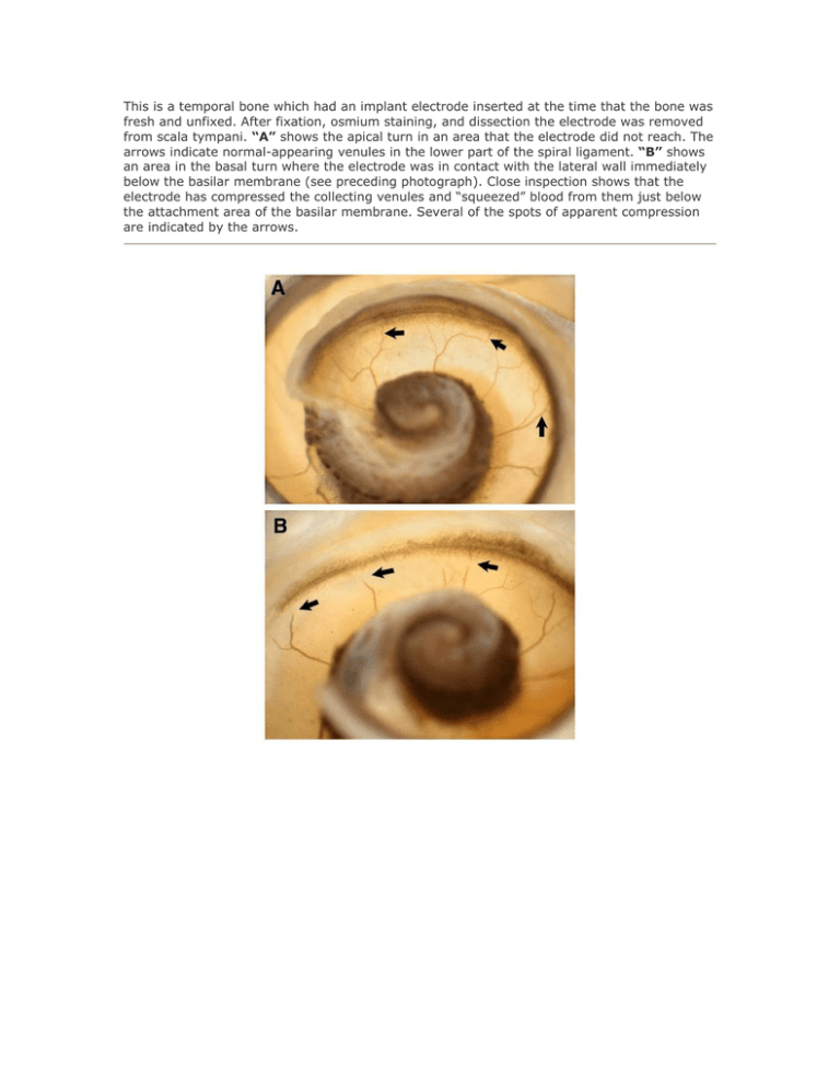
This is a temporal bone which had an implant electrode inserted at the time that the bone was fresh and unfixed. After fixation, osmium staining, and dissection the electrode was removed from scala tympani. “A” shows the apical turn in an area that the electrode did not reach. The arrows indicate normal-appearing venules in the lower part of the spiral ligament. “B” shows an area in the basal turn where the electrode was in contact with the lateral wall immediately below the basilar membrane (see preceding photograph). Close inspection shows that the electrode has compressed the collecting venules and “squeezed” blood from them just below the attachment area of the basilar membrane. Several of the spots of apparent compression are indicated by the arrows.
