A Classroom Demonstration of Protein Folding
advertisement

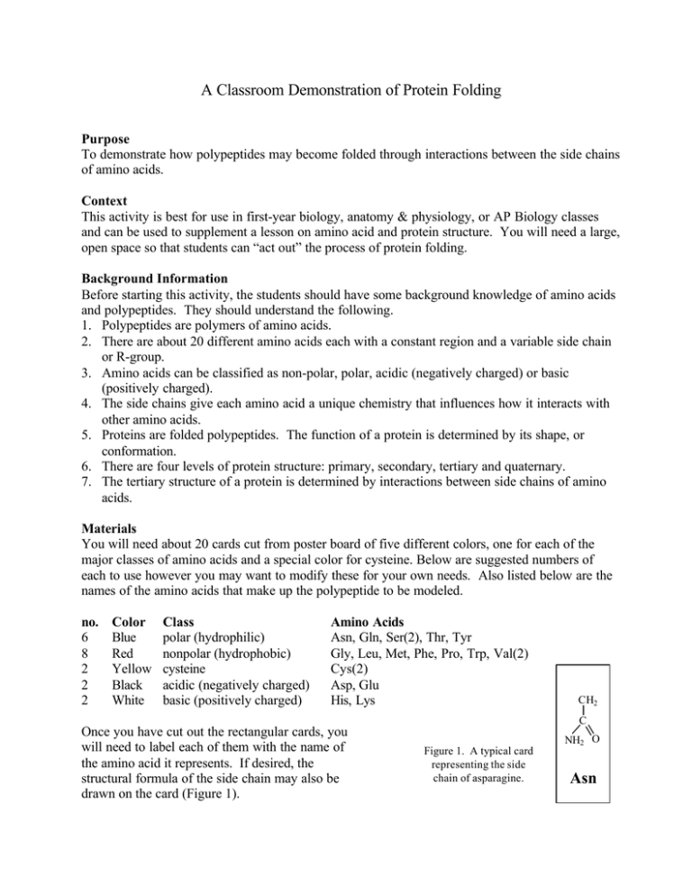
A Classroom Demonstration of Protein Folding Purpose To demonstrate how polypeptides may become folded through interactions between the side chains of amino acids. Context This activity is best for use in first-year biology, anatomy & physiology, or AP Biology classes and can be used to supplement a lesson on amino acid and protein structure. You will need a large, open space so that students can “act out” the process of protein folding. Background Information Before starting this activity, the students should have some background knowledge of amino acids and polypeptides. They should understand the following. 1. Polypeptides are polymers of amino acids. 2. There are about 20 different amino acids each with a constant region and a variable side chain or R-group. 3. Amino acids can be classified as non-polar, polar, acidic (negatively charged) or basic (positively charged). 4. The side chains give each amino acid a unique chemistry that influences how it interacts with other amino acids. 5. Proteins are folded polypeptides. The function of a protein is determined by its shape, or conformation. 6. There are four levels of protein structure: primary, secondary, tertiary and quaternary. 7. The tertiary structure of a protein is determined by interactions between side chains of amino acids. Materials You will need about 20 cards cut from poster board of five different colors, one for each of the major classes of amino acids and a special color for cysteine. Below are suggested numbers of each to use however you may want to modify these for your own needs. Also listed below are the names of the amino acids that make up the polypeptide to be modeled. no. 6 8 2 2 2 Color Blue Red Yellow Black White Class polar (hydrophilic) nonpolar (hydrophobic) cysteine acidic (negatively charged) basic (positively charged) Amino Acids Asn, Gln, Ser(2), Thr, Tyr Gly, Leu, Met, Phe, Pro, Trp, Val(2) Cys(2) Asp, Glu His, Lys CH2 C Once you have cut out the rectangular cards, you will need to label each of them with the name of the amino acid it represents. If desired, the structural formula of the side chain may also be drawn on the card (Figure 1). NH2 O Figure 1. A typical card representing the side chain of asparagine. Asn Procedure 1. Give each student one of the cards. They will play the role of the amino acid on their card. 2. On the board or overhead, display the following sequence: Met, Lys, His, Val, Ser, Leu, Asp, Glu, Cys, Asn, Tyr, Val, Phe, Trp, Pro, Ser, Thr, Gln, Cys, Gly. 3. Have students line up in the order of the sequence to form a polypeptide. They should line up front to back. Holding the side chain card in their left hand they should place their right hand on the right shoulder of the person in front of them. These represent the peptide bonds formed between the amino acids in the polypeptide. 4. Now you will demonstrate the folding process. The strongest interaction between the side chains is a covalent bond formed between cysteine molecules. Have the two students representing cysteine raise their cards into the air. Have the polypeptide chain bend so that the two side chains will be attracted to one another (Figure 2b). Students will need to move around to accommodate this procedure, however they should stay attached to one another so that the polypeptide doesn’t fall apart. 5. The slightly weaker ionic interactions will be demonstrated next. There are two oppositely charged regions near the N-terminus of the polypeptide. Fold the polypeptide so that the positively charged groups are near the negatively charged groups (Figure 2c). 6. Even weaker forces are at work in protein folding through hydrophobic interactions. Have the students with nonpolar side chains raise their cards. One region contains 4 amino acids with these side chains. The hydrophobic nature of these groups will cause them to clump together in an effort to “get away” from the water bathing the molecule and the protein gets another bend (Figure 2d). Discussion While this demonstration will give students a general understanding of the process of protein folding, there are many things left out which you may want to point out to them. First, this demo does not take into consideration the hydrogen bonding between repeating elements on the “backbone” of carbon and nitrogen that makes up the polypeptide chain. These important interactions give the protein its secondary structure and produce such elements as alpha helices and beta pleated sheets. Further, this demonstration only works in two dimensions. It is critical that students understand the three-dimensional nature of protein structure. Despite these shortcomings this model is very useful to help reinforce concepts covered in class and to visualize the process of protein folding. N-terminus n Met n + Lys + + His + n Val n p Ser p n Leu n - Asp n Disulfide Bridge - - Glu - p p S Cys n S p Asn p Tyr p n Val n p - + - + - + - n n n S S p S S n n p n n (c) n Hydrophobic Interactions (d) p Ser p Gln S Cys n Gly C-terminus (a) p p n n Pro p Thr p n n p p n Trp (b) n p p p p n + Ionic Bonding p n n Phe n n S p n n Figure 2. Steps in the protein folding demonstration. Each circle represents an amino acid. The symbols used within the circles indicate the grouping to which the amino belongs: n, nonpolar; p, polar; -, acidic; +, basic; S, sulfur containing cysteine. (a) Primary structure of the polypeptide. (b) Configuration following the formation of the covalently bonded disulfide bridge. (c) Configuration with ionic bonding between acidic and basic side groups. (d) Final form of protein with hydrophobic interactions included.


