17 Segmented Worms C H A P T E R Dividing the Body
C H A P T E R
17
Segmented Worms
Phylum Annelida
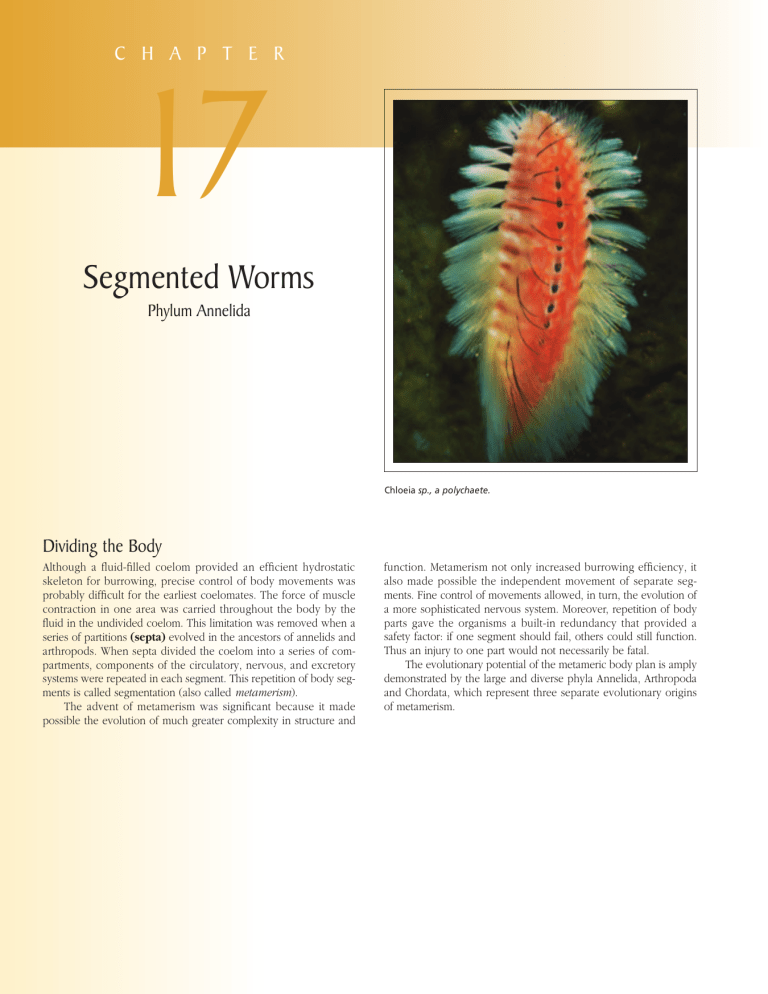
Chloeia sp., a polychaete.
Dividing the Body
Although a fluid-filled coelom provided an efficient hydrostatic skeleton for burrowing, precise control of body movements was probably difficult for the earliest coelomates. The force of muscle contraction in one area was carried throughout the body by the fluid in the undivided coelom. This limitation was removed when a series of partitions (septa) evolved in the ancestors of annelids and arthropods. When septa divided the coelom into a series of compartments, components of the circulatory, nervous, and excretory systems were repeated in each segment. This repetition of body segments is called segmentation (also called metamerism ).
The advent of metamerism was significant because it made possible the evolution of much greater complexity in structure and function. Metamerism not only increased burrowing efficiency, it also made possible the independent movement of separate segments. Fine control of movements allowed, in turn, the evolution of a more sophisticated nervous system. Moreover, repetition of body parts gave the organisms a built-in redundancy that provided a safety factor: if one segment should fail, others could still function.
Thus an injury to one part would not necessarily be fatal.
The evolutionary potential of the metameric body plan is amply demonstrated by the large and diverse phyla Annelida, Arthropoda and Chordata, which represent three separate evolutionary origins of metamerism.
w w w. m h h e . c o m / h i c k m a n i p z 1 3
P hylum Annelida (an-nel
′ i-da) (L. annelus, little ring, + ida, pl. suffix) consists of the segmented worms. It is a diverse phylum, numbering approximately 15,000 species, the most familiar of which are earthworms and freshwater worms
(class Oligochaeta) and leeches (class Hirudinea). However, approximately two-thirds of the phylum comprises of marine worms (class Polychaeta), which are less familiar to most people. Among the latter are many curious members; some are strange, even grotesque, whereas others are graceful and beautiful. They include clamworms, plumed worms, parchment worms, scaleworms, lugworms, and many others. Annelids are true protostome coelomates belonging to superphylum Lophotrochozoa, which exhibit spiral cleavage and mosaic development. They are a highly developed group in which the nervous system is more centralized and the circulatory system more complex than those of phyla we previously have studied.
Annelida are worms whose bodies are divided into similar rings, or segments, (also called metameres or somites) arranged in linear series and externally marked by circular rings called annuli (the name of the phylum refers to this characteristic). Body segmentation (metamerism) is a division of the body into a series of segments, each of which contains similar components of all major organ systems. In annelids the segments (metamerism) are delimited internally by septa. Segmentation has evolved independently in the annelids, arthropods, and vertebrates.
Annelids are sometimes called “bristle worms” because, with the exception of leeches, most annelids bear tiny chitinous bristles called setae (L. seta, hair or bristle). Short needlelike setae help anchor segments during locomotion and long, hairlike setae aid aquatic forms in swimming. Since many annelids burrow or live in secreted tubes, stiff setae also aid in preventing the worm from being pulled out or washed out of its home. Robins know from experience how effective earthworms’ setae are.
Annelids have a worldwide distribution, and a few species are cosmopolitan. Polychaetes are chiefly marine forms. Most are benthic, but some live pelagic lives in the open seas. Oligochaetes and leeches occur predominantly in fresh water or terrestrial soils.
Some freshwater species burrow in mud and sand and others among submerged vegetation. Many leeches are predators, specialized for piercing their prey and feeding on blood or soft tissues. A few leeches are marine, but most live in fresh water or in damp regions. Suckers are typically found at both ends of the body for attachment to the substratum or to their prey.
CHAPTER 17 Segmented Worms
Position in Animal Kingdom
1. Annelids belong to the lophotrochozoan protostome branch of the animal kingdom and have spiral cleavage and mosaic (determinate) development, characters shared with and indicating their relationship to molluscs.
2. Annelids as a group show a primitive metamerism with comparatively few differences between the different segments.
3. Characters shared with arthropods include an outer secreted cuticle and a similar nervous system.
Biological Contributions
1.
Metamerism represents the greatest innovation seen in this phylum. However, a more highly specialized metamerism is seen in arthropods.
2. A true coelomic cavity reaches a high stage of development in this group.
3. Specialization of the head region into differentiated organs, such as tentacles, palps, and eyespots of polychaetes, is more pronounced in some annelids than in other invertebrates so far considered.
4. There are modifications of the nervous system, with cerebral ganglia (brain), two closely fused ventral nerve cords with giant fibers running the length of the body, and various ganglia with their lateral branches.
5. The circulatory system is much more complex than any we have so far discussed. It is a closed system with muscular blood vessels and aortic arches (“hearts”) for pumping the blood.
6. The appearance of the fleshy parapodia, with their respiratory and locomotor functions, is likely an example of evolutionary convergence to appendages and specialized gills found in arthropods.
7. Well-developed nephridia in most of the segments have reached a differentiation that involves removal of waste from the blood as well as from the coelom.
8. Annelids are the most highly organized animals capable of complete regeneration. However, this ability varies greatly within the group.
363
BODY PLAN
The annelid body typically has an anterior prostomium, followed by a segmented body, and a terminal portion called the pygidium.
The prostomium and pygidium are not considered to be segments, but anterior segments often fuse with the prostomium to form a head. New segments differentiate during development just in front of the pygidium; thus the oldest segments are at the anterior end and the youngest segments are at the posterior end.
The body wall has strong circular and longitudinal muscles
(adapted for swimming, crawling, and burrowing) and is covered with epidermis and a thin, outer layer of nonchitinous cuticle (Figure 17-1).
In most annelids the coelom develops embryonically as a split in the mesoderm on each side of the gut (schizocoel), forming a pair of coelomic compartments in each segment. Peritoneum (a layer of mesodermal epithelium) lines the body wall of each compartment, forming dorsal and ventral mesenteries that cover all organs (Figure 17-1). Peritonea of adjacent segments meet to form septa, which are perforated by the gut and longitudinal blood vessels. Virtually every body system is affected in some way by this segmental arrangement.
364 PART THREE Diversity of Animal Life
Longitudinal muscle
Dorsal blood vessel
Segment Intestine
Circular muscle
Dorsal mesentery
Septum
Ventral mesentery
Ventral blood vessel
Septum
Visceral peritoneum
Parietal peritoneum
Intestine
Circular muscle
Longitudinal muscle
Ventral mesentery
Cuticle Ventral blood vessel
Figure 17-1
Annelid body plan.
Epidermis
Except in leeches, the coelom of most annelids is filled with fluid and serves as a hydrostatic skeleton.
Because the volume of fluid is essentially constant, contraction of the longitudinal body-wall muscles causes the body to shorten and become larger in diameter, whereas contraction of the circular muscles causes it to lengthen and become thinner. Separation of the hydrostatic skeleton into a series of coelomic cavities greatly increases its efficiency, because the force of local muscle contraction within one segment is not transferred and dampened throughout the length of the worm. For this reason, widening and elongation can occur in restricted areas, and crawling motions are produced by alternating waves of contraction passing down the body. Segments in which longitudinal muscles are contracted widen and anchor themselves against the substrate while other segments, in which circular muscles are contracted, elongate and stretch forward. Forces powerful enough for rapid burrowing as well as locomotion can thus be generated.
CLASS POLYCHAETA
The largest class of annelids is the Polychaeta with more than
10,000 species, most of them marine. Although most polychaetes are 5 to 10 cm long, some are less than 1 mm, and others may be as long as 3 m. They may be brightly colored in reds and greens, iridescent, or dull. Still others are picturesque, such as the “featherduster” worms (Figure 17-2).
Polychaetes differ from other annelids in having a welldifferentiated head with specialized sense organs; paired appendages, called parapodia, on most segments; and no clitellum (see p. 000) (Figure 17-3). As their name implies, they have many setae, usually arranged in bundles on the parapodia. Of all annelids they exhibit the most pronounced differentiation of body segments and specialization of sensory organs (see p. 000).
Many polychaetes are euryhaline and can tolerate a wide range of environmental salinity. The freshwater polychaete fauna is more diversified in warmer regions than in temperate zones.
Many polychaetes live under rocks, in coral crevices, or in abandoned shells. A number of species burrow into mud or sand and build their own tubes on submerged objects or in bottom
A B
Figure 17-2
Tube-dwelling sedentary polychaetes. A, One of the featherduster worms (called a Christmas-tree worm), Spirobranchus giganteus, has a double crown of radioles and lives in a calcareous tube. B, Sabellid polychaetes, Bispira brunnea, live in leathery tubes.
Jaw
Prostomial tentacles
Palp
Prostomium
Peristomium
Parapodia
A w w w. m h h e . c o m / h i c k m a n i p z 1 3 CHAPTER 17 Segmented Worms 365 sediment. Others adopt the tubes or homes of other animals, and some are planktonic. They are extremely abundant in some areas, for example, a square meter of mudflat may contain thousands of polychaetes. They play a significant part in marine food chains because they are eaten by fish, crustaceans, hydroids, and many other predators.
Polychaetes are often divided into two groups based on their activity: sedentary polychaetes and errant (free-moving) polychaetes. Sedentary polychaetes spend much or all of their time in tubes or permanent burrows. Many of them, especially those that live in tubes, have elaborate devices for feeding and respiration. Errant polychaetes (L. errare, to wander), include free-swimming pelagic forms, active burrowers, crawlers, and tube worms that only leave their tubes for feeding or breeding.
Most of these, like clam worms in the genus Nereis (Gr. name of a sea nymph) (Figure 17-3), are predatory and equipped with jaws or teeth. They have an eversible, muscular pharynx armed with teeth that can be thrust out with surprising speed to capture prey.
Form and Function
A polychaete typically has a “head,” or prostomium, which may or may not be retractile and which often bears eyes, tentacles, and sensory palps (Figure 17-3). The first segment (peristomium) surrounds the mouth and may bear setae, palps, or, in predatory forms, chitinous jaws. Ciliary feeders may bear a crown of tentacles that can be opened like a fan or withdrawn into the tube.
The polychaete trunk is segmented, and most segments bear parapodia, which may have lobes, cirri, setae, and other parts on them (Figure 17-3). Parapodia are used in crawling, swimming, or for anchoring the animal in its tube. They usually serve as the chief respiratory organs, although some polychaetes also have gills. Amphitrite (Gr. a mythical sea nymph), for example, has three pairs of branched gills and long extensible tentacles (Figure 17-4). Arenicola (L. arena, sand, + colo, inhabit), the burrowing lugworm (Figure 17-5), has paired gills on certain segments.
Everted pharynx
Eyes
Tentacles
(cirri)
B
Respiratory capillaries
Dorsal cirrus
Oblique muscle
Eggs
Dorsal vessel
Intestine Coelomic epithelium
Longitudinal muscle
Circular muscle
Notopodium
Parapodium
Setae
Neuropodium
Epidermis Aciculum Ventral cirrus
D
Nephridium Nerve cord
Ventral vessel
Figure 17-3
Nereis virens (A–D) and
Nereis diversicolor (E) are errant polychaetes.
A, Anterior end, with pharynx everted.
B, External structure.
C, Posterior end.
D, Generalized transverse section through region of the intestine. E, In this photo of a live N.
diversicolor, note the well-defined segments, the lobed parapodia, and the prostomium with tentacles.
C
Parapodia
Pygidium
Anus
Cirrus
E
366 PART THREE Diversity of Animal Life
Tentacle
Gills
Sand falling into shaft Water movement
A
B
C
D
Notopodia
Neuropodia
Proboscis Gills
Figure 17-5
Arenicola, the lugworm, lives in a U-shaped burrow in intertidal mudflats. It burrows by successive eversions and retractions of its proboscis. By peristaltic movements it keeps water filtering through the sand. The worm then ingests the food-laden sand.
Figure 17-4
Amphitrite, which builds its tubes in mud or sand, extends long grooved tentacles out over the mud to pick up bits of organic matter.
The smallest particles are moved along food grooves by cilia, larger particles by peristaltic movement. Its plumelike gills are blood red.
A, Section through exploratory end of tentacle. B, Section through tentacle in area adhering to substratum. C, Section showing ciliary groove. D, Particle being carried toward mouth.
Nutrition
A polychaete’s digestive system consists of a foregut, a midgut, and a hindgut. The foregut includes a stomodeum, a pharynx, and an anterior esophagus. It is lined with cuticle, and the jaws, where present, are constructed of cuticular protein. The more anterior portions of the midgut secrete digestive enzymes but absorption takes place toward the posterior end. A short hindgut connects the midgut to the exterior via the anus, which is on the pygidium.
Errant polychaetes are typically predators and scavengers.
Sedentary polychaetes feed on suspended particles, or they may be deposit feeders, consuming particles on or in the sediment.
Circulation and Respiration
Polychaetes show considerable diversity in both circulatory and respiratory structures. As previously mentioned, parapodia and gills serve for gaseous exchange in various species. However, in some polychaetes there are no special organs for respiration, and gaseous exchange takes place across the body surface.
The circulatory pattern varies greatly. In Nereis a dorsal longitudinal vessel carries blood anteriorly, and a ventral longitudinal vessel conducts it posteriorly (Figure 17-3D). Blood flows between these two vessels via segmental networks in the parapodia, septa, and around the intestine. In Glycera (Gr. Glykera, a feminine proper name) the circulatory system is reduced and joins directly with the coelom. Septa are incomplete, and thus the coelomic fluid assumes the function of circulation.
Many polychaetes have respiratory pigments such as hemoglobin, chlorocruorin, or hemerythrin (p. 000).
Excretion
Excretory organs consist of protonephridia and mixed proto- and metanephridia in some, but most polychaetes have metanephridia
(Figure 17-3). There is one pair per segment, with the inner end of each (nephrostome) opening into a coelomic compartment.
Coelomic fluid passes into the nephrostome, and selective resorption occurs along the nephridial duct (Figure 17-14).
Nervous System and Sense Organs
Organization of the central nervous system in polychaetes follows the basic annelid plan (Figure 17-15). Dorsal cerebral ganglia connect with a subpharyngeal ganglion via a circumpharyngeal connective. A double ventral nerve cord courses the length of the worm, with metamerically arranged ganglia.
Sense organs are highly developed in polychaetes and include eyes, nuchal organs, and statocysts. Eyes, when present, may range from simple eyespots to well-developed organs. Eyes are most conspicuous in errant worms. Usually the eyes are retinal cups, with rodlike photoreceptor cells (lining the cup wall) directed toward the lumen of the cup. The highest degree of eye development occurs in the family Alciopidae, which has large, image-resolving eyes similar in structure to those of some cephalopod molluscs (Figure 16-39, p. 000), with cornea, lens, retina, and retinal pigments. Alciopid eyes also have accessory retinas, a characteristic independently evolved by deep-sea fishes and some deep-sea cephalopods. The accessory retinas of alciopids are sensitive to different wavelengths. The eyes of these pelagic animals may be well adapted to function because penetration by the different wavelengths of light varies with depth. Studies with electroencephalograms show that they are
w w w. m h h e . c o m / h i c k m a n i p z 1 3 CHAPTER 17 Segmented Worms 367 sensitive to dim light of the deep sea. Nuchal organs are ciliated sensory pits or slits that appear to be chemoreceptive, an important factor in food gathering. Some burrowing and tube-building polychaetes have statocysts that function in body orientation.
Reproduction and Development
Polychaetes have no permanent sex organs, and they usually have separate sexes. Reproductive systems are simple: Gonads appear as temporary swellings of the peritoneum and shed their gametes into the coelom. The gametes are then carried to the outside through gonoducts, through the metanephridia, or by rupture of the body wall. Fertilization is external, and the early larva is a trochophore (see Figure 16-6).
Some polychaetes live most of the year as sexually immature animals called atokes, but during the breeding season a portion of the body becomes sexually mature and swollen with gametes (Figure 17-6).
An example is the palolo worm, which lives in burrows among coral reefs. During the swarming period, the sexually mature portions, now called epitokes, break off and swim to the surface. Just before sunrise, the sea is literally covered with them, and at sunrise they burst, freeing eggs and sperm for fertilization. Anterior portions of the worms regenerate new posterior sections. Swarming is of great adaptive value because the synchronous maturation of all the epitokes ensures the maximum number of fertilized eggs. However, this reproductive strategy is very hazardous; many types of predators have a feast on the swarming worms. In the meantime, the atoke remains safely in its burrow to produce another epitoke at the next cycle. In some polychaetes, epitokes arise from atokes by asexual budding (Figure 17-7) and become complete worms.
Characteristics of Phylum Annelida
1. Body segmented; symmetry bilateral
2. Body wall with outer circular and inner longitudinal muscle layers; outer transparent moist cuticle secreted by epithelium
3.
Chitinous setae often present; setae absent in leeches
4. Coelom (schizocoel) well developed and divided by septa, except in leeches; coelomic fluid functions as hydrostatic skeleton
5.
Circulatory system closed and segmentally arranged; respiratory pigments (hemoglobin, hemerythrin, or chlorocruorin) often present; amebocytes in blood plasma
6. Digestive system complete and not segmentally arranged
7. Respiratory gas exchange through skin, gills, or parapodia
8. Excretory system typically a pair of nephridia for each segment
9. Nervous system with a double ventral nerve cord and a pair of ganglia with lateral nerves in each segment; brain a pair of dorsal cerebral ganglia with connectives to ventral nerve cord
10. Sensory system of tactile organs, taste buds, statocysts (in some), photoreceptor cells, and eyes with lenses (in some)
11. Hermaphroditic or separate sexes; larvae, if present, are trochophore type; asexual reproduction by budding in some; spiral cleavage and mosaic development
Atoke
Epitoke
Clam Worms: Nereis
Clam worms (Figure 17-3), or sand worms as they are sometimes called, are errant polychaetes that live in mucous-lined burrows in or near low tide. Sometimes they are found in temporary hiding places, such as under stones, where they stay with their bodies covered and their heads protruding. They are most active at night, when they wiggle out of their hiding places and swim about or crawl over the sand in search of food.
The body, containing about 200 segments, may grow to 30 or 40 cm in length. The head is made up of a prostomium and a peristomium. The prostomium bears a pair of stubby palps, sensitive to touch and taste; a pair of short sensory tentacles; and two pairs of small dorsal eyes that are light sensitive. The peristomium bears the ventral mouth, a pair of chitinous jaws, and four pairs of sensory tentacles (Figure 17-3A).
Each parapodium has two lobes: a dorsal notopodium and a ventral neuropodium (Figure 17-3D) that bear setae with many blood vessels. Parapodia are used for both creeping and swimming and are controlled by oblique muscles that run from the midventral line to the parapodia in each segment. The worm swims by lateral undulatory wriggling of the body. It can dart through the water with considerable speed. These undulatory
Figure 17-6
Eunice viridis, the Samoan palolo worm. The posterior segments make up the epitokal region, consisting of segments packed with gametes.
Each segment has an eyespot on the ventral side. Once a year the worms swarm, and the epitokes detach, rise to the surface, and discharge their ripe gametes, leaving the water milky. By the next breeding season, the epitokes are regenerated.
368 PART THREE Diversity of Animal Life
Atoke
Epitokes
Figure 17-7
Rather than transforming a portion of its body into an epitoke, Autolytus prolifer asexually buds off complete worms from its posterior end that become sexual epitokes.
Figure 17-8
A scale worm, Hesperonoe adventor, normally lives as a commensal in the tubes of Urechis (phylum Echiura, p. 000).
movements can also be used to suck water into or pump it out of the burrow.
Clam worms feed on small animals, other worms, and a variety of larval forms. They seize food with their chitinous jaws, which they protrude through the mouth when they evert their pharynx. Food is swallowed as the worm withdraws its pharynx.
Movement of food through the alimentary canal is by peristalsis.
Other Interesting Polychaetes
Scale worms (Figure 17-8) are members of the family Polynoidae (Gr. Polynoe¯, daughter of Nereus and Doris, a sea god and goddess), one of the most diverse, abundant, and widespread of polychaete families. Their flattened bodies are covered with broad scales, modified from dorsal parts of the parapodia.
Most species are of modest size, but some are enormous (up to
190 mm long and 100 mm wide). They are carnivorous and feed on a wide variety of animals. Many are commensal, living in burrows of other polychaetes or in association with cnidarians, molluscs, or echinoderms.
Hermodice carunculata (Gr. herma, reef, + dex, a worm found in wood) (Figure 17-9) and related species are called fireworms because their hollow, brittle setae contain a poisonous secretion. When touched, the setae break off in the wound and cause skin irritation. Fireworms feed on corals, gorgonians, and other cnidarians.
Polychaete tube dwellers secrete many types of tubes. Some are parchmentlike or leathery (Figure 17-2B); some are firm, calcareous tubes attached to rocks or other surfaces (Figure 17-2A); and some are simply grains of sand or bits of shell or seaweed cemented together with mucous secretions. Many species burrow in sand or mud, lining their burrows with mucus (Figure 17-5).
Most sedentary tube and burrow dwellers are particle feeders, using cilia or mucus to obtain food, typically plankton and
Figure 17-9
A fireworm, Hermodice carunculata, feeds on gorgonians and stony corals. Its setae are like tiny glass fibers and serve to ward off predators.
detritus. Some, like Amphitrite (Figure 17-4), peep their heads out of the mud and send out long extensible tentacles over the surface to deposit feed. Cilia and mucus on the tentacles entrap particles found on the sea bottom and move them toward the mouth.
Lugworms, Arenicola, use an interesting combination of suspension and deposit feeding. They live in a U -shaped burrow through which, by peristaltic movements, they cause water to flow. Food particles are filtered out by the sand at the front of the burrow, and Arenicola then ingests the food-laden sand (Figure 17-5).
Fanworms, or “featherduster” worms, are beautiful tubeworms, fascinating to watch as they emerge from their secreted tubes and unfurl their lovely tentacular crowns to feed (Figure
w w w. m h h e . c o m / h i c k m a n i p z 1 3 CHAPTER 17 Segmented Worms 369
17-2). A slight disturbance, sometimes even a passing shadow, causes them to duck back quickly into the safety of the homes.
Food drawn to the feathery arms, or radioles, by ciliary action is trapped in mucus and is carried down ciliated food grooves to the mouth (Figure 17-10). Particles too large for the food grooves pass along the margins of the food grooves and drop off. Only small particles of food enter the mouth; sand grains are stored in a sac to be used later in enlarging the tube.
The parchment worm, Chaetopterus (Gr. chaite¯, long hair, + pteron, wing), feeds on suspended particles by an entirely different mechanism (Figure 17-11). It lives in a U -shaped, parchmentlike tube buried, except for the tapered ends, in sand or mud along the shore. The worm attaches to the side of the tube by ventral suckers. Fans (modified parapodia on segments 14 to 16) pump water through the tube by rhythmical movements. A pair of enlarged parapodia on segment 12 secretes a long mucous bag that reaches back to a small food cup just in front of the fans. All water passing through the tube is filtered through this mucous bag, the end of which is rolled into a ball by cilia in the cup.
When the ball is about the size of a BB shot (about 3 mm diameter), the fans stop beating and the ball of food and mucus is rolled forward by ciliary action to the mouth and swallowed.
Radiole
Direction of water flow
Water movement
Parchment like tube
Mouth
"Wing" (12th notopodia)
Mucous net
"Fans"
Food cup
Cilia
Pinnule section
Direction of food movement
Figure 17-11
Chaetopterus, a sedentary polychaete, lives in a U-shaped tube in the sea bottom. It pumps water through the parchmentlike tube (of which one-half has been cut away here) with its three pistonlike fans. The fans beat 60 times per minute to keep water currents moving. The winglike notopodia of the twelfth segment continuously secrete a mucous net that strains out food particles. As the net fills with food, the food cup rolls it into a ball, and when the ball is large enough
(about 3 mm), the food cup bends forward and deposits the ball in a ciliated groove to be carried to the mouth and swallowed.
Mouth
Ventral sac
(sand storage)
Tube
Food groove
B
Pinnule
Various sizes of particles sorted
CLASS OLIGOCHAETA
More than 3000 species of oligochaetes are found in a great variety of sizes and habitats. They include the familiar earthworms and many species that live in fresh water. Most are terrestrial or freshwater forms, but some are parasitic, and a few live in marine or brackish water.
With few exceptions, oligochaetes bear setae, which may be long or short, straight or curved, blunt or needlelike, or arranged singly or in bundles. Whatever the type, they are less numerous in oligochaetes than in polychaetes, as is implied by the class name, which means “few long hairs.” Aquatic forms usually have longer setae than do earthworms.
A
Proximal radiole sorting mechanism
Figure 17-10
Sabella, a polychaete ciliary feeder, extends its crown of feeding radioles from its leathery secreted tube, reinforced with sand and debris. A, Anterior view of the crown. Cilia direct small food particles along grooved radioles to mouth and discard larger particles. Sand grains are directed to storage sacs and later are used in tube building.
B, Distal portion of radiole showing ciliary tracts of pinnules and food grooves.
Earthworms
The most familiar of oligochaetes are earthworms (“night crawlers”), which burrow in moist, rich soil, emerging at night to explore their surroundings. In damp, rainy weather they stay near the surface, often with mouth or anus protruding from the burrow. In very dry weather they may burrow several feet underground, coil up in a slime chamber, and become dormant.
Lumbricus terrestris (L. lumbricum, earthworm), the form commonly studied in school laboratories, is 12 to 30 cm long (Figure
17-12). Giant tropical earthworms may have from 150 to 250 or
370 PART THREE Diversity of Animal Life
Brain
Buccal cavity
Prostomium
Pharynx Lateral nerve
Second left aortic arch
Seminal vesicles
Esophagus Epidermis Gizzard Circular muscle
Longitudinal muscle
Dorsal vessel
Crop
Intestine
A
Mouth
Left circumpharyngeal connective
Seminal receptacle
Testes Ovary
Sperm funnel
Egg funnel and oviduct
Nerve cord
Subneural vessel
Ventral vessel
Nephridium
Prostomium
B
Seminal receptacle openings
Genital openings Clitellum Setae
Anus
Circular muscle
Longitudinal muscle
Peritoneum
Epidermis Cuticle Dorsal vessel Mucous gland cell
Pores of gland
Sensory (receptor) cell of sense organ
Epithelial cell
Cuticle
Photoreceptor cell Typhlosole
Intestinal lumen
Chloragogen cells
Intestinal epithelium
Setae
Setal retractor muscle
Nephridium
Sensory fibers
D
C
Lateroneural vessel
Ventral vessel
Subneural vessel
Ventral nerve cord
Figure 17-12
Earthworm anatomy. A, Internal structure of anterior portion of worm. B, External features, lateral view. C, Generalized transverse section through region posterior to clitellum. D, Portion of epidermis showing sensory, glandular, and epithelial cells.
more segments and may grow to 4 m in length. They usually live in branched, interconnected tunnels.
Form and Function
The mouth of earthworms is overhung by a fleshy prostomium at the anterior end, and the anus is on the posterior end (Figure
17-12B). In most earthworms each segment bears four pairs of chitinous setae (Figure 17-12C), although in some oligochaetes each segment may have up to 100 or more. Each seta (a bristlelike rod set in a sac within the body wall) is moved by tiny muscles (Figure 17-13). The setae project through small pores in the cuticle to the outside. During locomotion and burrowing, setae anchor parts of the body to prevent slipping. Earthworms move by peristaltic movement: Contractions of circular muscles in the anterior end lengthen the body, pushing the anterior end forward where it is anchored by setae; contractions of longitudinal muscles then shorten the body, pulling the posterior end forward. As these waves of contraction pass along the entire body, it gradually moves forward.
w w w. m h h e . c o m / h i c k m a n i p z 1 3 CHAPTER 17 Segmented Worms 371
Aristotle called earthworms the “intestines of the soil.” Some 22 centuries later Charles Darwin published his observations in his classic The Formation of Vegetable Mould Through the Action of
Worms.
He showed how worms enrich soil by bringing subsoil to the surface and mixing it with topsoil. An earthworm can ingest its own weight in soil every 24 hours, and Darwin estimated that from
10 to 18 tons of dry earth per acre pass through their intestine annually, thus bringing potassium and phosphorus from the subsoil and also adding nitrogenous products to the soil from their own metabolism. They also drag leaves, twigs, and organic substances into their burrows closer to the roots of plants. Their activities are vitally important in aerating soil. Darwin’s views were at odds with his contemporaries, who thought earthworms were harmful to plants. But recent research has amply confirmed Darwin’s findings, and earthworm management is now practiced in many countries.
Nutrition
Most oligochaetes are scavengers. Earthworms feed mainly on decaying organic matter, bits of leaves and vegetation, refuse, and animal matter. After being moistened by secretions from the mouth, food is drawn inward by the sucking action of their muscular pharynx. The liplike prostomium aids in manipulating food into position. Calcium from soil swallowed with food tends to produce a high blood calcium level. Calciferous glands along the esophagus secrete calcium ions into the gut and so reduce the calcium ion concentration of their blood.
Calciferous glands also function in regulating acid-base balance of body fluids.
Leaving the esophagus, food is stored temporarily in the thin-walled crop before being passed on to the gizzard, which grinds food into small pieces. Digestion and absorption occur in the intestine.
The wall of the intestine is infolded dorsally to form a typhlosole, which greatly increases the absorptive and digestive surface (Figure 17-12C).
Surrounding the intestine and dorsal vessel and filling much of the typhlosole is a layer of yellowish chloragogen tissue (Gr.
chlo¯ros, green, + ago¯ge¯, a carrying away). This tissue serves as a center for synthesis of glycogen and fat, a function roughly equivalent to that of liver cells. When full of fat, chloragogen cells are released into the coelom where they float freely as cells called eleocytes (Gr. elaio, oil, + kytos, hollow vessel [cell]), which transport materials to the body tissues. Eleocytes can pass from segment to segment and may accumulate around wounds and regenerating areas, where they break down and release their contents into the coelom. Chloragogen cells also function in excretion.
Circulation and Respiration
Annelids have a double transport system: coelomic fluid and a closed circulatory system.
Food, wastes, and respiratory gases are carried by both coelomic fluid and blood in varying degrees. Blood circulates in a closed system of vessels, which includes a capillary systems in the tissues. Five main blood trunks run lengthwise through the body.
A single dorsal vessel runs above the alimentary canal from the pharynx to the anus. It is a pumping organ, provided with valves, and it functions as a true heart. This vessel receives blood from vessels of the body wall and digestive tract and pumps it anteriorly into five pairs of aortic arches.
The function of aortic arches is to maintain a steady pressure of blood in the ventral vessel.
A single ventral vessel serves as an aorta. It receives blood from the aortic arches and delivers it to the brain and rest of the body, providing segmental vessels to the walls, nephridia, and digestive tract.
Their blood contains colorless ameboid cells and a dissolved respiratory pigment, hemoglobin (p. 000). The blood of some annelids may have respiratory pigments other than hemoglobin, as noted on p. 000.
Earthworms have no special respiratory organs, but gaseous exchange takes place across their moist skin.
Excretion
Each segment (except the first three and the last one) bears a pair of metanephridia.
Each metanephridium occupies parts of two successive segments (Figure 17-14). A ciliated funnel, the nephrostome, lies just anterior to an intersegmental septum and leads by a small ciliated tubule through the septum into the segment behind, where it connects with the main part of the nephridium. Several complex loops of increasing size compose the nephridial duct, which terminates in a bladderlike structure leading to an opening, the nephridiopore.
The nephridiopore opens to the outside near the ventral row of setae. By means of cilia, wastes from the coelom are drawn into the nephrostome and tubule, where they are joined by salts and organic wastes transported from blood capillaries in the glandular part of the nephridium. Waste is discharged to the outside through a nephridiopore.
Aquatic oligochaetes excrete ammonia; terrestrial oligochaetes usually excrete the much less toxic urea. Lumbricus produces both, the level of urea depending somewhat on environmental conditions. Both urea and ammonia are produced by chloragogen cells, which may break off and enter the metanephridia directly, or their products may be carried by the blood. Some nitrogenous waste is eliminated through the body surface.
Oligochaetes are largely freshwater animals, and even such terrestrial forms as earthworms must exist in a moist environment. Osmoregulation is a function of the body surface and the
Formative cell
Longitudinal muscle
Circular muscle
Seta
Epidermis
Retractor muscle
Cuticle
Peritoneum
Protractor muscle
Figure 17-13
Seta with its muscle attachments showing relation to adjacent structures. Setae lost by wear are replaced by new ones, which develop from formative cells.
372 PART THREE Diversity of Animal Life nephridia, as well as the gut and dorsal pores. Lumbricus will gain weight when placed in tap water and lose it when returned to soil. Salts as well as water can pass across the integument; salts apparently being carried by active transport.
Nervous System and Sense Organs
The nervous system in earthworms (Figure 17-15) consists of a central system and peripheral nerves. The central system reflects the typical annelid pattern: a pair of cerebral ganglia (the “brain”) above the pharynx, a pair of connectives passing around the pharynx connecting the brain with the first pair of ganglia in the nerve cord; a ventral nerve cord, really double, running along the floor of the coelom to the last segment; and a pair of fused ganglia on the nerve cord in each segment. Each pair of fused ganglia provides nerves to the body structures, which contain both sensory and motor fibers.
Neurosecretory cells have been found in the brain and ganglia of both oligochaetes and polychaetes. They are endocrine in function and secrete neurohormones concerned with the regulation of reproduction, secondary sex characteristics, and regeneration.
For rapid escape movements most annelids have from one to several very large axons commonly called giant axons (Figure 17-16), or giant fibers, located in the ventral nerve cord.
Their large diameter increases rate of conduction (see p. 000) and makes possible simultaneous contractions of muscles in many segments.
Anterior
Gut
Bladder
Septum
Ciliated funnel
(nephrostome)
Nephridia
Posterior
Nephric tubule
Capillary network
Nephridiopore
Figure 17-14
Nephridium of earthworm. Wastes are drawn into the ciliated nephrostome in one segment, then passed through the loops of the nephridium, and expelled through the nephridiopore of the next segment.
Lateral nerves Pharynx Cerebral ganglia
Buccal cavity
In the dorsal median giant fiber of Lumbricus, which is 90 to 160
µm in diameter, speed of conduction has been estimated at 20 to
45 m/second, several times faster than in ordinary neurons of this species. This is also much faster than in polychaete giant fibers, probably because in earthworms the giant fibers are enclosed in myelinated sheaths, which insulates them.
Prostomium
Simple sense organs are distributed all over the body. Earthworms have no eyes but do have many lens-shaped photoreceptors in their epidermis. Most oligochaetes are negatively phototactic to strong light but positively phototactic to weak light.
Many single-celled sense organs are widely distributed in the epidermis. What are presumably chemoreceptors are most numerous on the prostomium. There are many free nerve endings in the integument, which are probably tactile in nature.
General Behavior
Earthworms are among the most defenseless of creatures, yet their abundance and wide distribution indicate their ability to thrive. Although they have no specialized sense organs, they are sensitive to many stimuli. They react positively to mechanical stimuli when such stimuli are moderate and negatively to a strong stimuli (such as footfall near them), which causes them to retire quickly into their burrows. They react to light, which they avoid unless it is very weak. Chemical responses aid them in the choice of food.
Chemical as well as tactile responses are very important to earthworms. They not only must sample the organic content of
Nerve cord Subpharyngeal ganglion
Circumpharyngeal connective
Figure 17-15
Anterior portion of earthworm and its nervous system. Note concentration of sensory endings in this region.
Sensory endings
Mouth the soil to find food, but also must sense its texture, acidity, and calcium content.
Experiments show that earthworms have some learning ability. They can be taught to avoid an electric shock, and thus can develop an association reflex. Darwin credited earthworms with a great deal of intelligence because they pulled leaves into their burrows by the narrow end, the easiest way for drawing a leaf-shaped object into a small hole. Darwin assumed that seizure of leaves by worms did not result from random handling or from chance but was deliberate. However, investigations since Darwin’s time have shown that the process is mainly one of trial and error, for earthworms often seize a leaf several times before getting it right.
w w w. m h h e . c o m / h i c k m a n i p z 1 3 CHAPTER 17 Segmented Worms 373
Median giant fiber
Lateral giant fiber
Lateral giant fiber connections
Nerve sheath
Lateral nerve
Figure 17-16
Portion of nerve cord of earthworm showing arrangement of simple reflex arc (in foreground) and the three dorsal giant fibers that are adapted for rapid reflexes and escape movements. Ordinary crawling involves a succession of reflex acts, the stretching of one segment stimulating the next to stretch. Impulses are transmitted much faster in giant fibers than in regular nerves so that all segments can contract simultaneously when quick withdrawal into a burrow is necessary.
Sensory neuron
Motor neuron
Ventral giant nerve cells Association neuron
Sensory cell
(receptor)
Muscle
(effector)
Reproduction and Development
Earthworms are monoecious (hermaphroditic); both male and female organs are found in the same animal (Figure 17-12A). In Lumbricus reproductive systems are found in segments 9 to 15. Two pairs of small testes and two pairs of sperm funnels are surrounded by three pairs of large seminal vesicles. Immature sperm from the testes mature in seminal vesicles, then pass into sperm funnels and down sperm ducts to the male genital pores in segment 15, where they are expelled during copulation. Eggs are discharged by a pair of small ovaries into the coelomic cavity, where ciliated funnels of the oviducts carry them outside through female genital pores on segment 14. Two pairs of seminal receptacles in segments 9 and
10 receive and store sperm from the mate during copulation.
Reproduction in earthworms may occur throughout the year as long as warm, moist weather prevails at night (Figure 17-17).
When mating, worms extend their anterior ends from their burrows and bring their ventral surfaces together (Figure 17-17).
Their surfaces are held together by mucus secreted by their clitellum (L. clitellae, packsaddle) and by special ventral setae, which penetrate each other’s bodies in the regions of contact.
After discharge, sperm travel to seminal receptacles of the other worm via its seminal grooves. After copulation each worm secretes first a mucous tube and then a tough, chitinlike band that forms a cocoon around its clitellum. As the cocoon passes forward, eggs from the oviducts, albumin from skin glands, and sperm from the mate (stored in the seminal receptacles) pour into it. Fertilization of eggs then takes place within the cocoon.
When the cocoon slips past the anterior end of the worm, its ends close, producing a sealed, lemon-shaped body. Embryogenesis occurs within the cocoon, and the form that hatches from the egg is a young worm similar to the adult. Thus development is direct with no metamorphosis. Juveniles do not develop a clitellum until they are sexually mature.
Freshwater Oligochaetes
Freshwater oligochaetes usually are smaller and have more conspicuous setae than earthworms. They are more mobile than earthworms and tend to have better-developed sense organs.
Most are benthic forms that creep about on the substrate or burrow in soft mud. Aquatic oligochaetes are an important food source for fishes. A few are ectoparasitic.
Some of the more common freshwater oligochaetes are the
1 mm long Aeolosoma (Gr. aiolos, quick-moving, + soma, body)
(Figure 17-18B); the 10 to 25 mm long Stylaria (Gr. stylos, pillar)
(Figure 17-18A); the 5 to 10 mm long Dero (Gr. dere, neck or throat) (Figure 17-18D); and the common 30 to 40 mm long
Tubifex (L. tubus, tube, + faciens, to make or do) (Figure 17-
18C), which is reddish and lives with its head in mud at the bottom of ponds and its tail waving in the water. Some oligochaetes, such as Aeolosoma, may asexually form chains of zooids by transverse fission (Figure 17-18B). Tubifex is an alternate host necessary in the life cycle of Myxobolus cerebralis, a parasite that causes a very serious condition in rainbow trout called whirling disease in North America.
CLASS HIRUDINEA: LEECHES
Leeches occur predominantly in freshwater habitats, but a few are marine, and some have even adapted to terrestrial life in warm, moist places. They are more abundant in tropical countries than in temperate zones. Some leeches attack human beings and are a nuisance to outdoor enthusiasts.
Most leeches are between 2 and 6 cm in length, but some, including “medicinal” leeches, reach 20 cm. The giant of all is the Amazonian Haementeria (Gr. haimateros, bloody) (Figure
17-19), which reaches 30 cm.
374 PART THREE Diversity of Animal Life
Clitellum
Sperm duct
Testis
Seminal vesicles
Ovary
Sperm
(in red)
Seminal receptacle
A
Eggs
Egg sac
Sperm exchange (copulation) in earthworms
Oviduct
G
Mating earthworms
B
Slime tube
Deposition of eggs in mucous sac
Seminal receptacle
(with sperm)
Fertilization
Fertilized eggs
C
MATING AND REPRODUCTION
IN EARTHWORMS
Worm emerging
Cocoon
E
D
Figure 17-17
Earthworm copulation and formation of egg cocoons. A, Mutual insemination; sperm from genital pore (segment 15) pass along seminal grooves to seminal receptacles (segments 9 and 10) of each mate. B and C, After worms separate, a slime tube formed over the clitellum passes forward to receive eggs from oviducts and sperm from seminal receptacles. D, As cocoon slips off over anterior end, its ends close and seal. E, Cocoon is deposited near burrow entrance. F, Young worms emerge in 2 to 3 weeks. G, Two earthworms in copulation.
Their anterior ends point in opposite directions as their ventral surfaces are held together by mucous bands secreted by the clitella.
Cocoon slipping off
B
Leeches are usually flattened dorsoventrally and exhibit a variety of patterns and colors: black, brown, red, or olive green.
Some can force their pharynx or proboscis into soft tissues such as the gills of fish. The most specialized leeches, however, have sawlike chitinous jaws with which they cut through tough skin.
Many leeches live as carnivores on small invertebrates; some are temporary parasites; and some are permanent parasites, never leaving their host.
Like oligochaetes, leeches are hermaphroditic and have a clitellum, which appears only during breeding season. The clitellum secretes a cocoon for reception of eggs. Leeches are more highly specialized than oligochaetes. They have lost the setae used by oligochaetes in locomotion and have developed suckers for attachment while sucking blood (their gut is specialized for storage of large quantities of blood).
Aeolosoma
A
D
Dero
Stylaria
Tubifex
C
F
Figure 17-18
Some freshwater oligochaetes.
A, Stylaria has the prostomium drawn out into a long snout.
B, Aeolosoma uses cilia around the mouth to sweep in food particles, and it buds off new individuals asexually.
C, Tubifex lives head down in long tubes.
D, Dero has ciliated anal gills.
w w w. m h h e . c o m / h i c k m a n i p z 1 3 CHAPTER 17 Segmented Worms 375 extended to ingest small invertebrates or to take blood from cold-blooded vertebrates. Some freshwater leeches are true bloodsuckers, preying on cattle, horses, humans, and other mammals. Some terrestrial leeches feed on insect larvae, earthworms, and slugs, which they hold by an oral sucker while using a strong sucking pharynx to ingest food. Other terrestrial forms climb bushes or trees to reach warm-blooded vertebrates such as birds or mammals.
Most leeches are fluid feeders. Many prefer to feed on tissue fluids and blood pumped from open wounds. True bloodsuckers, which include the so-called medicinal leech, Hirudo medicinalis (L. hirudo, a leech) (Figure 17-21), have cutting plates, or
“jaws,” for cutting tissues. Some parasitic leeches leave their hosts only during the breeding season, and certain fish parasites are permanently parasitic, depositing their cocoons on their host fish.
For centuries “medicinal leeches” (Hirudo medicinalis) were used for bloodletting because of the mistaken idea that a host of bodily disorders and fevers were caused by an excess of blood. A 10- to 12-cmlong leech can extend to a much greater length when distended with blood, and the amount of blood it can suck is considerable. Leech collecting and leech culture in ponds were practiced in Europe on a commercial scale during the nineteenth century. Wordsworth’s poem
“The Leech-Gatherer” was based on this use of leeches.
Leeches are once again being used medically. When fingers, toes, or ears are severed, microsurgeons can reconnect arteries but not all the more delicate veins. Leeches are used to relieve congestion until the veins can grow back into the healing appendage.
Figure 17-19
The world’s largest leech, Haementeria ghilianii, on the arm of Dr. Roy
K. Sawyer, who found it in French Guiana, South America.
Form and Function
Unlike other annelids, leeches have a fixed number of segments
(usually 34; 15 or 30 in some groups), but they appear to have many more because each segment is marked by transverse grooves to form superficial rings (Figure 17-20).
Unlike other annelids, leeches lack distinct coelomic compartments. In all but one species the septa have disappeared, and the coelomic cavity is filled with connective tissue and a system of spaces called lacunae.
The coelomic lacunae form a regular system of channels filled with coelomic fluid, which in some leeches serves as an auxiliary circulatory system.
Most leeches creep with looping movements of the body, by attaching first one sucker and then the other and pulling the body along the surface. Aquatic leeches swim with a graceful undulatory movement.
Nutrition
Leeches are popularly considered to be parasitic, but many are predaceous. Even the true bloodsuckers rarely remain on the host for a long period of time. Most freshwater leeches are active predators or scavengers equipped with a proboscis that can be
Respiration and Excretion
Gas exchange occurs only through the skin except in some fish leeches, which have gills. There are 10 to 17 pairs of nephridia, in addition to coelomocytes and certain other specialized cells that also may be involved in excretory functions.
Nervous and Sensory Systems
Leeches have two “brains”: one is anterior and composed of six pairs of fused ganglia (forming a ring around the pharynx), the other is posterior and composed of seven pairs of fused ganglia.
An additional 21 pairs of segmental ganglia occur along the double nerve cord. In addition to free sensory nerve endings and photoreceptor cells in the epidermis, there is a row of sense organs, called sensillae, in the central annulus of each segment.
Pigment-cup ocelli also are present in many species.
Leeches are highly sensitive to stimuli associated with the presence of a prey or host. They are attracted by and will attempt to attach to an object smeared with appropriate host substances, such as fish scales, oil secretions, or sweat. Those that feed on the blood of mammals are attracted by warmth; terrestrial haemadipsids of the tropics will converge on a person standing in one place.
376 PART THREE Diversity of Animal Life
Eyes
Segment
Anterior sucker
Mouth
Nephridiopore
Proboscis
Salivary gland
Male gonopore
Seminal vesicle
Female gonopore
Testis
Ovary
Crop
Sperm duct
Intestine
Ceca
Sensillae
Anus
Posterior sucker
Posterior sucker
A B
Figure 17-20
Structure of a leech, Placobdella.
A, External appearance, dorsal view.
B, Internal structure, ventral view.
Figure 17-21
Hirudo medicinalis feeding on blood from human arm.
Reproduction
Leeches are hermaphroditic but cross-fertilize during copulation.
Sperm are transferred by a penis or by hypodermic impregnation (a spermatophore is expelled from one worm and penetrates the integument of the other). After copulation their clitellum secretes a cocoon that receives eggs and sperm. Leeches may bury their cocoons in mud, attach them to submerged objects, or, in terrestrial species, place them in damp soil. Development is similar to that of oligochaetes.
Circulation
The coelom of leeches has been reduced by the invasion of connective tissue and, in some, by a proliferation of chloragogen tissue, to a system of coelomic sinuses and channels. Some orders of leeches retain a typical oligochaete circulatory system, and in these the coelomic sinuses act as an auxiliary blood-vascular system. In other orders the traditional blood vessels are lacking and the system of coelomic sinuses forms the only blood-vascular system. In those orders contractions of certain longitudinal channels provide propulsion for the blood (the equivalent of coelomic fluid).
EVOLUTIONARY SIGNIFICANCE
OF METAMERISM
No truly satisfactory explanation has yet been given for the origins of segmentation and the coelom, although the subject has stimulated much speculation and debate. All classical explanations of the origin of segmentation and the coelom have had important arguments leveled against them, and more than one may be correct, or none, as suggested by R. B. Clark.* Recent comparative DNA analyses indicate that the coelom and segmentation evolved independently in more than one group of animals: once in chordates and twice in protostomes. Clark stressed the functional and evolutionary significance of these features to the earliest animals that possessed them. He argued forcefully that the adaptive value of a coelom, at least in protostomes, was as a hydrostatic skeleton in a burrowing animal.
Thus contraction of muscles in one part of the animal could act antagonistically on muscles in another part by transmission of the force of contraction through the enclosed constant volume of fluid in the coelom.
Although the original function of the coelom may have been served to facilitate burrowing in the substrate, certain other advantages accrued to its possessors. For example, coelomic fluid would have acted as a circulatory fluid for nutrients and wastes, making large numbers of flame cells distributed throughout the tissues unnecessary. Gametes could be stored in the spacious coelom for release simultaneously with other individuals in the population (thus enhancing chances of fertilization) and synchronous release of gametes would have selected for greater nervous and endocrine control. Finally, separation of the coelom into a series of compartments by septa (segmentation) would have increased burrowing efficiency and made possible independent and separate movements by separate segments, as mentioned in the prologue to this chapter. Independent movements of segments in different parts of the body would have placed selective value on a more sophisticated nervous system for control of movements, thereby leading to elaboration of the central nervous system.
*Clark, R. B. 1964. Dynamics in metazoan evolution. The origin of the coelom and segments. Oxford, U.K., Clarendon Press.
w w w. m h h e . c o m / h i c k m a n i p z 1 3 CHAPTER 17 Segmented Worms 377
Classification of Phylum Annelida
Classification of annelids is based primarily on the presence or absence of parapodia, setae, and other morphological features.
Because both oligochaetes and hirudineans (leeches) bear a clitellum, these two groups are often placed under the heading
Clitellata (cli-tel-la ′ ta) and members are called clitellates.
Alternatively, because both Oligochaeta and Polychaeta possess setae, some authorities place them together in a group called
Chaetopoda (ke-top
′ o-da) (N.L. chaeta, bristle, from Gr. chaite¯, long hair, + pous, podos, foot).
Class Polychaeta (pol
′ e-ke
′ ta) (Gr. polys, many, + chaite¯, long hair). Mostly marine; head distinct and bearing eyes and tentacles; most segments with parapodia (lateral appendages) bearing tufts of many setae; clitellum absent; sexes usually separate; gonads transitory; asexual budding in some; trochophore larva usually present. Examples: Nereis, Aphrodita, Glycera, Arenicola,
Chaetopterus, Amphitrite.
Class Oligochaeta (ol ′ i-go-ke ′ ta) (Gr. oligos, few, + chaite¯, long hair). Body with conspicuous segmentation; number of segments variable; setae few per segment; no parapodia; head absent; coelom spacious and usually divided by intersegmental septa; hermaphroditic; development direct, no larva; chiefly terrestrial and freshwater. Examples: Lumbricus, Stylaria, Aeolosoma, Tubifex.
Class Hirudinea (hir ′ u-din ′ e-a) (L. hirudo, leech, + ea, characterized by): leeches. Body with fixed number of segments
(normally 34; 15 or 30 in some groups) with many annuli; oral and posterior suckers usually present; clitellum present; no parapodia; setae absent (except in Acanthobdella ); coelom closely packed with connective tissue and muscle; development direct; hermaphroditic; terrestrial, freshwater, and marine. Examples:
Hirudo, Placobdella, Macrobdella.
Branchiobdellida, a group of small annelids that are parasitic or commensal on crayfish and show similarities to both oligochaetes and leeches, are here placed with oligochaetes, but they are considered a separate class by some authorities. They have 15 segments and bear a head sucker.
One genus of leech, Acanthobdella, has some characteristics of leeches and some of oligochaetes; it is sometimes separated from other leeches into a special class, Acanthobdellida, that characteristically has 30 segments, setae on the first five segments, and no anterior sucker.
PHYLOGENY AND ADAPTIVE
RADIATION
Phylogeny
There are so many similarities in early development of molluscs, annelids, and primitive arthropods that few biologists have doubted their close relationship. These three phyla were considered the sister group of flatworms. Many marine annelids and molluscs have an early embryogenesis typical of protostomes, in common with some marine flatworms, and that developmental pattern is probably a shared ancestral trait (p. 000). Annelids share with arthropods a similar body plan and nervous system, as well as similarities in development. The most important resemblance probably lies in the segmented plans of annelid and arthropod body structures. These long-accepted evolutionary relationships are not supported, however, by a recent hypothesis based on analysis of the base sequence in the gene encoding small-subunit ribosomal RNA (p. 000), which places annelids and molluscs in a superphylum Lophotrochozoa and arthropods in another protostome superphylum, Ecdysozoa.
Regardless of its relationship to other phyla, Annelida remains a well-accepted monophyletic group. What can we infer about the common ancestor of annelids? Most hypotheses of annelid origin have assumed that segmentation arose in connection with development of lateral appendages (parapodia) resembling those of polychaetes. However, the oligochaete body is adapted to vagrant burrowing in a substratum with a peristaltic movement that is highly benefited by a segmented coelom. On the other hand, polychaetes with well-developed parapodia are generally adapted to swimming and crawling in a medium too fluid for effective peristaltic locomotion. While parapodia do not prevent such locomotion, they do little to further it, and they seem likely to have evolved as an adaptation for swimming.
Although polychaetes have the most primitive reproductive system, some authorities argue that ancestral annelids were more similar to oligochaetes in overall body plan and that those of polychaetes and leeches are more evolutionarily derived.
Leeches are closely related to oligochaetes but have diverged from them by having a swimming existence and no burrowing.
This relationship is shown by the cladogram in Figure 17-22.
Adaptive Radiation
Annelids are an ancient group that has undergone extensive adaptive radiation. The basic body structure, particularly of polychaetes, lends itself to almost endless modification. As marine worms, polychaetes have a wide range of habitats in an environment that is not physically or physiologically demanding. Unlike earthworms, whose environment imposes strict physical and physiological demands, polychaetes have been free to experiment and thus have achieved a wide range of adaptive features.
A basic adaptive feature in evolution of annelids is their septal arrangement, resulting in fluid-filled coelomic compartments.
Fluid pressure in these compartments is used to create a hydrostatic skeleton, which in turn permits precise movements such as burrowing and swimming. Powerful circular and longitudinal muscles can flex, shorten, and lengthen the body.
Feeding adaptations show great variation, from the sucking pharynx of oligochaetes and the chitinous jaws of carnivorous polychaetes to the specialized tentacles and radioles of particle feeders.
378 PART THREE Diversity of Animal Life
Annelida
Clitellata
Hirudinida
Polychaeta Oligochaeta Acanthobdellida Branchiobdellida Hirudinea
34 segments
Loss of remaining setae
Anterior body sucker apo dia
Superficial annuli
Posterior body sucker
Tendency to reduced septal walls
Tendency to fixed number of metameres
Distinct, fixed reproductive system
Clitellum
Direct development
Hermaphroditism
Paired epidermal setae
Annelid head
Common ancestor of annelid-arthropod clade
(wormlike, metameric, with mesoderm from 4d embryonic cell)
Figure 17-22
Cladogram of annelids, showing the appearance of shared derived characters that specify the five monophyletic groups (based on Brusca and
Brusca, 1990). The Acanthobdellida and the Branchiobdellida are two small groups discussed briefly in the note on p. 000. Brusca and Brusca place both groups, together with the Hirudinea (“true” leeches), within a single taxon, the Hirudinida. This clade has several synapomorphies: tendency toward reduction of septal walls, the appearance of a posterior sucker, and the subdivision of body segments by superficial annuli. Note also that, according to this scheme, the Oligochaeta have no defining synapomorphies; that is, they are defined solely by retention of plesiomorphies
(retained primitive characters, p. 000), and thus might be paraphyletic.
Source: Modified from R. C. Brusca and G. J. Brusca, Invertebrates.
Sinauer Associates, Inc., Sinderland, MA, 1990.
In polychaetes the parapodia have been adapted in many ways and for a variety of functions, chiefly locomotion and respiration.
In leeches many adaptations (such as suckers, cutting jaws, pumping pharynx, and distensible gut) relate to their predatory and bloodsucking habits.
S U M M A R Y
Phylum Annelida is a large, cosmopolitan group containing marine polychaetes, earthworms and freshwater oligochaetes, and leeches.
Certainly the most important structural innovation underlying diversification of this group is metamerism (segmentation), a division of the body into a series of similar segments, each of which contains a repeated arrangement of many organs and systems. The coelom also is highly developed in annelids, and this, together with the septal arrangement of fluid-filled compartments and a well-developed body-wall musculature, is an effective hydrostatic skeleton for precise burrowing and swimming movements. Further segmented specialization occurs in arthropods, the subjects of Chapters 18, 19, and 20.
w w w. m h h e . c o m / z o o l o g y CHAPTER 17 Segmented Worms 379
Polychaetes, the largest class of annelids, are mostly marine.
On each segment they have many setae, which are borne on paired parapodia. Parapodia show a wide variety of adaptations among polychaetes, including specialization for swimming, respiration, crawling, maintaining position in a burrow, pumping water through a burrow, and accessory feeding. Some polychaetes are mostly predaceous and have an eversible pharynx with jaws. Other polychaetes rarely leave the burrows or tubes in which they live. Several types of deposit and filter feeding are known among members of this group. Polychaetes are dioecious, have a primitive reproductive system, no clitellum, external fertilization, and a trochophore larva.
Class Oligochaeta contains earthworms and many freshwater forms; they have a small number of setae per segment (compared with Polychaeta) and no parapodia. They have a closed circulatory system, and the dorsal blood vessel is the main pumping organ.
Paired nephridia occur in most segments. Earthworms contain the typical annelid nervous system: dorsal cerebral ganglia connected to a double, ventral nerve cord with segmental ganglia running the length of the worm. Oligochaetes are hermaphroditic and practice cross-fertilization. The clitellum plays an important role in reproduction, including secretion of mucus to surround the worms during copulation and secretion of a cocoon to receive eggs and sperm and in which embryonation occurs. A small, juvenile worm hatches from the cocoon.
Leeches (class Hirudinea) are mostly freshwater, although a few are marine and a few are terrestrial. They feed mostly on fluids; many are predators, some are temporary parasites, and a few are permanent parasites. The hermaphroditic leeches reproduce in a fashion similar to that of oligochaetes, with cross-fertilization and cocoon formation by the clitellum.
Embryological evidence places annelids with molluscs and arthropods in the Protostomia. Recent molecular evidence suggests that annelids and molluscs are more closely related to each other (in superphylum Lophotrochozoa) than either phylum is to arthropods
(in superphylum Ecdysozoa).
R E V I E W Q U E S T I O N S
1. What characteristics of phylum Annelida distinguish it from other phyla?
2. Distinguish among the classes of phylum Annelida.
3. Describe the annelid body plan, including body wall, segments, coelom and its compartments, and coelomic lining.
4. Explain how the hydrostatic skeleton of annelids helps them to burrow. How is the efficiency for burrowing increased by segmentation?
5. Describe three ways that various polychaetes obtain food.
6. Define each of the following: prostomium, peristomium, pygidium, radioles, parapodium, neuropodium, notopodium.
7. Explain functions of each of the following in earthworms: pharynx, calciferous glands, crop, gizzard, typhlosole, chloragogen tissue.
8. Compare the main features of each of the following in each class of annelids: circulatory system, nervous system, excretory system.
9. Describe the function of the clitellum and cocoon.
10. How are freshwater oligochaetes generally different from earthworms?
11. Describe the ways in which leeches obtain food.
12. What are the main differences in reproduction and development among the three classes of annelids?
13. What was the evolutionary significance of segmentation and the coelom to its earliest possessors?
14. What are the phylogenetic relationships among the molluscs, annelids, and arthropods? Give evidence for these relationships.
S E L E C T E D R E F E R E N C E S
Fischer, A., and U. Fischer. 1995. On the life-style and life-cycle of the luminescent polychaete Odontosyllis enopla (Annelida: Polychaeta).
Invert. Biol. 114: 236–247. If epitokes of this species survive their spawning swarm, they can return to a benthic existence.
Lent, C. M., and M. H. Dickinson. 1988. The neurobiology of feeding in leeches. Sci. Am. 258: 98–103 (June). Feeding behavior in leeches is controlled by a single neurotransmitter (serotonin).
McClintock, J. 2001. Blood suckers. Discover 22: 56–61 (Dec.). Describes modern medical uses for leeches.
Mirsky, S. 2000. When good hippos go bad. Sci. Am. 282: 28 (Jan.).
Placobdelloides jaegerskioeldi is a parasitic leech that breeds only in the rectum of hippopotomuses.
Pernet, B. 2000. A scaleworm’s setal snorkel. Invert. Biol. 119: 147–151.
Sthenelais berkeleyi is an apparently rare but large (20 cm) polychaete that buries its body in sediment and communicates with water above just by its anterior end. Ciliary movement on parapodia pumps water into the burrow for ventilation. The worm remains immobile for long periods, except when prey comes near; it then rapidly everts its pharynx to capture prey.
Rouse, G. W., and K. Fauchald. 1998. Recent views on the status, delineation and classification of the Annelida. Am. Zool. 38: 953–964. A discussion of analyses that make Polychaeta paraphyletic, as well as other groups that should/could go into phylum Annelida. These authors conclude that sequence analysis is “clearly no panacea.”
Winnepenninckx, B. M. H., Y. Van de Peer, and T. Backeljau. 1998. Metazoan relationships on the basis of 18S rRNA sequences: A few years later . . .
Am. Zool. 38: 888–906. Their calculations and analysis support monophyly of Clitellata but cast doubt on monophyly of Polychaeta.
Z O O L O G Y L I N K S T O T H E I N T E R N E T
Visit the textbook’s Online Learning Center at www.mhhe.com/hickmanipz13 to find live Internet links for each of the topics listed here.
Phylum Annelida
Class Polychaeta
Class Oligochaeta
Class Hirudinea
