HEART – STRUCTURE
advertisement
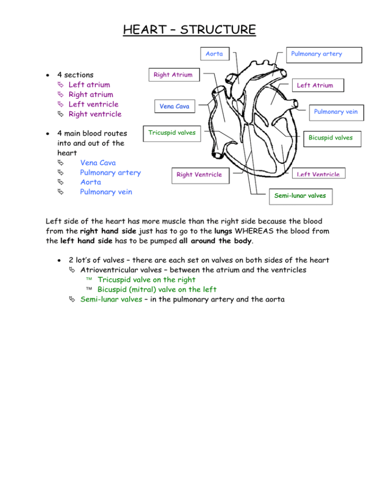
HEART – STRUCTURE Aorta • • 4 sections Left atrium Right atrium Left ventricle Right ventricle Pulmonary artery Right Atrium Left Atrium Vena Cava Tricuspid valves 4 main blood routes into and out of the heart Vena Cava Pulmonary artery Right Ventricle Aorta Pulmonary vein Pulmonary vein Bicuspid valves Left Ventricle Semi-lunar valves Left side of the heart has more muscle than the right side because the blood from the right hand side just has to go to the lungs WHEREAS the blood from the left hand side has to be pumped all around the body. • 2 lot’s of valves – there are each set on valves on both sides of the heart Atrioventricular valves – between the atrium and the ventricles Tricuspid valve on the right Bicuspid (mitral) valve on the left Semi-lunar valves – in the pulmonary artery and the aorta CARDIAC CYCLE Deoxygenated blood enters from the body into the right atrium via the Vena Cava Vena Cava Once all the blood has entered into the right atrium the blood is pushed down into the right ventricle Right Atrium Right Ventricle The blood is then pumped up through the Pulmonary artery to the lungs where gaseous exchange occurs ( CO2 diffuses out of the blood and O2 is diffuses in from the lungs Pulmonary Artery The newly oxygenated blood is returned to the heart via the Pulmonary Artery into the left atrium Left Atrium The blood is pumped down into the left Ventricle Left Ventricle The blood is pumped from the left ventricle up through the Aorta and is pumped around the body to provide oxygen to organs Aorta Miss G’s input (I hope you don’t mind Claire and Matilda) Pressure changes in the heart and how the valves open and close The only way blood moves from the atrium and into the ventricles, is due to pressure changes. When the atriums are relaxed and at diastole, they fill with blood causing the pressure to increase. The atrium will sino-atrial node (SAN) then receive impulses (from the SA node) to contract (systole) and atrio-ventricular node (AVN) the pressure increases even more, and is higher than the pressure in the ventricles, causing the atrioventricular valves (AV valves) to Bundle of His open, forcing blood to enter the ventricles. As blood fills the Purkinje fibres ventricles, the pressure increases, and become higher in the ventricles than in the atrium. This causes the AV valves to close. At this same time, the AV node (stimulated by the impulses across the top two atria) will send impulses over the ventricles (bundle of His and purkinji fibres), causing the ventricles to contract. Whilst the ventricles have contracted, the atriums have been relaxed and have filled with blood. Once the ventricles have contracted, both the atrium and ventricles will relax and be at diastole, and will wait for another impulse to reach the SA node. The noise of the heart beat (‘lub dub’), is only due the AV valves opening and closing. The ‘lub’ noise is due the AV valves opening and the ‘dub’ occurs when the valves close Atrial Systole Ventricular Systole Diastole atria contract blood enters ventricles ventricles contract blood enters arteries atria and ventricals both relax blood enters atria and ventricles Events Name semilunar valves open Pressure (kPa) 20 0 15 0.1 0.2 semilunar valves close 0.3 0.4 0.5 0.6 artery artery atrium atrium 0.7 0.8 0.7 0.8 10 5 0 ventrical ventrical atrioventricular valves open atrioventricular valves close PCG ECG Time (s) 0 0.1 0.2 0.3 0.4 0.5 0.6

