Marfan Syndrome - faculty at Chemeketa
advertisement
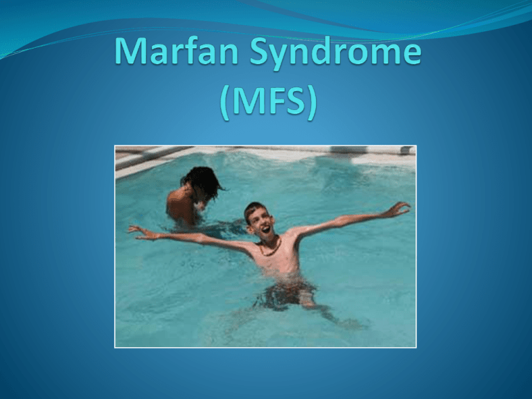
Description Heritable condition that affects connective tissue. Connective tissue affects: Heart Lungs Blood vessels Nervous system Skin Skeleton Eyes Not related to sex, race, ethnic groups. 1 in 5,000 people in the US have this disorder History is fun Antoine Marfan (1858-1942) It was in the course of his clinical studies in 1896 that Marfan described the main features of a syndrome that later was given his name. Marfan's patient was a five year old girl, who was thin, and had long limbs and abnormally long fingers and toes. It starts with the Fibrillin Gene Marfan syndrome develops before you are born. Mutation on FBN1 Located on chromosome 15 Encodes the protein fibrillin Fibrillin protein Glycoprotein essential for the formation of elastic fibers found in connective tissue. Connect with other Fibrillin proteins to make microfibrils, which become connective tissue. Defective Fibrillin-1 Protein Reduction of the amount of fibrillin-1 protein produced by cells Structure and stability of protein affected Transport of fibrillin-1 protein impaired Decreased production and quality of connective tissue Genetics Autosomal Dominant Variable expression Caused by over 500 different mutations on FBN1 50% chance of inheritance Unaffected couples have a 1 in 10,000 chance of having a child with Marfan syndrome 25% caused by spontaneous mutation of gene How is the body affected? Skeleton Affects the long bones: arms, fingers, legs, toes disproportionately long. Tall, slender and loose jointed Long, narrow face Protruding or indented sternum Pigeon Chest (pectus caranatum) Funnel Chest (pectus excavatum) May impair cardiac and respiratory function Curvature of the spine Scoliosis - side to side curvature Lordosis - inner curvature of lower back Kyphosis – outward curvature on the spine of upper back Arched palate, crowded teeth, receding mandible Eyes Dislocation of lenses Slightly higher or lower, or shifted to one side More than half of those affected with marfan syndrome Nearsightedness Extremely common Retinal Detachment Holes or tears in the inner lining of the eye Early development of Glaucoma or cataracts Heart and blood vessels Abnormally large mitral valve leaflets Causes prolapse causing mitral regurgitation Present in 75% of cases Mitral valve regurgitation Backflow of blood into left atrium Heart murmurs Breathlessness, exhaustion, irregular pulse Heart and blood vessels cont. Stretched aortic valve leaflets Aortic regurgitation Leak from aorta into left ventricle Left ventricle must compensate, left ventricular hypertrophy Chest pain, heart failure Aortic dissection Faulty connective tissue weakens and stretches the wall of the aorta. Tears in inner and middle aortic layers Life threatening – sudden onset of chest pain, pain in back, or abdomen Sweaty, vomiting, faint, weak pulse. Nervous System Weakening and stretching of dura membrane Connective tissue around vertebrae Wear away bone surrounding spinal cord Radiating pain in the abdomen, pain/numbness or weakness of the legs, loss of bowel function. Dural ectasia Increased chance of learning disabilities such as ADHD Skin Stretch marks Appear at sites subject to stress: lower back, buttocks, shoulders, breasts, thighs, abdomen Increased risk for abdominal or inguinal hernia Lungs Restrictive lung disease, primarily due to pectus abnormalities or scoliosis, occurs in 70 percent of people with MFS. Diminished alveoli elasticity Susceptible to asthma, bronchitis, pneumonia Swollen aviolies may lead to spontaneous pneumothorax Sleep apnea Looseness of the connective tissues in the airways Assessment No specific laboratory tests Observation/Medical history Family history Eye examination by an ophthalmologist, who uses a slit lamp to look for lens dislocation after fully dilating the pupil. Arm/Leg to trunk size ratio Echocardiogram Assessment cont. If patient has a family history must have at least 2 of the body systems known to be affected to be diagnosed If patient has no family history must have three body systems affected 2 systems must show clear signs specific for Marfan syndrome Treatment There is no cure for Marfan syndrome Treatment is symptomatic Skeleton Annual evaluations Particularly important during periods of rapid growth Pain clinics Loose joints Orthopedic Braces Back Ankles Surgery Pectus excavatum Eyes Regular examinations Glasses/Contact lenses Surgery Removal or replacement of lenses Retina reattachment Cataract surgery Heart Regular echocardiograms Medical bracelets Go to the hospital on first sign of chest pain Reduce stress on aorta Enlargement of the aorta Aortic dissection Aortic dilation Aortic valve regurgitation Mitral valve regurgitation Drugs to lower blood pressure and decrease the forcefulness of the heartbeat are often recommended. Beta blockers Calcium-channel blockers Physical activity kept minimal Dural Ecstasia Identified through MRI Mild cases left alone Extreme pain Treated with Medication or spinal shunting Lungs Surgery to correct pectus abnormalities No smoking!! Pneumothorax Chest tube Supplemental oxygen Emphysema Bronchodilater Mortality and Morbidity Cardiovascular disease Aortic dissection Chronic aortic regurgitation Infant mortality Mitral regurgitation combined with tricuspid prolapse and regurgitation If untreated the average age of death is 30-40 years Outlook Marfan syndrome is a life long condition With early identification, life expectancy is similar to that of the average person 70-80 years. Sources http://www.mayoclinic.com/health/marfan-syndrome/ http://www.americanheart.org/presenter.jhtml?identifier=4672 http://www.marfan.org/ http://www.medicinenet.com/marfan_syndrome/ http://en.wikipedia.org/wiki/Marfan_syndrome http://emedicine.medscape.com/article/946315-overview http://www.emsresponder.com/print/Emergency--Medical-Services/MarfanSyndrome--Aortic-Dissection-and-the-EMS-Provider/1$1705 Any questions?
