File - Ms. G's Classroom
advertisement
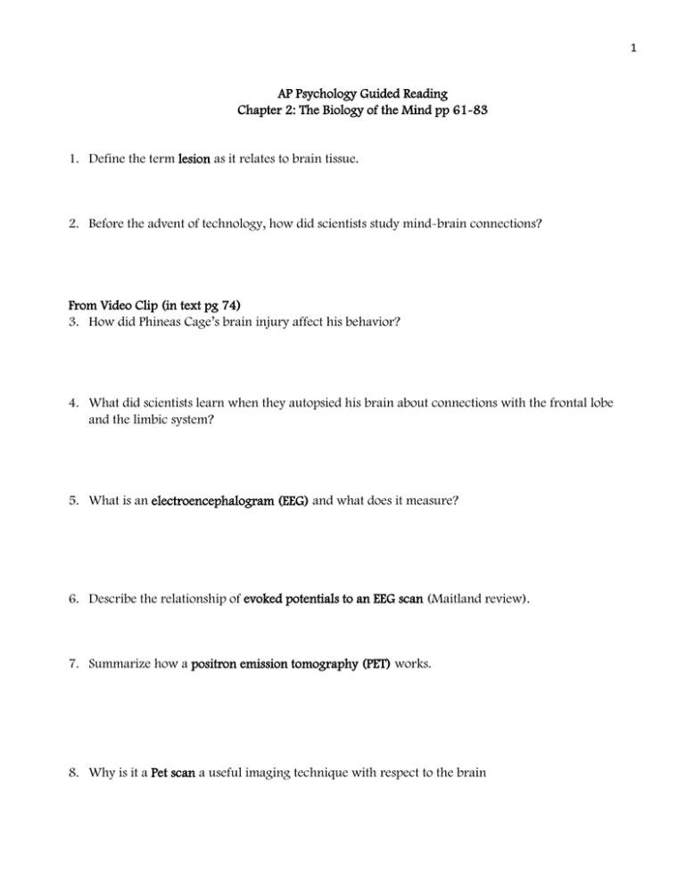
1 AP Psychology Guided Reading Chapter 2: The Biology of the Mind pp 61-83 1. Define the term lesion as it relates to brain tissue. 2. Before the advent of technology, how did scientists study mind-brain connections? From Video Clip (in text pg 74) 3. How did Phineas Cage’s brain injury affect his behavior? 4. What did scientists learn when they autopsied his brain about connections with the frontal lobe and the limbic system? 5. What is an electroencephalogram (EEG) and what does it measure? 6. Describe the relationship of evoked potentials to an EEG scan (Maitland review). 7. Summarize how a positron emission tomography (PET) works. 8. Why is it a Pet scan a useful imaging technique with respect to the brain 2 9. Describe the technique of magnetic resonance imaging (MRI). 10. How is an MRI helpful is viewing the brain? 11. Explain how an fMRI (functional MRI) is different from a regular MRI with respect to brain imaging. 12. Describe the importance of fMRI technology with respect to understanding the functions of the brain. 13. Describe the triune brain theory with respect to evolution (in PP). 14. Label the three parts of the triune brain below. 3 15. Describe the location and the function of the brainstem. 16. Label the following parts of the brainstem below: pons, reticular formation, medulla oblongata 17. Describe the effects of contralateral “wiring” of nerves that occurs in the brainstem. 18. Describe the location and function of the medulla oblongata. 19. Describe the location and function(s) of the pons. 20. Where is the reticular formation located? 21. What are the functions of the reticular formation? 4 22. Describe what happens to a sleeping cat if its reticular formation is severed. 23. Although not part of the brain stem, the thalamus is often pictured in diagrams of the brain stem. Label the thalamus on the brain stem diagram in # 16 AND explain its function! 24. Describe the location and function(s) of the cerebellum. Label the cerebellum in the brain diagram in #26. 25. Describe the location of the limbic system. 26. Label the amydgala, hippocampus, hypothalamus and pituitary gland on the diagram below. 27. Describe the function of the hippocampus. 5 28. Describe the shape of the amygdala and its functions. 29. Where is the hypothalamus located? 30. Describe the brain function(s) of the hypothalamus. 31. Where is the nucleus accumbens located? 32. What are the pleasure (reward) centers of our brain? 33. What structures and neurotransmitters are involved in reward pathways? 34. How does the Reward Deficiency Syndrome explain addictions? 35. Describe the structure of the cerebrum. 36. What is the corpus collosum and indicate its function. 37. Describe the overall function of the cerebrum. 6 38. In the diagram below, label the four lobes of the brain, then in the chart, indicate each lobes function. Cerebral Cortex Lobe Function Frontal Lobes Parietal Lobes Occipital Lobes Temporal Lobes 39. Why is the cerebral cortex compared to “bark on a tree”? 40. What is the function of the convolutions found on the surface of the cerebral cortex? 41. Differentiate among gyri, sulci, and fissures. 42. Describe the function of glial cells with respect to the nerve cells found in the cerebral cortex. 7 43. Discuss the differences between the cerebral cortex and the cerebrum. 44. Specific where is the motor cortex located and what is its function? 45. From what side of the body does the motor cortex of the right hemisphere receive sensory input? 46. Describe what happens when researchers stimulate specific parts of the motor cortex on the left hemisphere? 47. Where is the sensory (somatosensory) cortex located and what is its function? 48. Describe the relationship between sensitivity of a body region and the size of the area in the sensory cortex devoted to the body region. 49. Describe what is meant by the brain homunculus and relate your answer to the sensory and motor cortex 50. Where are visual and auditory stimuli processed in the cortex? 8 51. Label the sensory, motor, visual, and auditory cortexes (arrows) AND label the four lobes the cerebellum, pons, and medulla in the diagram below. 52. Describe what is meant by a brain-computer interface (BCI)? 53. Describe the function of the association regions of the cerebral cortex. 54. It is true that we only use 10% of our brain? Explain your answer. 55. Describe what would happen if a person had damage to the association area of the frontal lobe. 56. Discuss the functions tied to association areas in the parietal lobes. 9 57. Explain what may result if there was damage to the underside of the temporal lobe. 58. Define the term plasticity with respect to the brain. 59. How does plasticity relate to the brain after cerebral illness or injury? 60. What is neurogenesis? 61. Why would a young child be in a better position to recover from a brain trauma than an adult? 62. What is meant by a “split brain” and when would doctors conduct such a surgical procedure? 63. Explain how the visual information highway from the eye to the brain works. 10 64. Describe what happened when split brain patients were the word HEART flashed across their visual field. 65. Define the term lateralization. 66. Studies on split brain patients have confirmed the functions of the both the left and the right hemispheres. What are the main functions of these two hemispheres? 67. After studying the brain, do you believe that people are left or right brain dominate? Explain your answer. 68. Describe the relationship between handedness and brain organization (inset at end of chapter) 69. One more time, label the entire brain!
