File - Paxson Science
advertisement
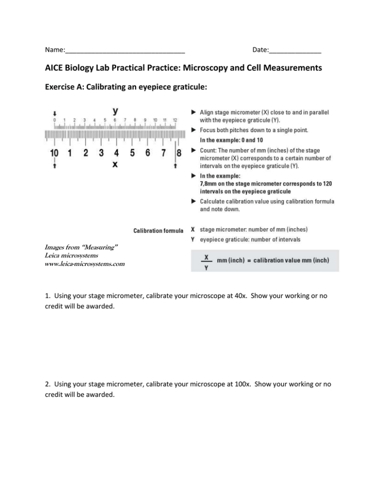
Name:________________________________ Date:______________ AICE Biology Lab Practical Practice: Microscopy and Cell Measurements Exercise A: Calibrating an eyepiece graticule: Images from “Measuring” Leica microsystems www.leica-microsystems.com 1. Using your stage micrometer, calibrate your microscope at 40x. Show your working or no credit will be awarded. 2. Using your stage micrometer, calibrate your microscope at 100x. Show your working or no credit will be awarded. Exercise B: Plan Drawing and Measurement Practice 1. Select your first slide (stem, root, or leaf):_________________________________________ 2. Place your slide on the stage and do a plan drawing of the stem, root, or leaf at 40x. Refer to your tips for drawing plan diagrams from the “Introduction to Microscopy” Activity. 3. Measure the diameter of the stem, root, or leaf in micrometers at the widest point. Show your working. Diameter in mm:____________ Diameter in μm:____________ 4. Change your objective lens so that you are on the low magnification at 100x. Draw one vascular bundle using a plan diagram (ask Ms. Paxson if you cannot locate one or are unsure of what a vascular bundle is—it looks a bit like a circle with three circles inside of it. It may be stained dark blue and red). 3. Measure the diameter of the vascular bundle on your diagram at its widest point. Show your working. Diameter in mm:____________ Diameter in μm:____________ 4. Calculate the magnification of your drawing. (Hint—Use a ruler!) Show your working: Exercise C: Onion Epidermal Cells Epidermal cells cover the surfaces of plant organs such as leaves. The bulb of an onion is made up of fleshy, modified leaves. Procedure: 1. With a scalpel, strip a small, think, transparent, layer of cells from the inside of a fresh onion leaf. 2. Place it gentle on a clean, dry slide, and add a drop of methylene blue. THIS WILL STAIN YOUR CLOTHES AND FINGERS, so be careful. Cover the slide. 3. Observe under the microscope. 4. Count the number of onion cells that line up end to end in a single low power field:________ Create a plan drawing of your onion cells on first scanning, then low power: 5. Calculate the width of a single onion cell. Show your working. 6. Calculate the length of a single onion cell. Show your working. Exercise D: Human cheek cells 1. Using a clean cotton swab found at the back of the room, swab your cheek and transfer the inner cells to a clean, glass slide. 2. Stain the slide using a single drop of methylene blue, and cover it. 3. Observe under the microscope. 4. Complete a high-power drawing (draw cells) of at least three cells. 5. What are some similarities and differences between the onion cells and human cheek cells? You don’t need to use exact terms, but describe what you see.
