Pharynx-2015-Case-Scenario
advertisement
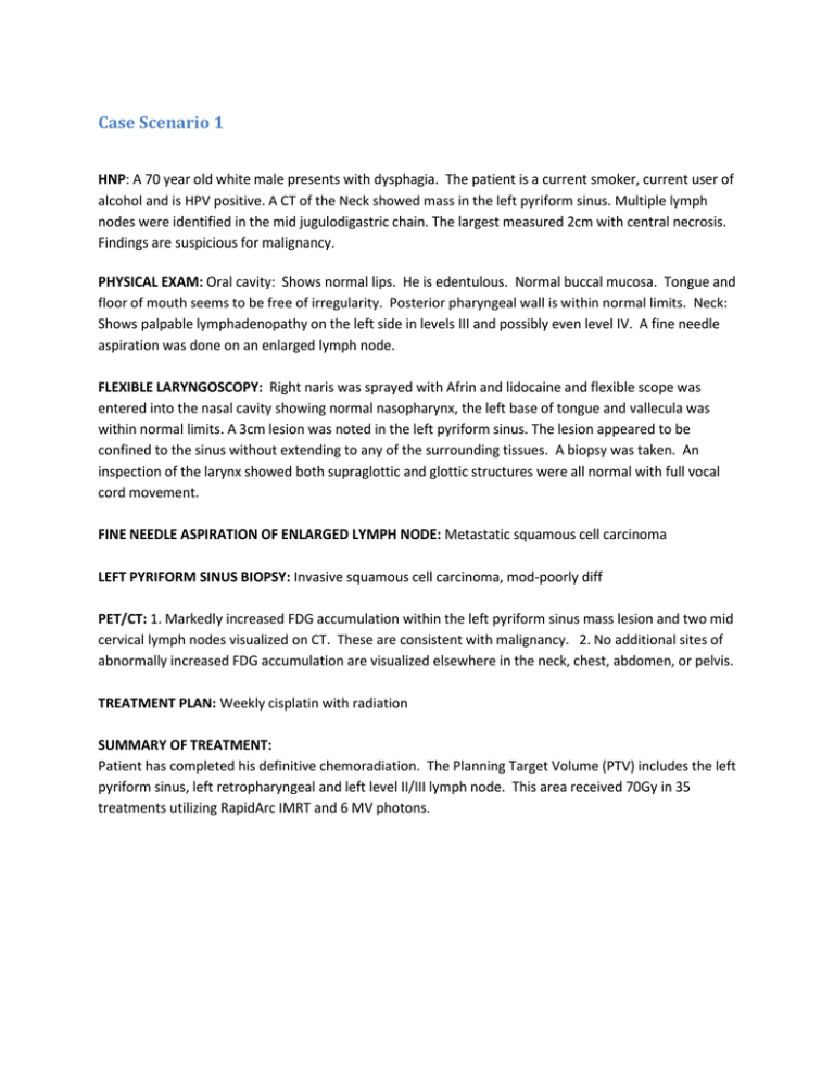
Case Scenario 1 HNP: A 70 year old white male presents with dysphagia. The patient is a current smoker, current user of alcohol and is HPV positive. A CT of the Neck showed mass in the left pyriform sinus. Multiple lymph nodes were identified in the mid jugulodigastric chain. The largest measured 2cm with central necrosis. Findings are suspicious for malignancy. PHYSICAL EXAM: Oral cavity: Shows normal lips. He is edentulous. Normal buccal mucosa. Tongue and floor of mouth seems to be free of irregularity. Posterior pharyngeal wall is within normal limits. Neck: Shows palpable lymphadenopathy on the left side in levels III and possibly even level IV. A fine needle aspiration was done on an enlarged lymph node. FLEXIBLE LARYNGOSCOPY: Right naris was sprayed with Afrin and lidocaine and flexible scope was entered into the nasal cavity showing normal nasopharynx, the left base of tongue and vallecula was within normal limits. A 3cm lesion was noted in the left pyriform sinus. The lesion appeared to be confined to the sinus without extending to any of the surrounding tissues. A biopsy was taken. An inspection of the larynx showed both supraglottic and glottic structures were all normal with full vocal cord movement. FINE NEEDLE ASPIRATION OF ENLARGED LYMPH NODE: Metastatic squamous cell carcinoma LEFT PYRIFORM SINUS BIOPSY: Invasive squamous cell carcinoma, mod-poorly diff PET/CT: 1. Markedly increased FDG accumulation within the left pyriform sinus mass lesion and two mid cervical lymph nodes visualized on CT. These are consistent with malignancy. 2. No additional sites of abnormally increased FDG accumulation are visualized elsewhere in the neck, chest, abdomen, or pelvis. TREATMENT PLAN: Weekly cisplatin with radiation SUMMARY OF TREATMENT: Patient has completed his definitive chemoradiation. The Planning Target Volume (PTV) includes the left pyriform sinus, left retropharyngeal and left level II/III lymph node. This area received 70Gy in 35 treatments utilizing RapidArc IMRT and 6 MV photons. What is the primary site? C12.9 Pyriform sinus What is the histology? 8070/3 Squamous cell carcinoma What is the grade/differentiation? 3 poorly differentiated Stage/ Prognostic Factors CS Tumor Size CS Extension CS Tumor Size/Ext Eval 030 100 1 CS SSF 9 CS SSF 10 CS SSF 11 998 999 988 CS Lymph Nodes CS Lymph Nodes Eval Regional Nodes Positive Regional Nodes Examined CS Mets at Dx CS Mets Eval CS SSF 1 CS SSF 2 CS SSF 3 CS SSF 4 CS SSF 5 CS SSF 6 CS SSF 7 CS SSF 8 Summary Stage 220 1 95 95 00 0 020 988 011 000 000 000 988 988 3 Regional to Lymph Nodes cT2 cN2b cM0 Stage IVA CS SSF 12 CS SSF 13 CS SSF 14 CS SSF 15 CS SSF 16 CS SSF 17 CS SSF 18 CS SSF 19 CS SSF 20 CS SSF 21 CS SSF 22 CS SSF 23 CS SSF 24 CS SSF 25 988 988 988 988 988 988 988 988 988 988 988 988 988 988 Path Stage T N M Stage 99 Clinical Stage Treatment Diagnostic Staging Procedure Surgery Codes Surgical Procedure of Primary Site Scope of Regional Lymph Node Surgery Surgical Procedure/ Other Site Systemic Therapy Codes Chemotherapy Hormone Therapy Immunotherapy Hematologic Transplant/Endocrine Procedure Systemic/Surgery Sequence 02 00 01 0 02 00 00 00 3 Radiation Codes Radiation Treatment Volume Regional Treatment Modality 05 31 Regional Dose Boost Treatment Modality Boost Dose Number of Treatments to Volume Reason No Radiation Radiation/Surgery Sequence 07000 00 00000 35 0 3 Case Scenario 2 68 year old black male presents with mass on lateral wall of oropharynx found during a dental procedure. Patient is a smoker and has a history of alcohol use. Physical Exam: Pink lips with intact gingiva and soft floor of mouth. The tongue demonstrates unrestricted movement and is soft diffusely including the base of tongue. Tumor on right lateral wall of oropharynx appears to involve the posterior tonsillar pillar. The anterior tonsillar pillar is intact. Direct laryngoscopy: A McIntosh laryngoscope blade was placed in his oral cavity and allowed visualization of the base of tongue and oropharyngeal region. The base of the tongue was soft as well, with no apparent abnormalities as was the posterior pharyngeal wall. The soft palate was unremarkable and the uvula was singular and demonstrated no mucosal masses, lesions or ulcerations. However, the right lateral wall of the oropharynx appeared to have a broad centrally ulcerated lesion with heaped up mucosal edges around it circumferentially. The lesion appeared to involve the posterior tonsillar pillar. Biopsy: Invasive, poorly differentiated squamous cell carcinoma CT Larynx/Neck: There is thickening along the right lateral wall of the oropharynx which likely correspond to the clinically known lateral wall cancer. The lesion is relatively sessile, limiting assessment of its true size. As can be visualized on these CT images, it measures up to 2.4 x 1.9 cm in cross section, and 2.2 cm craniocaudal. Two enlarged retropharyngeal lymph nodes were visualized. The largest measured 2.3 cm (image 72 of 165). These lymph nodes most likely represent metastasis from the known oropharyngeal primary. Operative Report: 1. TRANSORAL RESECTION OF ORO0PHARYNGEAL LESION ON THE RIGHT LATERAL WALL 2. RIGHT SELECTIVE NECK DISSECTION OF RETROPHARYNGEAL AND LEVELS 1-4. Pathology: A) LYMPH NODES RIGHT RETROPHARYNGEAL (3) TWO LYMPH NODES POSITIVE FOR METASTATIC SQUAMOUS CELL CARICNOMA. o THE LARGEST METASTIC FOCUS IN THE LYMPH NODE WAS 2.1 CM. IN GREATEST DIMENSION WITH TUMOR INVASION THROUGH THE LYMPH NODE CAPSULE. o THE LARGEST METASTIC FOCUS IN THE SECOND LYMPH NODE WAS 1.3 CM. IN GREATEST DIMENSION WITHOUT TUMOR INVASION THROUGH THE LYMPH NODE CAPSULE o THE THIRD LYMPH NODE WAS NEGATIVE FOR TUMOR. B) LYMPH NODES RIGHT NECK LEVEL II, EXCISION: MULTIPLE, 6, LYMPH NODES ARE NEGATIVE FOR TUMOR. C) LYMPH NODES RIGHT NECK LEVEL III, EXCISION: MULTIPLE, 2, LYMPH NODES ARE NEGATIVE FOR TUMOR. D) LYMPH NODES RIGHT NECK LEVEL IV, EXCISION: MULTIPLE, 3, LYMPH NODES ARE NEGATIVE FOR TUMOR. E) OROPHARYNX, RIGHT LATERAL WALL: INVASIVE MODERATELY DIFFERENTIATED SQUAMOUS CELL CARCINOMA FORMING A 2.6 X 2.3 CM. ULCERATED MASS. THE TUMOR INVADES INTO POSTERIOR TONSILLAR PILLAR TO A DEPTH OF 1.1 CM. PERINEURAL INVASION IS PRESENT. LYMPHOVASCULAR INVASION IS IDENTIFIED. THE SURGICAL MARGINS ARE NEGATIVE FOR TUMOR. Treatment Summary: Patient completed his concurrent chemo/radiotherapy. He received 60 Gy in 30 sessions to initial neck lymph node region utilizing 6 MV photons, 3D conformal radiotherapy and opposing lateral fields. Patient concurrently received cisplatin. What is the primary site? C10.2 Lateral wall of oropharynx What is the histology? 8070/3 Squamous Cell Carcinoma What is the grade/differentiation? 3 – poorly differentiated Stage/ Prognostic Factors CS Tumor Size CS Extension CS Tumor Size/Ext Eval 026 150 3 CS SSF 9 CS SSF 10 CS SSF 11 040 999 988 CS Lymph Nodes CS Lymph Nodes Eval Regional Nodes Positive Regional Nodes Examined CS Mets at Dx CS Mets Eval CS SSF 1 CS SSF 2 CS SSF 3 CS SSF 4 CS SSF 5 CS SSF 6 CS SSF 7 CS SSF 8 Summary Stage Clinical Stage 200 3 02 14 00 0 021 988 000 001 000 000 988 988 3-Regional lymph T2 N2b M0 Stage IVA CS SSF 12 CS SSF 13 CS SSF 14 CS SSF 15 CS SSF 16 CS SSF 17 CS SSF 18 CS SSF 19 CS SSF 20 CS SSF 21 CS SSF 22 CS SSF 23 CS SSF 24 CS SSF 25 988 988 988 988 988 988 988 988 988 988 988 988 988 988 Path Stage T2 N2b M0 Stage IVA Treatment Diagnostic Staging Procedure Surgery Codes Surgical Procedure of Primary Site Scope of Regional Lymph Node Surgery Surgical Procedure/ Other Site Systemic Therapy Codes Chemotherapy Hormone Therapy Immunotherapy Hematologic Transplant/Endocrine Procedure Systemic/Surgery Sequence 02 27 5 0 02 00 00 00 3 Radiation Codes Radiation Treatment Volume Regional Treatment Modality 05 32 Regional Dose Boost Treatment Modality Boost Dose Number of Treatments to Volume Reason No Radiation Radiation/Surgery Sequence 06000 00 00000 030 0 3
