Tissues-Membranes
advertisement
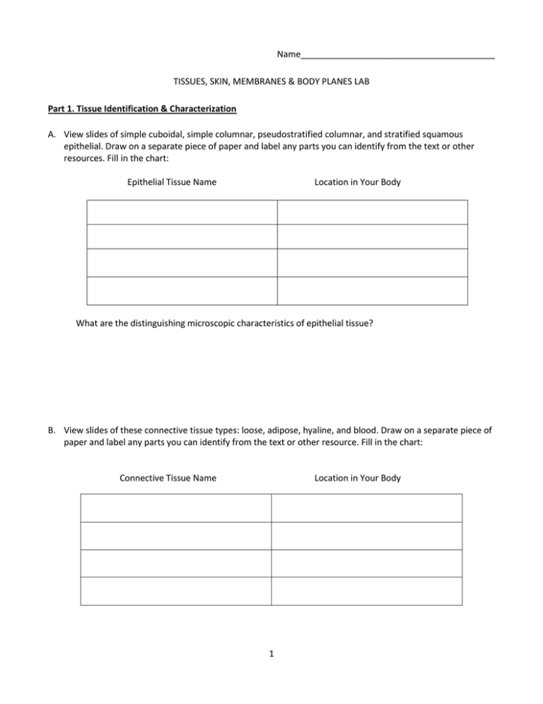
Name________________________________________ TISSUES, SKIN, MEMBRANES & BODY PLANES LAB Part 1. Tissue Identification & Characterization A. View slides of simple cuboidal, simple columnar, pseudostratified columnar, and stratified squamous epithelial. Draw on a separate piece of paper and label any parts you can identify from the text or other resources. Fill in the chart: Epithelial Tissue Name Location in Your Body What are the distinguishing microscopic characteristics of epithelial tissue? B. View slides of these connective tissue types: loose, adipose, hyaline, and blood. Draw on a separate piece of paper and label any parts you can identify from the text or other resource. Fill in the chart: Connective Tissue Name Location in Your Body 1 What are the distinguishing microscopic characteristics of most connective tissue? C. View slides of skeletal muscle, smooth muscle, and cardiac muscle. Draw on a separate piece of paper and label any parts you can identify. 1. Describe the appearance of the muscle tissues. Skeletal Smooth Cardiac 2. Would adipose tissue have more or less mitochondria than a skeletal muscle? 3. List the four types of special connective tissue. 2 D. View a slide or slides of nervous tissue. Draw on a separate piece of paper and label any parts you can identify. 1. Describe the appearance of the nervous tissue(s). 2. What are the neuroglial cells and what is their function? 3. How does the structure of a nerve cell match its function? Part 2. Skin Structure and Function The skin is an organ system called the integumentary system. It does much more than cover the body exterior. The skin has several functions, most, but not all concerned with protection. Basic skin structure The skin has two distinct regions, the upper epidermis composed of epithelial cells and underlying connective tissue called the dermis. The layers are firmly cemented together along a wavy border. Deep to the dermis is the subcutaneous tissue, also known as hypodermis. The hypodermis is composed mostly of adipose cells and is not considered part of the skin. Although you might not know it, your epidermis is in constant motion. The stratum corneum is peeling and flaking away. The keratinocytes are moving toward the surface and will eventually die and become part of the stratum corneum. The basal cells of the stratum basale are creating new keratinocytes, which immediately begin their journey upward. 3 Layers of the dermis. Dense connective tissue makes up the bulk of the dermis. Collagen and elastic fibers are found throughout the dermis along with adipose cells, fibroblasts, and phagocytes. The dermal blood supply allows the skin to regulate temperature. There are two principal regions of the dermis. a. Papillary layer b. Reticular layer Exercise 1—Discovering Distribution of Cutaneous Glands Appendages of the skin include hair, nails, and cutaneous glands. These are all derived from the epidermis but reside in the dermis. Cutaneous glands fall into two categories: sebaceous (oil) glands and sweat (sudoriferous) glands. Sebum, a combination of oils and fragmented cells, is the product of the sebaceous glands. It acts like a natural skin cream. Sweat glands are of two types, eccrine producing clear perspiration (containing water, salts, and urea), and apocrine, producing milky protein and fat rich substance (including water, salts, and urea). Apocrine glands occur in the axillary and genital areas. Sweat glands are controlled by the nervous system and are part of the body’s heat regulating system. Materials: two squares of paper (1cm x 1cm), adhesive tape, and a betadine swab 1. Using the betadine solution, paint an area of the medial aspect of your left palm (avoid the crease lines) and a region of your left forearm. Allow the solution to dry thoroughly. The painted area in each case should be slightly larger than the paper squares. 2. Mark one piece of ruled bond paper with an “H” (for hand) and the other “A” (for arm). Have your lab partner securely tape the appropriate square of paper letter side up over each iodine-painted area. Leave the squared in place for 10 minutes. While waiting to determine the results, continue with other sections of this lab. 3. Remove the squares and count the number of blue-black dots on each square. The appearance of a blueblack dot indicates an active sweat gland. The iodine in the pore dissolves in the sweat and reacts with the starch in the bond paper to produce the blue-black color producing “sweat maps.” Number of dots on paper from hand Number of dots on paper from arm __________ __________ Exercise 3. Body Membranes Body membranes cover surfaces, line body cavities, and form protective sheets around organs. Body membranes fall into one of two categories; epithelial membranes or connective tissue membranes. There are three types of epithelial membranes, one being cutaneous membranes. Cutaneous membranes form the skin, also known as the integumentary system. The skin serves to protect underlying organs and tissues in the body. In general, we can use the term “membrane” to refer to any thin layer or layers covering or surrounding something. You might be familiar with the term “plasma membrane” as the phospholipid layer surrounding every cell. Now we have tissue membranes consisting of several layers of tissue sandwiched together that surround body cavities, organs, or entire organ systems. A. Describe these membranes, including a location for each. 4 B. Sketch each. 1. Serous membranes 2. Mucous membrane 3. Synovial membrane 4. Cutaneous membrane 5
