Gel Electrophoresis
advertisement

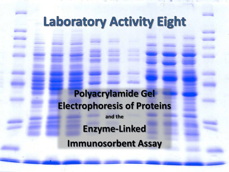
Polyacrylamide Gel Electrophoresis of Proteins and the Enzyme-Linked Immunosorbent Assay 1 Introduction to the theory & application of electrophoresis. Use SDS-PAGE to separate proteins in crude spinach leaf extracts & NH4SO4 fractions from lab 6. Observe large & small subunits of Rubisco (55 & 14 kD). Estimate/verify the MW’s of BSA vs. Lysozyme (67 & 14 kD). Introduction to the theory & application of ELISA (EnzymeLinked Immuno Sorbent Assays). Perform a simple (hypothetical) pregnancy test (simulated detection of human chorionic gonadotropin hormone). 2 An analytical technique that separates mixtures of charged biomolecules on the basis of their relative mobilities in a gelatinous matrix when an electrical field is applied. Types of charged biomolecules include . . . Nucleic Acids (DNA, RNA, nucleotides). Proteins Some drugs or metabolites. 3 Power Supply – source of direct current; provides electromotive force (voltage) to drive migration of charged solute molecules. Gel – a stationary matrix that provides differential physical resistance to migration of charged solute molecules. Buffer – controls pH; dissolved ions help conduct electricity. Electrodes – conduct / deliver current into buffer & gel. Gel Box / Container – holds gel & buffer in place; insulates against short-circuits. 4 1. 2. 3. Starch Agarose Polyacrylamide. 5 One of the first gel matrices. A mixture of two -1,4 linked glucose polymers: 25% Amylose: coiled, unbranched starch. 75% Amylopectin: -1,6 branched globular starch. Heating in water causes hydration, swelling & disorganization of amylose. Cooling causes cross-linking of amylopectin globules. Often used for the separation & analysis of “isozymes”. 6 A linear polymer of galactose and 3,6-anhydrogalactose: -1,4-(3,6-anhydro)-galactopyranosyl--1,3-galactopyranan. Isolated from seaweed (sulfated form is “Agar”). Molten solutions spontaneously form double-helical chains that coalesce into a porous matrix as they cool. Pore size is controlled by the concentration of agarose. 7 A linear polymer of acrylamide crosslinked with N,N’-methylene-bisacrylamide. Pore size is controlled by the amount of cross-linking (concentration of N,N’methylene-bis-acrylamide). Linear polymerization and cross-linking is carried out in a single reaction. Separation technique is known as “PAGE” (PolyAcrylamide Gel Electrophoresis). N,N’-methylenebis-acrylamide Acrylamide Monomers Free Radicals Cross-Linked Polyacrylamide Ammonium Persulfate 8 A soft, fragile gel. Typically run horizontally. Used for separation of nucleic acid fragments. Polyacrylamide A stiffer, more resilient gel. Typically run vertically. Used for separation of proteins, polypeptides. Agarose 9 10 V= Where: V E q f Exq f = velocity of charged molecule migration. = potential difference (voltage) of electric field. = charge of the molecule. = frictional coefficient (physical resistance). Thus: Molecules with a greater charge will move faster. Gel resistance tends to slow the velocity of migration. Larger molecules experience greater resistance, and thus migrate slower than smaller molecules. 11 Movement through gel is promoted by mobile phase. Small molecules enter/exit pores in gel; large molecules by-pass pores. Movement through gel is promoted by electric field. All molecules must penetrate gel & pass through pores. 12 Increases density of sample mix; making it sink to bottom of well. Very low MW, charged, colored molecule. Migrates very fast through gel. Denotes migration position of smallest colorless protein. Bromophenol blue commonly used. Mixture of known proteins/MW’s. Span the separation range of gel, encompass proteins of interest. Used in estimating MW of unknown proteins. Generally binds to all proteins (but not to gel). Helps to locate protein positions in gel. Coomassie brilliant blue commonly used. 13 Proteins are zwitterions (they can carry both + / - charges at the same time). Charge : Mass ratio varies from one protein species to the next. Potentially problematic – proteins with same mass, but different charges could migrate at different velocities. Many proteins are comprised of different subunits. Potentially problematic – a mixture of proteins could migrate in both directions at once. Potentially problematic – difficult to handle different degrees of subunit association vs. dissociation. Problems are overcome with “ ”. 14 Protein samples are treated with a mixture of SDS, -mercaptoethanol at high temperatures (90 – 95 °C). Sodium Dodecyl Sulfate SDS destabilizes hydrophobic interactions. -ME reduces disulfide bridges. -mercaptoethanol Potent Denaturation Agent (Subunits dissociate, polypeptides unfold) SDS binds to linear polypeptide at a ration of 1.4 g SDS/g protein – gives proteins a constant charge:mass ratio (i.e. charge is directly MW). Protein migration in gel is inversely MW (smaller proteins migrate farther than larger proteins). 15 - Log10MW kDa + MW Markers Rf If an unknown protein moves with an Rf of 0.5, log10 of its MW = 4.62. Thus, MW = 104.62 = 41,687. 16 Prepare lab 6 samples for electrophoresis (add glycerol, SDS, ME & heat). Need 2 mg/mL Add “2X” treatment buffer Assemble electrophoresis apparatus. Load samples onto gel & run. Perform ELISA pregnancy test simulation. Stain/destain gels. TA’s take photographs. 17 Protein ladder BSA/lyzozyme Cell-pellet Cell supernatant ( very light band) Cell-free extract 18 Enzyme-Linked Immunosorbent Assay. A diagnostic tool used to immunologically detect the presence of antigenic substances. Relies on the specificity of antibodies. 19 An enzyme-antibody conjugate that still recognizes it’s antigen and is able to carry out a colorimetric reaction. Antibody (with attached enzyme) binds to antigen. Colorless reactants are provided; colored products are produced. Location & intensity of color reaction indicates antigen detection. 20 Typically performed in 96-well polystyrene microtiter plates. Polystyrene tightly adsorbs many biomolecules, especially proteins. After antigen binding, wells are rinsed and treated with a series of reagents to detect antigen. Antigen is dispensed Antigen binds to plastic Primary antibody binds to antigen Secondary enzymeantibody binds to primary antibody Substrate is added, color reaction occurs 21 ELISA assays can be extremely sensitive ( ng amounts). Dilution series of samples are normally analyzed. Positive & negative controls must always be included. ELISA results are generally “relative” (i.e. more or less vs. yes or no). Absolute (quantitative) results are difficult. Color development is determined spectrophotometrically. Color development is continuous (i.e. colors continue to darken). Color development can be limited by availability of enzyme substrate. 22 23


