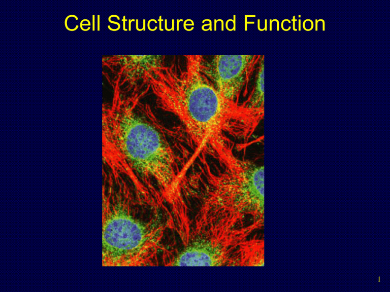
Cell Structures and Function
advertisement

Cell Structure and Function 1 Microscopes • Anton Leeuwenhoek invented the microscope in the late 1600’s, which first showed that all living things are composed of cells. Also, he was the first to see microorganisms. 2 Cell Structure • In 1655, the English scientist Robert Hooke coined the term “cellulae” for the small box-like structures he saw while examining a thin slice of cork under a microscope. 3 Cell Theory • • In 1838 – 1839, German scientists Schleiden and Schwann, proposed the first 2 principles of the cell theory: • All organisms are composed of one or more cells. • Cells are the smallest living units of all living organisms. 15 years later, the German physician Rudolf Virchow proposed the third principle: • Cells arise only by division of a previously existing cell. 4 Basic Cell Structure • • • • • All cells have the following basic structure: A thin, flexible plasma membrane surrounds the entire cell that regulates the passage of materials between the cell and its surrounding The interior is filled with a semi-fluid material called the cytoplasm. At some point, all cells contain DNA, the heritable material that directs the cell’s activities Also inside some cells are specialized structures called organelles. 5 Cell Theory • The cell is the lowest level of structure that is capable of performing all the activities of life. • Regulate its internal environment. • Take in and use energy. • Respond to its local environment. • Develop and maintain its complex organization. • Divide to form new cells. 6 Cell Characteristics • Two major kinds of cells - prokaryotic cells and eukaryotic cells - can be distinguished by their structural organization. • • • • Eukaryotic cells - Contain membrane-enclosed organelles, including a DNA-containing nucleus Prokaryotic cells - Lack such organelles The cells of the microorganisms called bacteria and archaea are prokaryotic. All other forms of life have the more complex eukaryotic cells 7 The Cell Nucleus (contains DNA) Prokaryotic cell Eukaryotic cell DNA (no nucleus) 25,000 Organelles 8 Multicellular Organisms • • Some organisms consist of a single cells, others are multicellular aggregates of specialized cells. Multicellular Organisms exhibit three major structural levels above the cell: • Similar cells are grouped into tissues • Several tissues coordinate to form organs • Several organs form an organ system. 9 Generalized Eukaryotic Cell 10 Cell Size Limit • Most cells are relatively small because as size increases, volume increases much more rapidly than surface area. • longer diffusion time • limit to the volume of cytoplasm that can be effectively controlled by genes. 11 Cell Size Limit • A cell must exchange materials with its environment. Cell volume determines the amount of materials that must be exchanged, while surface area limits how fast exchange can occur. In other words, as cells get larger the need for materials increases faster than the ability to absorb them. 12 Visualizing Cells 13 Prokaryotic Cells • Simplest organisms • Cytoplasm is surrounded by plasma membrane and encased in a rigid cell wall composed of peptidoglycan. • No distinct interior compartments • Some use flagellum for locomotion, threadlike structures protruding from cell surface 14 Eukaryotic Cells • Characterized by compartmentalization by an endomembrane system, and the presence of membrane-bound organelles. • central vacuole • vesicles • chromosomes • cytoskeleton • cell walls 15 Copyright © The McGraw-Hill Companies, Inc. Permission required for reproduction or display. Infolding of the plasma membrane Plasma membrane DNA Cell wall Prokaryotic cell Prokaryotic ancestor of eukaryotic cells Endoplasmic reticulum (ER) Nuclear envelope Nucleus Plasma membrane Eukaryotic cell 16 Endosymbiosis • Endosymbiotic theory suggests engulfed prokaryotes provided hosts with advantages associated with specialized metabolic activities. Copyright © The McGraw-Hill Companies, Inc. Permission required for reproduction or display. Internal membrane system Aerobic bacterium Mitochondrion Ancestral eukaryotic cell Eukaryotic cell with mitochondrion 17 Animal Cell 18 Plant Cell 19 Cell Membrane Fluid Mosaic Model Glycoprotein Extracellular fluid Carbohydrate Cholesterol Peripheral protein Cytoplasm Extracellular matrix protein Glycolipid Transmembrane proteins Filaments of cytoskeleton 20 Nucleus • • • • • Repository for genetic material Chromatin: DNA and proteins Nucleolus: Chromatin and ribosomal subunits - region of intensive ribosomal RNA synthesis Nuclear envelope: Surface of nucleus bound by two phospholipid bilayer membranes Double membrane with pores Nucleoplasm: semifluid medium inside the nucleus 21 Nucleus 22 Chromosomes • DNA of eukaryotes is divided into linear chromosomes. • Exist as strands of chromatin, except during cell division • Histones associated packaging proteins 23 The Nucleus And The Nuclear Envelope 24 Nucleolus • • • Protein synthesis occurs at tiny organelles called ribosomes. Ribosomes are composed of a large subunit and a small subunit. Ribosomes can be found alone in the cytoplasm, in groups called polyribosomes, or attached to the endoplasmic reticulum. 25 Ribosomes • Ribosomes are RNA-protein complexes composed of two subunits that join and attach to messenger RNA. • site of protein synthesis • assembled in nucleoli 26 Endomembrane System • Compartmentalizes cell, channeling passage of molecules through cell’s interior. • Endoplasmic reticulum • Rough ER - studded with ribosomes • Smooth ER - few ribosomes 27 Rough ER • • Rough ER is especially abundant in cells that secrete proteins. • As a polypeptide is synthesized on a ribosome attached to rough ER, it is threaded into the cisternal space through a pore formed by a protein complex in the ER membrane. • As it enters the cisternal space, the new protein folds into its native conformation. • Most secretory polypeptides are glycoproteins, proteins to which a carbohydrate is attached. • Secretory proteins are packaged in transport vesicles that carry them to their next stage. Rough ER is also a membrane factory. • Membrane-bound proteins are synthesized directly into the membrane. • Enzymes in the rough ER also synthesize phospholipids from precursors in the cytosol. • As the ER membrane expands, membrane can be transferred as transport vesicles to other components of the endomembrane system. 28 Smooth ER • • • • • The smooth ER is rich in enzymes and plays a role in a variety of metabolic processes. Enzymes of smooth ER synthesize lipids, including oils, phospholipids, and steroids. These include the sex hormones of vertebrates and adrenal steroids. In the smooth ER of the liver, enzymes help detoxify poisons and drugs such as alcohol and barbiturates. Smooth ER stores calcium ions. • Muscle cells have a specialized smooth ER that pumps calcium ions from the cytosol and stores them in its cisternal space. • When a nerve impulse stimulates a muscle cell, calcium ions rush from the ER into the cytosol, triggering contraction. 29 The Golgi apparatus • The Golgi apparatus is the shipping and receiving center for cell products. • Many transport vesicles from the ER travel to the Golgi apparatus for modification of their contents. • The Golgi is a center of manufacturing, warehousing, sorting, and shipping. • The Golgi apparatus consists of flattened membranous sacs— cisternae—looking like a stack of pita bread. • The Golgi sorts and packages materials into transport vesicles. 30 Functions Of The Golgi Apparatus Golgi apparatus cis face (“receiving” side of Golgi apparatus) 1 Vesicles move 2 Vesicles coalesce to 6 Vesicles also form new cis Golgi cisternae from ER to Golgi transport certain Cisternae proteins back to ER 3 Cisternal maturation: Golgi cisternae move in a cisto-trans direction 5 Vesicles transport specific proteins backward to newer Golgi cisternae 0.1 0 µm 4 Vesicles form and leave Golgi, carrying specific proteins to other locations or to the plasma membrane for secretion trans face (“shipping” side of Golgi apparatus) TEM of Golgi apparatus 31 Membrane Bound Organelles Nucleus • • • Lysosomes – vesicle containing digestive enzymes that break down food/foreign particles Vacuoles – food storage and water regulation Peroxisomes - contain enzymes that catalyze the removal of electrons and associated hydrogen atoms 1 µm Lysosome Lysosome contains active hydrolytic enzymes Food vacuole fuses with lysosome Hydrolytic enzymes digest food particles Digestive enzymes Lysosome Plasma membrane Digestion Food vacuole (a) Phagocytosis: lysosome digesting food 32 Energy Organelles • • • Mitochondria • bounded by exterior and interior membranes • interior partitioned by cristae Chloroplasts • have enclosed internal compartments of stacked grana, containing thylakoids • found in photosynthetic organisms Both organelles house energy in the form of ATP 33 Mitochondria • • • • • Mitochondria are found in plant and animal cells. Sites of cellular respiration, ATP synthesis Bound by a double membrane surrounding fluid-filled matrix. The inner membranes of mitochondria are cristae The matrix contains enzymes that break down carbohydrates and the cristae house protein complexes that produce ATP 34 Chloroplasts • • • A chloroplast is bounded by two membranes enclosing a fluid-filled stroma that contains enzymes. Membranes inside the stroma are organized into thylakoids that house chlorophyll. Chlorophyll absorbs solar energy and carbohydrates are made in the stroma. 35 Cytoskeleton • • The eukaryotic cytoskeleton is a network of filaments and tubules that extends from the nucleus to the plasma membrane that support cell shape and anchor organelles. Protein fibers • Actin filaments - cell movement • Microtubules - centrioles • Intermediate filaments 36 Centrioles • • Centrioles are short cylinders with a 9 + 0 pattern of microtubule triplets. Centrioles may be involved in microtubule formation and disassembly during cell division and in the organization of cilia and flagella. 37 Cilia and Flagella • • • Contain specialized arrangements of microtubules Are locomotor appendages of some cells Cilia and flagella share a common ultrastructure Outer microtubule doublet Dynein arms 0.1 µm Central microtubule Outer doublets cross-linking proteins inside Microtubules Radial spoke Plasma membrane Basal body Plasma membrane (b) 0.5 µm (a) 0.1 µm Triplet (c) Cross section of basal body 38 Cilia and Flagella • • • Cilia (small and numerous) and flagella (large and single) have a 9 + 2 pattern of microtubules and are involved in cell movement. Cilia and flagella move when the microtubule doublets slide past one another. Each cilium and flagellum has a basal body at its base. 39 Cilia and Flagella (a) Motion of flagella. A flagellum usually undulates, its snakelike motion driving a cell in the same direction as the axis of the flagellum. Propulsion of a human sperm cell is an example of flagellatelocomotion (LM). Direction of swimming 1 µm (b) Motion of cilia. Cilia have a backand-forth motion that moves the cell in a direction perpendicular to the axis of the cilium. A dense nap of cilia, beating at a rate of about 40 to 60 strokes a second, covers this Colpidium, a freshwater protozoan (SEM). 15 µm 40 Cell Junctions • Long-lasting or permanent connections between adjacent cells, 3 types of cell junctions: TIGHT JUNCTIONS Tight junction Tight junctions prevent fluid from moving across a layer of cells 0.5 µm At tight junctions, the membranes of neighboring cells are very tightly pressed against each other, bound together by specific proteins (purple). Forming continuous seals around the cells, tight junctions prevent leakage of extracellular fluid across A layer of epithelial cells. DESMOSOMES Desmosomes (also called anchoring junctions) function like rivets, fastening cells Together into strong sheets. Intermediate Filaments made of sturdy keratin proteins Anchor desmosomes in the cytoplasm. Tight junctions Intermediate filaments Desmosome Gap junctions Space between Plasma membranes cells of adjacent cells 1 µm Extracellular matrix Gap junction 0.1 µm GAP JUNCTIONS Gap junctions (also called communicating junctions) provide cytoplasmic channels from one cell to an adjacent cell. Gap junctions consist of special membrane proteins that surround a pore through which ions, sugars, amino acids, and other small molecules may pass. Gap junctions are necessary for communication between cells in many types of tissues, including heart muscle and animal embryos. 41 Tight Junctions • Connect cells into sheets. Because these junctions form a tight seal between cells, in order to cross the sheet, substances must pass through the cells, they cannot pass between the cells. Tight junction 42 Anchoring Junctions • Attach the cytoskeleton of a cell to the matrix surrounding the cell, or to the cytoskeleton of an adjacent cell. Plasma membranes Intracellular attachment proteins Cell 1 Cell 2 Cytoskeletal filament Intercellular space Transmembrane linking proteins Extracellular matrix 43 Communicating Junctions • Link the cytoplasms of 2 cells together, permitting the controlled passage of small molecules or ions between them. Two adjacent connexons form a gap junction Connexon Adjacent plasma membranes Intercellular space 44 Prokaryotes Eukaryotes Organisms Monera (bacteria) All other organisms Size Very small (1 – 5 μm) Much larger (10 – 100 μm) Complexity Relatively simple Complex Cell wall Usually present (contains peptidoglycan) Sometimes present (lacks peptidoglycan) 45 Prokaryotes Eukaryotes Plasma membrane Always present Always present Internal membranes May contain infoldings of the plasma membrane but usually lack internal membranes Complex system of internal membranes divides cell into specialized compartments 46 Prokaryotes Eukaryotes Membranebound organelles Absent Present Ribosomes Smaller and free in the cytoplasm Larger and may be bound to ER Cytoskeleton Absent Present Flagella Solid flagellin; rotate Microtubules; bend 47 Prokaryotes Eukaryotes Structure of genetic material Single, naked, circular DNA molecule Many linear chromosomes, each made of 1 DNA molecule joined with protein Location of genetic material In an area of the cytoplasm called the nucleoid Inside a membrane-bound nucleus 48


