Vernier EMG of masseter revised
advertisement
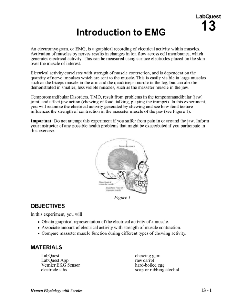
LabQuest Introduction to EMG 13 An electromyogram, or EMG, is a graphical recording of electrical activity within muscles. Activation of muscles by nerves results in changes in ion flow across cell membranes, which generates electrical activity. This can be measured using surface electrodes placed on the skin over the muscle of interest. Electrical activity correlates with strength of muscle contraction, and is dependent on the quantity of nerve impulses which are sent to the muscle. This is easily visible in large muscles such as the biceps muscle in the arm and the quadriceps muscle in the leg, but can also be demonstrated in smaller, less visible muscles, such as the masseter muscle in the jaw. Temporomandibular Disorders, TMD, result from problems in the temporomandibular (jaw) joint, and affect jaw action (chewing of food, talking, playing the trumpet). In this experiment, you will examine the electrical activity generated by chewing and see how food texture influences the strength of contraction in the masseter muscle of the jaw (see Figure 1). Important: Do not attempt this experiment if you suffer from pain in or around the jaw. Inform your instructor of any possible health problems that might be exacerbated if you participate in this exercise. Figure 1 OBJECTIVES In this experiment, you will Obtain graphical representation of the electrical activity of a muscle. Associate amount of electrical activity with strength of muscle contraction. Compare masseter muscle function during different types of chewing activity. MATERIALS LabQuest LabQuest App Vernier EKG Sensor electrode tabs Human Physiology with Vernier chewing gum raw carrot hard-boiled egg soap or rubbing alcohol 13 - 1 Introduction to EMG PROCEDURE Part I Conscious Clenching of the Jaw Select one person from your lab group to be the subject. 1. Connect the EKG Sensor to LabQuest and choose New from the File menu. If you have an older sensor that does not auto-ID, manually set up the sensor. 2. On the Meter screen, tap Length. Change the data-collection length to 30 seconds. Select OK. 3. Instruct the subject to be seated. Remove excess oil from the skin with soap and water or alcohol to improve the adhesion of the electrode tabs to the skin. Position the upper electrode tab facing the ear so that the electrode wire may be looped over the ear (see Figure 2 Figure 2). Position the lower tab so it faces downward and the wire hangs down. Attach the EKG electrodes to the tabs; in this experiment red and green leads are interchangeable. Place a third electrode tab on some other area of the body, such as the left or right forearm, and attach the black EKG electrode to this tab. 4. Have the student sit with his or her jaw relaxed. Start data collection. If your graph has a stable baseline for 5 s (see Figure 3), stop data collection and continue to Step 4. If your graph has an unstable baseline, stop data collection and try again until you have a stable baseline for 5 s. 5. Start data collection. After recording 5 s of stable baseline with the jaw relaxed, instruct the subject to clench his/her jaw for 5 s, then relax. Repeat this process of clenching for 5 second and relaxing for 5 s to obtain several events. Data collection will end after 30 s. Figure 3 6. Determine the potential during the time when the subject’s jaw was relaxed. a. Tap and drag over the first period during which the subject’s jaw was relaxed (approximately 0–5 s). b. Choose Statistics from the Analyze menu. c. Record the minimum and maximum values in Table 1, rounding to the nearest 0.01 mV. d. Choose Statistics from the Analyze menu to turn off statistics. 7. Determine the maximum and minimum potential when the subject was clenching his/her jaw. a. Tap and drag over the next 5 s interval (approximately 5–10 s), during which the subject was clenching his/her jaw. b. Choose Statistics from the Analyze menu. c. Record the minimum and maximum values in Table 1, rounding to the nearest 0.01 mV. Human Physiology with Vernier 13 - 2 Introduction to EMG Part II Comparison of Muscle Action in the Chewing of Different Foods 8. Start data collection. If your graph has a stable baseline for 5 s (see Figure 3), stop data collection and continue to Step 9. If your graph has an unstable baseline, stop and try again until you have a stable baseline for 5 s. 9. Start data collection. After recording 5 s of stable baseline, instruct the subject to take a bite of hard-boiled egg and chew for the next 15−20 s. After chewing and swallowing has been accomplished, have the subject relax his/her jaw to return to baseline for the last 5−10 s of data collection. 10. Determine the potential during the time when the subject’s jaw was relaxed. a. Tap and drag over the first period during which the subject’s jaw was relaxed (approximately 0–5 s). b. Choose Statistics from the Analyze menu. c. Record the minimum and maximum values in Table 1, rounding to the nearest 0.01 mV. d. Choose Statistics from the Analyze menu to turn off statistics. 11. Determine the maximum heart rate over the entire run. a. Tap and drag over the data recorded during the chewing interval of Run 2. b. Choose Statistics from the Analyze menu. c. Record the minimum and maximum values in Table 1, rounding to the nearest 0.01 mV. 12. Repeat Steps 8–11 with the subject chewing on a raw carrot. If chewing and swallowing has not been completed by 25 s, subject should cease chewing and relax the jaw to return to baseline for the final 5 s of data collection. 13. Repeat Steps 8–11 with the subject chewing on a piece of gum. The subject should cease chewing and relax the jaw to return to baseline for the final 5 s of data collection. 14. Calculate the difference between each minimum and maximum value and record this value in the data table under the column marked ∆ mV. Human Physiology with Vernier 13 - 3 Introduction to EMG DATA Table 1 Condition Interval Minimum mV Maximum mV mV 0–5 s Jaw clenching 5–10 s 0–5 s Chewing hard-boiled egg chewing interval 0–5 s Chewing raw carrot chewing interval 0–5 s Chewing gum chewing interval DATA ANALYSIS 1. Rank, in order (from greatest to least), the amplitude of EMG electrical activity for each of the items tested: carrot, egg, chewing gum. 2. Compare the frequency of muscle activation during mastication (chewing) of the three food items tested. Is there a significant difference in the number of similar spikes generated during a 5 s interval of data collection for each of the items tested? 3. Compare rates of chewing within your lab group/class. Are there significant differences? 4. On the basis of the findings in this experiment what recommendation would you make to a friend with a temporomandibular disorder (TMD) regarding his/her food choices? Human Physiology with Vernier 13 - 4 Introduction to EMG 5. The “Iron Jaw Trick” is a popular circus act in which a performer (or two performers) hang from a trapeze by his/her teeth. What exercises might someone do to strengthen the masseter muscles of the jaw so that a trick of this type could be performed successfully? 6. Chronic headaches and temperomandibular disorders (TMD) may be the result of unconscious clenching of the jaw. What are some ways to reduce/prevent jaw clenching? EXTENSION Test other food items, such as beef jerky, gummi bears, pudding, various meats, various fruits, etc. Human Physiology with Vernier 13 - 5
