The Respiratory System
advertisement
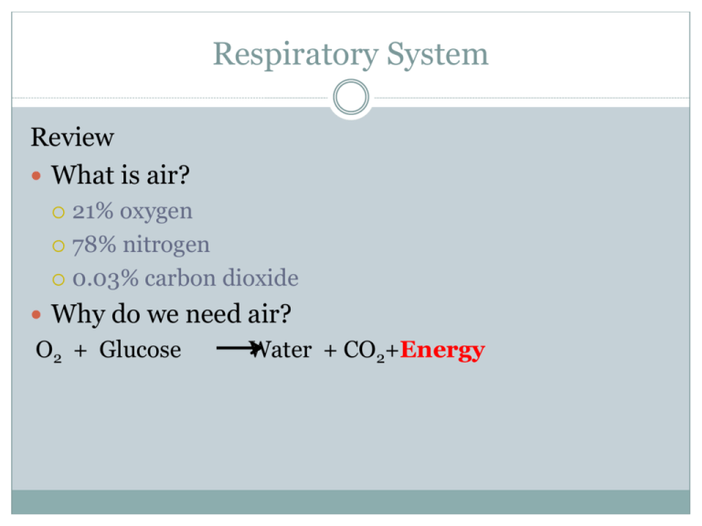
Respiratory System Review What is air? 21% oxygen 78% nitrogen 0.03% carbon dioxide Why do we need air? O2 + Glucose Water + CO2+Energy The Respiratory System Functions 1. Take oxygen from the atmosphere and deliver it to the bloodstream. 2. Take carbon dioxide from the bloodstream and expel it from the body. This system works closely with the circulatory system. The Respiratory System Nasal Cavity: 2 cavities that contain hairs, cilia, mucous and a rich blood supply Function: • Hairs and cilia filter particles • Mucous traps particles and moistens the air • Blood vessels warm air up to body temperature The Respiratory System Pharynx: muscular region at back of mouth Function: • Passageway for air and food Trachea: long tube lined with cilia (tiny hairs) and mucous : made up of cartilage rings Function: • Allows air to travel to and from the lungs • Cilia help filter out and remove dirt and bacteria from entering the lungs. • Dirt and bacteria are pushed up into the esophagus Cilia cells in the trachea The Respiratory System Epiglottis: flap of skin at the top of the larynx Function: • Prevents a bolus of food from entering the trachea Larynx: box shaped part, made of cartilage and elastic folds called the vocal cords. Function: • Produces sound larynx video The Respiratory System Bronchi: 2 branches of trachea that contain carilage rings, mucous, and cilia Function: • Delivers oxygen to each lung Bronchiole: smaller branches of bronchi without cartilage and lined with smooth muscle Function: • Deliver oxygen to alveoli The Respiratory System Alveoli: (alveolus = singular) : small grape like sacs at the end of the bronchioles to maximize surface area : measures 0.2 um in diameter Function: • Allows for the diffusion of gases (O2 and CO2) between the air and blood. The Respiratory System Alveoli: Do Not Copy • wall of alveolus is one cell layer thick • 150 million per lung • Have a combine surface area of 90 m2 • Surrounded by a network of capillaries (circulatory system) This is where the Circulatory System and the Respiratory System meet and exchange gases! The alveolus is covered by capillaries, which carry the blood to the body. O2 moves from the alveolus to capillaries through diffusion - movement from high concentration to low concentration. aka Inhaling CO2 moves into alveolus by diffusion, collected from all the alveoli in the lungs and pushed back up and out the trachea to the mouth and nose. aka Exhaling Video Pig lungs Question: How do you know when to inhale and exhale? Breathing is involuntary, it is controlled by a part of the brain (Medulla Oblongata) that detects the concentration of CO2. If CO2 is high, the brain increases breathing. If CO2 is low, the brain decreases breathing. Homeostasis!!! Mechanics of Breathing (How do we breathe?) Inhaling: rib muscles pull out and up, while diaphragm muscle contracts pulling down Chest cavity volume increases resulting in a decreased air pressure Air moves from area of high pressure (atmosphere) to low pressure (in the lungs) Mechanics of Breathing (How do we breathe?) Exhaling: Rib muscles and diaphragm relax Chest cavity volume decreases resulting in an increased air pressure Air moves from area of high pressure (in the lungs) to low pressure (atmosphere) Breathing: Animation Animation 2 Respiratory Diseases: •Asthma •Chronic Bronchitis •Emphysema •Pneumonia •Influenza •Tuberculosis Asthma Bronchi are temporarily swollen and mucus restricts air flow. Person finds it hard to breath Chronic Bronchitis Bronchi are permanently swollen and have too much mucus. Person often coughs and breathless due to less O2 Emphysema Alveoli (air sacs) collapse and/or are destroyed permanently. Less gas exchange = harder to get O2 Pneumonia Alveoli fill with fluid caused by bacteria or viruses causing coughing, chest pain, fever, etc.
