Sheep Brain
advertisement
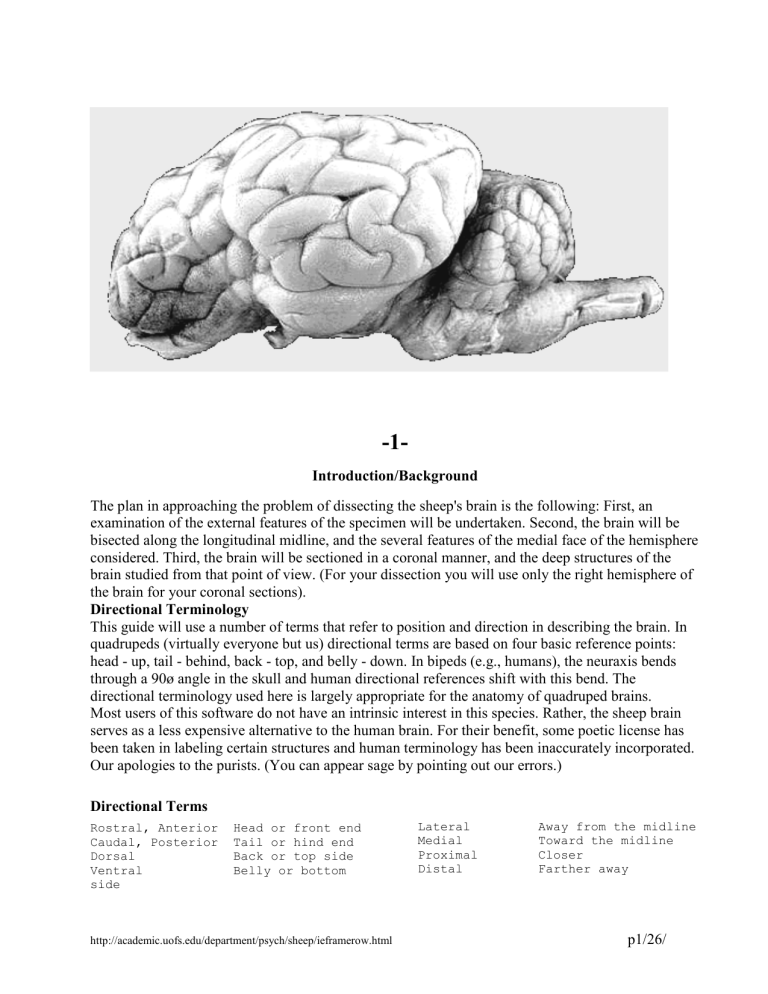
-1Introduction/Background The plan in approaching the problem of dissecting the sheep's brain is the following: First, an examination of the external features of the specimen will be undertaken. Second, the brain will be bisected along the longitudinal midline, and the several features of the medial face of the hemisphere considered. Third, the brain will be sectioned in a coronal manner, and the deep structures of the brain studied from that point of view. (For your dissection you will use only the right hemisphere of the brain for your coronal sections). Directional Terminology This guide will use a number of terms that refer to position and direction in describing the brain. In quadrupeds (virtually everyone but us) directional terms are based on four basic reference points: head - up, tail - behind, back - top, and belly - down. In bipeds (e.g., humans), the neuraxis bends through a 90ø angle in the skull and human directional references shift with this bend. The directional terminology used here is largely appropriate for the anatomy of quadruped brains. Most users of this software do not have an intrinsic interest in this species. Rather, the sheep brain serves as a less expensive alternative to the human brain. For their benefit, some poetic license has been taken in labeling certain structures and human terminology has been inaccurately incorporated. Our apologies to the purists. (You can appear sage by pointing out our errors.) Directional Terms Rostral, Anterior Caudal, Posterior Dorsal Ventral side Head or front end Tail or hind end Back or top side Belly or bottom http://academic.uofs.edu/department/psych/sheep/ieframerow.html Lateral Medial Proximal Distal Away from the midline Toward the midline Closer Farther away p1/26/ Three directional planes exist in the brain: rostral/caudal, dorsal/ventral, and medial/lateral. When sectioning (cutting) the brain, which planes are visible is determined by the type of section and two of these are used in this guide. In the sagittal section (which is made parallel to the midline, dorsal to ventral) the rostral/caudal and dorsal/ventral planes can be seen. In the coronal or cross section (made perpendicular to the midline, dorsal to ventral) the medial/lateral and dorsal/ventral planes can be seen. Subdivisions of the Brain Forebrain Telencephalon: cerebral cortex, corpora striata (caudate nucleus, internal capsule, putamen), and rhinencephalon (olfactory brain, e.g., olfactory bulb, hippocampus, amygdala, septal region, and cingulate cortex) Diencephalon: thalamus, hypothalamus, pineal gland, and posterior lobe of the pituitary (a migrated portion of the hypothalamus) Midbrain Mesencephalon: corpora quadrigemia (tectum--inferior and superior colliculi), tegmentum Hindbrain Metencephalon: cerebellum, pons Myelencephalon: medulla oblongata The Brain Stem is composed of the thalamus, hypothalamus, midbrain, pons and medulla. \ 1 http://academic.uofs.edu/department/psych/sheep/ieframerow.html p2/26/ Dissection Instructions 2: 2 On the ventral side of the brain, about midway between rostral and caudal ends, locate the pituitary gland. It is a midline mass of tissue lying caudal to the optic chiasm. If your brain still has all of its membranes intact, a number of types of tissue should be visible. For example, the grayish, semitransparent, tough membrane is the dura mater, while the dark brown, spongy material on either side of the pituitary are capillary beds. Rostrally, you may also find large globs of yellowish fat that are associated with the eyes. Buried in all this is the bulbous pituitary itself. 2A http://academic.uofs.edu/department/psych/sheep/ieframerow.html p3/26/ Dissection Instructions 3: Carefully dissect connective tissues from the caudal aspect of the pituitary and gently lift the pituitary mass from its caudal end. You should be able to see the III rd (oculomotor) cranial nerve pair attached to the ventral surface of the brain, on either side of the midline. These nerves are fairly broad, but quite flat, and may be difficult to see if they are lying down directly on the brain. Directly on the midline, anterior to the oculomotor nerves, you may find the thin stalk of the pituitary (the infundibulum) which connects the 3 body of the pituitary to the base of the brain. Keeping the pituitary lifted away from the ventral surface of the brain, use a pair of small scissors to sever the two nerves (III) and the infundibulum as far away from the brain as you can. Carefully interrupt any other connective tissue present, lift the pituitary away, mark the caudal or rostral aspect of the pituitary (you'll forget), and set it aside. You can examine it later to see the difference between its anterior (rostral) and posterior (caudal) lobes. Continuing with the ventral aspect of your specimen, at its very rostral limit, locate the two light colored pad-like flaps of tissue which are the olfactory bulbs. Caudal to the olfactory bulbs, with a little removal of dura and fat, you should be able to find the cut stumps of the optic nerves (II). Follow these back and you'll see that they blend into an "X" on the midline. The fused part of the X is the optic chiasm. Caudal to the chiasm are the optic tracts, which are part of the ventral surface of the brain. Stay on the Ventral Surface 4-- 4 http://academic.uofs.edu/department/psych/sheep/ieframerow.html Just caudal to the optic tracts, two very large bundles of fibers form the ventral surface of the brain. These are the cerebral peduncles. On the medial part of each you should find the oculomotor nerve (III) that we saw earlier. While carefully removing the membranes between the cerebral and cerebellar cortices, look up between them to find to see the fine spaghettilike trochlear nerve (IV) as it p4/26/ 4B proceeds ventralwards from its exit point on the dorsal aspect of the brain stem. The large trigeminal nerve (V) runs rostrally through this region. It is usually quite difficult to remove its protective membrane, consequently, it is often severed fairly close to the brain stem, leaving only a stump on the side of the pons. Stay on the Ventral Surface 5-Several cranial nerves can be seen exiting from the medulla. At its rostral end, you may be able to see the thin (and easily ripped off, see "Other View") abducens nerve (VI). The abducens exits from the trapezoid body (a thin transverse band of fibers on the surface of the brain just caudal to the pons, see Plate 8) at a point just lateral to the longitudinally coursing pyramidal tracts (see Plate 8). Lateral to the abducens is the more substantial facial nerve (VII). Lateral to this, tucked up just under the cerebellum, you may see the tuft 5 5B http://academic.uofs.edu/department/psych/sheep/ieframerow.html that forms the vestibulocochlear nerve (VIII). Caudal to the vestibulocochlear nerve you may see another light tuft of nerves that are made up of the glossopharyngeal nerve(IX) blending into the more caudal vagus nerve (X). Caudal to this you may see the spinal accessory nerve (XI) which appears as a line of connected filaments running in a rostralcaudal direction (they may take on the appearance of a broad yard rake). The hypoglossal nerve (XII) exits in several branches from the lateral edge of the caudal medulla (see "5B"). p5/26/ A New Perspective 6-Place the brain on its ventral surface. Look down from the top at the most ventrocaudal point of the cerebellum. If your thin arachnoid membrane is still in place (it isn't in Plate 6), note how the arachnoid forms the roof of a space, the cisterna magna, as it sweeps caudally from the cerebellum to the medulla. Carefully separate the caudal part of the cerebellum from the medulla; as you lift the cerebellum the arachnoid will rupture, and you should be able to see yet another membrane (or fragments of it); this is the tela chorioidea, forming the 6 posterior roof of the 4th ventricle. Separate the cerebellum from the medulla until that membrane ruptures; the internal space revealed by this maneuver is the 4th ventricle. The caudal point at which the two sides of the tela choroidea come together is called the obex. This can be seen on the dorsal surface of the medulla and forms the caudal boundary of the 4th ventricle. Looking into the 4th ventricle, you may see some dark spongy tufts; these are pieces of choroid plexus. Another Perspective 7-- 7 http://academic.uofs.edu/department/psych/sheep/ieframerow.html Leaving the brain on its ventral surface, we will now look down over the rostral end of the cerebellum. Looking down from the top, careful bending of the cerebellum in a caudal direction will reveal a second cistern, the superior cistern, under the rostral surface of the cerebellum (not visible in this Plate). Looking further down the brain stem, you may be able to see the white membrane forming the rostral roof of the 4th ventricle, the anterior medullary velum. Rostrally, this membrane attaches to the caudal inferior colliculus. Caudally, it continues under the cerebellum. p6/26/ Back to the Ventral Surface 8-- 8 9 After you have located the cranial nerves, and the cisterns just described, carefully strip away any remaining dura and arachnoid from the medulla. On the ventral aspect of the medulla a number of surface features can be readily seen: Note the longitudinal ridges coursing immediately on either side of the midline (marked by the ventral median sulcus); these are the pyramidal tracts. At the rostral end of the medulla, locate the band of transverse fibers paralleling the pons that form the trapezoid body. Ventral and Lateral Perspectives 9 The very prominent swelling just anterior to the trapezoid body; this is the pons (L. bridge). Three cerebellar peduncles attach the Cerebellum to the brain stem. The large middle cerebellar peduncle (brachium pontis) can be seen from the lateral perspective. http://academic.uofs.edu/department/psych/sheep/ieframerow.html p7/26/ Another look between Cerebral and Cerebellar Cortices 10 Looking down from the top, once again, gently pull back on the cerebellum to reveal the dorsal surface of the brain stem. Here you should be able to easily see the corpora quadrigemina, that is, the superior (rostral) and inferior (caudal) colliculi. The superior colliculi are considerably larger than the inferior. Collectively, this area is also called the tectum. 10 Ventral-Rostral Features 11 Just rostral to the pons, the ventral surface of the brain is formed by the cerebral peduncles, which run up to the optic tracts (seen earlier). The IIIrd cranial nerves can be seen exiting from the peduncles. The interpeduncular cistern lies in the "V" on the midline where the caudal-medial aspects of the peduncles meet. 11 http://academic.uofs.edu/department/psych/sheep/ieframerow.html p8/26/ 12 Ventral-Rostral Features 12 Just anterior to the interpeduncular cistern, locate the small, but distinct, protuberance lying on the midline; this is the mammillary body, and it marks the caudal limit of the hypothalamus, as seen from the ventral approach. The rostral border of the hypothalamus is marked by the optic chiasm, and the lateral boundaries are the medial edges of the cerebral peduncles. The general outline of the hypothalamus from the ventral aspect, takes on something of a diamond configuration. The remainder of the diencephalon (i.e., the thalamus) cannot be seen without sectioning the brain. http://academic.uofs.edu/department/psych/sheep/ieframerow.html p9/26/ Ventral-Rostral Features 13 Still viewing the brain from the ventral aspect, notice the fairly large, relative smooth masses of cortical tissue just lateral to the cerebral peduncles, extending, at the caudal limit, from the lateralmost part of the pons rostrally to include the olfactory bulbs. This mass of tissue is the rhinencephalon. The large rhinal fissure marks the lateral boundary of this region. The larger part, beginning at about the rostral/caudal level of the optic chiasm and proceeding caudally is 13 the hippocampal gyrus. Within this gyrus resides the amygdala and part of the hippocampus (not visible without sectioning the brain). Rostral to the hippocampal gyrus you will find the lateral olfactory gyrus. Running along the medial surface of the lateral olfactory gyrus, locate the lateral olfactory stria, a band of fibers originating in the olfactory bulbs and coursing caudally. The medial olfactory stria is seen as a fiber bundle coursing medially, disappearing into the longitudinal fissure just anterior to the optic chiasm. 13B http://academic.uofs.edu/department/psych/sheep/ieframerow.html p10/26/ Dorsal-Rostral Perspective-The two cerebral hemispheres are separated by the longitudinal fissure. Each hemisphere is divided into four major lobes. The frontal lobes are limited caudally by the cruciate fissure. The area caudal to the cruciate fissure is the parietal lobe whose line of separation from the more posteriorly placed occipital lobe is ill-defined. The temporal lobe in the sheep is very little developed in contrast to primates. In the sheep, it is represented by a slight bulge 14 superior to the hippocampal gyrus. The cruciate fissure is somewhat variable in the sheep, and sometimes difficult to locate. However, the superior frontal sulcus is easily located. Parallelling the longitudinal fissure, the superior frontal sulcus divides the frontal poles into approximately equal left- and right-halves, and, if traced caudally, it is seen to "T-end" into the cruciate fissure. 14B http://academic.uofs.edu/department/psych/sheep/ieframerow.html p11/26/ 15 The Medial Face-- If you are very fortunate, you may be able to see the central canal of the caudal medulla and spinal cord as it moves rostrally and opens up under the cerebellum, becoming the 4th ventricle (cerebrospinal fluid actually tends to run caudally in the ventricular system). Under the rostral cerebellum, the relatively thick anterior medullary velum should be easily located, It forms the anterior roof of the 4th ventricle and the rostral limit of the 4th ventricle is marked by the attachment of the anterior medullary velum to the caudal inferior colliculus. The 4th ventricle is continuous with the cerebral aqueduct of the midbrain. Tissue dorsal to the middle of the aqueduct is the tectum, and that ventral to its middle, excluding the cerebral peduncles, is the tegmentum. The tectum consists of the corpora quadrigemina and the underlying lamina quadrigemina; the latter can be seen as the cut surface immediately superior to the aqueduct, appearing somewhat like the end of a piece of plywood (which is a laminated structure). The cerebral aqueduct opens up into the 3rd ventricle (discussed further in Plate 16) which in turn is continuous with the two lateral ventricles that run out into each cerebral hemisphere. 16 Rostral Medial Face-- The relationship between the 3rd ventricle and the thalamus is somewhat unusual. Most of the medial portions of the two thalami are fused in a structure called the massa intermedia (appropriately named, if you remember your Latin roots). Where this fusion exists, of course, there can be no ventricular space. Consequently, the 3rd ventricle must run around the massa intermedia. In the vicinity of the massa intermedia, the lateral walls of the 3rd ventricle are formed by the unfused medial nuclei of the two thalami. Ventrally the lateral walls of the 3rd ventricle are formed by the medial nuclei of the hypothalami. At about 10 o'clock from the massa intermedia (assuming the brain is horizontal with the ventral side up), locate http://academic.uofs.edu/department/psych/sheep/ieframerow.html p12/26/ the small piece of tissue that is just outside the 3rd ventricle. This is the pineal body. Looking at the most ventral part of the 3rd ventricle, you should be able to appreciate how it extends down into the hollow stalk attaching the pituitary and hypothalamus (the infundibulum). Just caudal to this region, you will find the cut surface of the mammillary body. Just rostral to this region, you will find the cut surface of the optic chiasm. The corpus callosum is the very prominent collection of axons that 17 extends for some distance along the medial face of the cerebral hemispheres. At the rostral end, it curves ventrally and caudally, virtually making a 180ø turn. The area of turning is called the genu, and if you look carefully (with a perfectly cut brain), you will see that the caudal extending portion of this bend comes to a point and ends; this is the rostrum. At the caudal end of the corpus callosum it can be seen that a similar 180ø turn is made; this bend is the splenium. The main "body" of the corpus callosum runs between these two turns. Immediately dorsal to the body of the corpus callosum (i.e., that part between the splenium and genu) locate the crease that forms the callosal sulcus. The cortical outfolding just dorsal to the callosal sulcus is the cingulate gyrus, which, in turn, is bounded dorsally by a crease, the cingulate sulcus. Follow the cingulate gyrus caudally, and you will find that it courses ventrally and laterally, eventually becoming continuous with the hippocampal gyrus that we saw on the ventral surface of the rostral brain. Just inferior to the genu and rostrum of the corpus callosum is a region of medial face cortex called the septal area. As you will see in cross-sections, this region does not extend very deeply from the midline. At the caudal edge of the septal area, you should be able to locate the small, light-colored dot which is a cross-section of the anterior commissure. Just inferior to the attachment point of the pineal body, there is a much smaller (you won't be able to see it here, but it may appear in cross-section) bundle of decussating fibers called the posterior commissure. If the bisection of the brain was exactly on-center, there will be a membrane extending ventralwards from the body of the corpus callosum, the septum pellucidum, which separates the two lateral ventricles from each other. The fornix is a bundle of fibers that parallels the corpus callosum through much of its course. You can find the body of the fornix lying about midway between the massa intermedia and the body of the corpus callosum. You'll get a better look at the fornix in Plate 18. http://academic.uofs.edu/department/psych/sheep/ieframerow.html p13/26/ 18 Time for a Little Digging-- You are about to scrape away part of the medial face of one of your brain halves. Choose your half wisely. If your cut is off center, be certain that the structures we are interested in are still in the half you choose to scrape, they don't lie far from the midline. The most caudal part of the fornix is made up, in part, of decussating fibers called the hippocampal commissure (also known as the commissure of the fornix), which interconnect the two hippocampi. The more rostral part of the structure is made up of fibers largely originating in the hippocampi, paralleling the midline, and these are called the body of the fornix. Just dorsal to the anterior commissure, the fibers of the body of the fornix separate into two distinct bundles of fibers which move just lateral to the midline, forming the columns of the fornix. Some cell bodies in the mammillary body send their axons to the dorsal anterior thalamus through the mammillothalamic tract, which lies just beneath the medial face of the hemisphere. Use a scalpel, and, holding the blade at a right angle to the medial face of the hemisphere, very carefully scrape away the tissue between the anterior commissure and the mammillary body in order to uncover the column of the fornix, which will appear as a distinct white line arcing ventrocaudally from the body of the fornix. Next, do the same thing in the area between the mammillary body and the dorsal anterior part of the massa intermedia, uncovering the mammillothalamic tract. The Tree of Life (Internal Structure of the Cerebellum)-Note the internal structure of the cerebellum. The thick core of myelinated axons branching out into separate folia, reminiscent of a tree (arbor vitae), frosty window pane (for those of you in northern latitudes), or cauliflower florets (any analogy to food is a good analogy) 19 http://academic.uofs.edu/department/psych/sheep/ieframerow.html p14/26/ Dissection Instructions 20: Now you get to do some serious slicing. You will be making a series of coronal sections using one of your hemi-brains. Typically, you should use the half that you haven't scraped to see the mammillothalamic tract and columns of the fornix . Once again, you should minimize cutting artifacts by making one clean pass through the tissue, rather than sawing through it. You might also consider the following: As you are asked to make sections of the brain, rather than attempting to make the section at the exact level in one single cut, make a number of thinner cuts until the cut-face of your specimen resembles the 20B relevant figure. If you are skilled at cutting you can end up with a deck of brain slices each about as thick as a robust slice of bologna (don't shuffle this deck). Plate 20 shows the results of a cut that passes through the genu of the corpus callosum. Try to have the angle of your cuts match that of the sagittal section that is shown prior to each coronal section. Substantial deviations from this angle will make it somewhat challenging for you to match our plates with your sections. Looking at the cut surface of your first section, the darker material is mostly cell bodies, and the lighter tissue is mostly axons. Working from the ventral aspect of your section, look laterally until you find the first infolding, this is the rhinal fissure. To appreciate the full extent of this fissure, either look at the ventral lateral surface of your uncut hemi- brain or look back at Plate 13. Obviously, you should be able to see the rhinal fissure in many of your coronal sections, we will only label it here, however. Impress your friends by finding it elsewhere. Just medial to the rhinal fissure you may be able to locate the lateral olfactory stria. Moving to the medial face of the ventral part of your section, the gray matter here is the septal region. You should have no trouble locating the corpus callosum lying under the ventral surface of the medial cerebral cortex. As you did for the rhinal fissure, either look at the uncut hemi- brain (medial face) or look back at Plate 16 to 20 http://academic.uofs.edu/department/psych/sheep/ieframerow.html p15/26/ appreciate the full extent of the corpus callosum. Obviously, the corpus callosum will show up in many of your coronal sections, we have only labeled it here. You may also see part of the rostrum of the corpus callosum as it extends ventrally and caudally from the genu. The thin space above the corpus callosum is the callosal sulcus, the cortex above that is the cingulate gyrus , and the space above that is the cingulate sulcus. We have only labeled them here, but, as you would expect, these structures will occur in many of your crossections (Examine either the medial face of the uncut hemi-brain or Plate 17). Off the midline, the space below the corpus callosum is lateral ventricle. The caudate nucleus forms the lateral wall of the lateral ventricle. Just lateral and ventral to the caudate nucleus, find the stippled band of white and gray matter. The white matter is the internal capsule. Ventral and lateral to the internal capsule you should be able to see the putamen (a nuclear group). This arrangement of nuclei and fibers (caudate nucleus, internal capsule, and putamen) gives the area a striated appearance, and thus led to terming this group the corpus striatum. Immediately lateral to the putamen, you should be able to see a solid streak of white matter, the external capsule. Just lateral to this, typically, you can see the triangularly shaped claustrum. The caudate, internal capsule, putamen, external capsule, claustrum and septal area will appear in subsequent sections of this guide, however they will not always be labelled. 21 Notice how the septal area has become larger on the ventral medial face of the brain. The caudate is still prominent and the internal capsule should be easier to see. You may be able to see the beginning of the globus pallidus, which is contiguous with the putamen on its ventral medial face. The globus pallidus has a lighter and more homogeneous coloration than the putamen. If you had trouble seeing the claustrum in the last plate, it should be clear here. http://academic.uofs.edu/department/psych/sheep/ieframerow.html p16/26/ Anterior Optic Chiasm 22 In this section you can see the anterior part of the optic chiasm, which is stuck to the ventral surface of the brain. You can also see the decussating fibers of the anterior commissure running across the midline of the brain. You may also see some fibers of the columns of the fornix as they form 22 http://academic.uofs.edu/department/psych/sheep/ieframerow.html p17/26/ 23 Caudal Optic Chiasm 23 On the midline, immediately superior to the chiasm, you will find a vertical space; this is the most rostral part of the 3rd ventricle. Here, the medial face of the anterior hypothalamus forms the lateral wall of the 3rd ventricle. Note that the dorsal part of the 3rd ventricle extends out under the body of the fornix. (The "T" shape isn't clear here because the left side of the fornix has twisted down from its natural positioning.) The lateral ventricles take on the shape of horns, extending out and up over the caudate. Here too, you can see the effects of the "C" shape of the fornix, with the body visible dorsally and the columns visible ventrally. The gray area just ventral and medial to the caudate is your first glimpse of the thalamus in crossection; this is the most dorsal and rostral region. The medial face of the thalamus forms the lateral walls of the dorsal third ventricle (see Plates 15-17). 24 Just caudal to the Optic Chiasm 24 On the ventral surface of the brain you will see that the optic chiasm no longer extends across the midline (the ventral hypothalamus has taken its place) and the optic tracts are proceeding laterally. The massa intermedia of the thalamus makes its appearance here, obstructing the middle of the 3rd ventricle. Other subdivisions of the thalamus can be seen lateral to the massa intermedia. Our old friend the caudate nucleus is still visible here, but is much reduced from rostral sections. At the junction between the caudate and dorsal thalamus you may be able to spot a streak of white matter, this is the stria terminalis. Like the fornix, the stria terminalis takes on a modified "C" structure as it courses between the amygdala and hypothalamus. The fornix is fairly flattened here because we are seeing its central body as well as lateral fibers of the fimbria that are joining the body. You'll get a better feel for the origin of the http://academic.uofs.edu/department/psych/sheep/ieframerow.html p18/26/ fimbria in Plate 28. Notice that, in addition to the preceding, you can still see the columns of the fornix. 25 The optic tracts have moved more laterally and, lateral to them and buried in the cortex, you should be able to see the amygdala (more appropriately, amygdaloid complex). It has an elongated teardrop shape. There may still be a hint of the putamen located above the dorsal end of the amygdala, with the external capsule and claustrum just lateral to the putamen. The caudate is still visible as well. The thalamus makes up most of the medial section. The hypothalamus extends through about the bottom third of the diencephalon at this point. Four white dots are visible. The top two are the mammillothalamic tracts and the ventral are the columns of the fornix (approaching their endpoint in the mammillary bodies). Look back at Plate 18 to get a perspective on these pathways. 26 Rostral Mammillary Body 26 Not much has changed from the last section. We have indicated some tufts of choroid plexus in the third and lateral ventricles. At this point, you should have fibers crossing between the two hippocampi, forming the commisure of the fornix (hippocampal commisure). They aren't distinctly visible because they are blending with fimbria and fornix fibers. The internal capsule is very prominent. The ventral aspect of the 3rd ventricle is fairly small. http://academic.uofs.edu/department/psych/sheep/ieframerow.html p19/26/ 27 Caudal Mammillary Body-The most interesting development here is that you can see another demonstration of the modified "C" we've encountered previously. Here you can see dorsal and ventral aspects of the lateral ventricle and the hippocampus as they are cut twice through their "C" course. The optic tracts are meeting up with their major target, the lateral geniculate nucleus of the thalamus. You can also see the cerebral peduncles forming on the ventral surface of the brain. 28 http://academic.uofs.edu/department/psych/sheep/ieframerow.html Here you can more clearly see the "C" shape of the hippocampus as well as the white fibers of the fimbria. The habenula may also be seen on the dorsal surface of the diencephalon. The pulvinar of the thalamus may be seen just medial to the lateral geniculate body. p20/26/ 29 Right Through the Pineal Body 29 Here we have captured the pineal gland sitting in the notch between the two lobes of the thalamus. The thin 3rd ventricle can be seen (we have passed caudal to the massa intermedia). The medial geniculate nucleus may be visible in your section. The Superior Colliculus 30 30 This section catches the rostral part of the superior colliculi. If you look carefully, you should be able to see that they have a subtle striated (striped) appearance. The cerebral aqueduct can be see here on the midline (no other ventricular space can be seen at this point). It is fairly small at this point, it will broaden out more caudally. Surrounding the cerebral aqueduct is the periaqueductal gray region (check those latin roots). The cerebral cortex that is visible at this point is the occipital cortex. Note that there is no interconnecting corpus callosum at this level. http://academic.uofs.edu/department/psych/sheep/ieframerow.html p21/26/ 31 Just Rostral to the Cerebellum 31 Here you can see the caudal part of the superior colliculus (notice the striations) and the beginning of the inferior colliculus (which will not appear striated). The cerebral aqueduct has become much larger. The rostral part of the pons can be seen at the ventral edge of the section. The unsectioned rostral part of the cerebellum can be seen behind the cut brain stem (this is a caudally directed view). 32 Immediately Rostral to the Cerebellum 32 This section through the pons shows the pontine fibers running up the lateral edge of the pons forming the brachium pontis. You can see both the 4th ventricle and the anterior medullary velum that forms the roof of rostral part of this ventricular space (this is a caudally directed view). http://academic.uofs.edu/department/psych/sheep/ieframerow.html p22/26/ 33 34 At the Level of the Trapezoid Body 33 This section shows the massive peduncular attachments of the cerebellum as well as some of the nuclei that are found deep within the cerebellum. You should be able to see that the majority of the lateral medulla is covered by a thin sheet of fibers, the trapezoid body. You may also see some striations on the ventral midline as these trapezoid fibers decussate. Caudal to the Cerebellar Peduncles 34 In the ventral lateral medulla, you should be able to see the inferior cerebellar peduncle. In this caudally directed view, you can see that the 4th ventricle is beginning to close down as the ventricle approaches the obex. http://academic.uofs.edu/department/psych/sheep/ieframerow.html p23/26/ 35 Caudal Cerebellum— In this caudally directed view, you can see the 4th ventricle closing down at its caudal end, and the black dot at the floor of this ventricle which is the beginning of the central canal. In the ventral medial medulla you may be able to see a zebra pattern created by the inferior olivary nuclei. In this section, you can see a tuft of glossopharyngeal (IX) fibers on the lateral edge of the medulla. Note-The lateral cerebellar cortices appear to curve away from you, they do not. These are actually flat cut surfaces that have fallen back because they do not have underlying cortical tissue to hold them up. We're Not in Kansas Anymore— This section through the cervical spinal cord shows the classic butterfly shape formed by the gray matter of the cord. (We know, it looks white, but it's gray, trust us.) You can also see dorsal and ventral rootlets on the outside of the cord. The central canal is very small and is not readily visible here. 36 http://academic.uofs.edu/department/psych/sheep/ieframerow.html p24/26/ Glossary Abducens (Cranial Nerve VI): An efferent nerve which connects to the abductor muscles of the eye. Amygdala: A division of the basal ganglia which is located in the temporal lobe. Amygdala 25 26 Anterior Commissure: A bundle of nerve fibers which crosses the median plane in the upper part of the lamina terminalis and which contains connections between the olfactory bulbs, amygdaloid nuclei, and parts of the temporal lobe cortices. Anterior Medullary Velum: A thin layer of tissue between the cerebellar peduncles which forms the roof of the rostral part of the fourth ventricle. Body of the Fornix: The segment of the fornix which projects rostrally and ventrally in the edge of the septum pellucidum from the hippocampal commissure to a point just above the anterior commissure. Brachium Pontis (Middle Cerebellar Peduncle): A bundle of nerve fibers connecting the pons and cerebellum. Brain Stem: Collectively, the midbrain, pons, and medulla. Callosal Sulcus: The sulcus on the medial surface of the cerebral hemisphere which separates the corpus callosum from the cingulate gyrus. Caudate Nucleus: One of the basal ganglia, it consists of a head, which forms the anterior lateral wall of the lateral ventricle, a body, lying above the dorso-lateral thalamus, and a tail, which is located above the lateral ventricle's inferior horn. Central Canal: A narrow channel which extends the length of the spinal cord from the caudal tip of the fourth ventricle. Cerebellar Cortex: The outer layer of the cerebellum, it consists of three layers cells: molecular (the outermost), Purkinje, and granular (the innermost). Cerebellum: From the Latin for "little brain," the cerebellum is located behind the cerebral hemispheres. Its functions include coordination, equilibrium, and muscle tone. The cerebellum consists of a medial part, the vermis, and two cerebellar hemispheres. Cerebral Aqueduct: A narrow channel through the midbrain which connects the third and fourth ventricles. Cerebral Cortex: The outer layer (gray matter)of the cerebral hemispheres. It consists of six layers: the molecular(outermost; layer I); external granular (layer II); external pyramidal (III); internal granular (IV); internal pyramidal (V); and multiform (innermost; layer VI). Cerebral Peduncle: A peduncle is a large band of nerve fibers in the brain, usually composed of multiple tracts. The cerebral peduncle is the ventral part of each half of the midbrain (not including the tectum). Choroid Plexus: These membranes are the primary source of cerebrspinal fluid. They can be found in the lateral, third, and fourth ventricles. Cingulate Gyrus (Collosal Gyrus): A gyrus is a ridge (outfolding) of the surface of the cerbrum. The cingulate gyrus is located on the medial surface of the cerebral hemisphere between the cingulate sulcus and the sulcus of the corpus collosum. Cingulate Sulcus: A sulcus (plural sulci) is s groove (infold) of the surface of the brain or spinal cord. The cingulate sulcus is on the medial surface of the cerebrum. Claustrum: A thin sheet of gray matter located between the external capsule and putamen.(medially; between the insula and extreme capsule laterally) http://academic.uofs.edu/department/psych/sheep/ieframerow.html p25/26/ Column of the Fornix: (See Fornix) The column of the fornix is the segment which turns backward and downward behind the anterior commissure into the hypothalamus. It ends mainly in the mamillary body. Commissure of the Fornix (Hippocampal Commissure): (See Fornix) A commissure is a bundle of nerve fibers which crosses the median plane, sometimes, but not always, connecting similar structures on the two sides of the brain. The commissure of the fornix is just beneath the splenium of the corpus callosum. Corpus Callosum: A large band of nerve fibers which mainly connects corresponding areas of the two cerebral hemispheres. It consists of four parts which form a rostrally-projecting “hook” shape: the rostrum, genu, body, and splenium. External Capsule: Layer of nerve fibers between the putamen and the claustrum. Facial Nerve (Cranial Nerve VII): The facial nerve arises from the ventrolateral surface of the pons at its junction with the medulla. It is composed mostly of motor fibers which supply the muscles of the face, including those that close the eyes, parasympathetic relays to the lacrimal gland (which produces tears) and the salivary glands, taste fibers from the anterior two-thirds of the tongue, and some cutaneous fibers from the external ear. Fimbria: The main bundle of efferent nerve fibers arising from the hippocampus. Frontal Lobe: The part of the cerebral hemisphere anterior to the central sulcus. Genu: From the Latin for knee, genu refers to a bend. The most anterior part of the corpus callosum is the genu. Globus Pallidus: Part of the basal ganglia Glossopharyngeal Nerve (Cranial Nerve IX): This cranial nerve arises from the ventrolateral surface of the medulla, just caudal to the facial nerve. It contains several afferent nerve fibers important to the regulation of blood pressure and respiration, taste fibers from the back of the tongue, and sensory fibers from the palate and pharynx. It also contains parasympathetic fibers for several glands, and motor fibers for the stylopharyngeus muscle. http://academic.uofs.edu/department/psych/sheep/ieframerow.html p26/26/
