Skeletal Muscle - PROFESSOR AC BROWN
advertisement
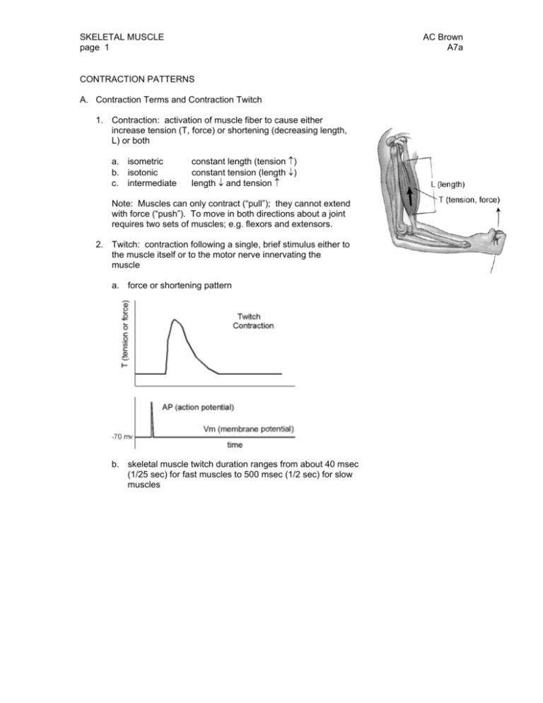
SKELETAL MUSCLE page 1 AC Brown A7a CONTRACTION PATTERNS A. Contraction Terms and Contraction Twitch 1. Contraction: activation of muscle fiber to cause either increase tension (T, force) or shortening (decreasing length, L) or both a. isometric b. isotonic c. intermediate constant length (tension ↑) constant tension (length ↓) length ↓ and tension ↑ Note: Muscles can only contract (“pull”); they cannot extend with force (“push”). To move in both directions about a joint requires two sets of muscles; e.g. flexors and extensors. 2. Twitch: contraction following a single, brief stimulus either to the muscle itself or to the motor nerve innervating the muscle a. force or shortening pattern b. skeletal muscle twitch duration ranges from about 40 msec (1/25 sec) for fast muscles to 500 msec (1/2 sec) for slow muscles SKELETAL MUSCLE page 2 AC Brown A7a CONTRACTION PATTERNS (continued) B: Response to Multiple Stimuli (Summation) Two stimuli; second stimulus delivered while the muscle is still contracting A series (“train”) of stimuli 1. Phenomenon: since the electrical refractory period of nerve and muscle is much less than the twitch duration, a second stimulus can be given during the twitch 2. Characteristics a. second twitch begins before the first twitch is complete (contraction is not all-or-none) b. peak tension is greater than with a single twitch (mechanical summation) c. the smaller the delay between the two stimuli, the greater the peak tension (up to a certain point) C. Fusion and Tetanus 1. Apply continuous train of stimuli 2. Result Low frequency Intermediate frequency High frequency Series of separate twitches Summation but incomplete fusion (a & b) above Complete fusion and maximum tension (Tetanus) SKELETAL MUSCLE page 3 AC Brown A7a MECHANISM OF CONTRACTION A. Functional Anatomy of Skeletal Muscle 1. Muscle fiber (muscle cell, myocyte) a. excitable membrane (sarcolema); generates and propagates action potentials, similar to neuron axon (conduction velocity in order of 5 m/sec) b. composed of myofibrils, sarcoplasmic reticulum system, mitochondria, etc. 2. Myofibril a. length same as length of fiber b. divided lengthwise into sarcomeres, repeating units, with characteristic striation (band) pattern (striated muscle) c. composed of interdigitated filaments 3. Filaments: two types a. thick filament: myosin b. thin filament: actin, troponin, tropomyosin Note: The striation pattern is due to the arrangement of the filaments: I band: thin filaments only, appears light A band: both thick and thin filaments, appears dark H zone: thick filament only, intermediate density 4. Sarcoplasmic reticulum system (SR) a. transverse (T) tubules 1) penetrate into the fiber 2) electrically continuous with the cell membrane b. longitudinal tubules, with enlargements (terminal cisternae) adjacent to the ttubules 1) sequester (store) calcium ions by active transport; energy for the active transport is furnished by ATP Note: as a result, at rest the normal Ca the remaining cytoplasm is very low 2+ concentration in SKELETAL MUSCLE page 4 AC Brown A7a MECHANISM OF CONTRACTION B. Sliding Filament Model 1. Muscle length change occurs by the sliding of the interdigitated thick and thin filaments 2. Actin and myosin are capable of forming cross bridges that tend to lock the filaments in place relative to each other a. in the resting state, tropomyosin blocks the myosin-binding site on actin b. Ca++ can bind to thin filament troponin, which causes reconfiguration of tropomyosin, permitting formation of cross bridges between actin and myosin c. ATP can bind to myosin and be degraded into ADP and Pi (inorganic phosphate); the rate of this reaction is greatly increased in the presence of cross bridges d. the energy released from ATP hydrolysis can be used to change the angle of the cross bridge by rotation of the myosin heads, leading to fiber shortening and/or increased fiber tension 3. In the absence of cross bridges, filaments slide freely C. Sequence of Contraction Events 1. Spread of action potential along the muscle fiber 2. Depolarization of the transverse tubule system 3. Transverse tubule depolarization causes Ca++ channels in the lateral sacs of the sarcoplasmic reticulum (SR) to open; the increase in P-Ca leads to rapid passive diffusion from the SR into the fiber cytoplasm 4. Some of the Ca++ ions bind to troponin, permitting the formation of actin-myosin cross bridges 5. Using the energy provided by ATP → ADP conversion at the bridge site, the cross bridge reorients, leading to muscle contraction 6. A new ATP replaces the ADP at the bridge site, permitting the bridge to break 7. A new bridge is formed at the next site, etc., leading to continued contraction SKELETAL MUSCLE page 5 AC Brown A7a MECHANISM OF CONTRACTION (continued) C. Sequence of Contraction Events (continued) 8. When the fiber repolarizes, the Ca++ channels close and the cytoplasmic Ca++ is resequestered into the lateral sacs 9. Cross bridges cease forming and muscle relaxes D. Basis of Mechanical Summation 1. Twitch: Ca++ released by AP resequestered before 2nd AP arrives 2. Summation: part of the Ca++ released by 1st AP still present in the cytoplasm when 2nd AP arrives, so the second contraction begins from a higher baseline 3. Tetanus: train of APs at a frequency high enough to keep Ca++ at a cytoplasmic concentration enabling bonds to continue to form MUSCLE FIBER TYPES A. Classes 1. Fast glycolytic fibers 2. Slow oxidative fibers 3. Fast oxidative fibers (intermediate characteristics intermediate between "fast glycolytic" and "slow oxidative" types) B. Characteristics Contraction time Color Myoglobin concentration ATP source (major) FAST FIBERS GLYCOLYTIC fast white (pale) low FAST FIBERS OXIDATIVE intermediate red intermediate SLOW FIBERS OXIDATIVE slow red (dark) high glycolysis glycolysis, but can become more oxidative with use intermediate Intermediate; depends partly on use fatigue resistant intermediate oxidative phosphorylation standing, walking posture and tone ATPase activity Vascularization high less Fatigability Motor unit contraction force Role easily fatigued high abrupt movements; e.g. jumping low more fatigue resistant lower SKELETAL MUSCLE page 6 CONTROL OF MUSCLE FORCE A. Cross-Section The larger the cross section, the more sites are available for parallel bond formation, so the larger the maximum force; depends on amount of protein (myosin, actin, etc.) in muscle B. Length 1. Length-tension diagram 2. Basis of length-tension relation: interaction between thick and then filaments A minimum overlap of thick and thin filaments B-C optimum overlap of thick and thin filaments D-E interference with bond formation due to adjacent thin filaments AC Brown A7a SKELETAL MUSCLE page 7 CONTROL OF MUSCLE FORCE (continued) D. Motor Unit Recruitment 1. Definition of Motor Unit: a single motor axon and all of the skeletal muscle fibers it innervates Innervation Ratio = muscle fibers per nerve axon Mean value of innervation ratio ranges from about 3 (extraoccular muscles) to 150 (large, fast muscles) A given muscle generally contains a range of motor units, from those with low innervation ratio ("small": few muscle fibers per axon, low tension) to high innervation ratio ("large": many muscle fibers per axon, larger tension) 2. Recruitment pattern As the motor nerve activity for a given muscle increases progressively, first the smallest motor units (lowest innervation ratio) are activated, next the intermediate size motor units are recruited, and finally the large motor units are activated (size principle) E. Contraction Gradation 1. firing frequency of individual motor unit 2. recruitment of motor units, from small to large EFFECT OF TRAINING AND AGEING A. Training 1. Muscle fiber hypertrophy (more protein) 2. Increase efficiency of oxidative energy utilization 3. Conversion on intermediate muscle cells from glycolytic to oxidative 4. Capillary proliferation (increase maximum circulation) Note: (1) above leads to increase maximum strength; (2) and (3) and (4) above lead to increase endurance (capacity for aerobic work) B. Ageing 1. Decrease number of fibers (leads to loss of maximum strength) Note: Skeletal muscle fibers, like cardiac muscle fibers and neurons, do not regenerate if lost. SKELETAL MUSCLE PATHOPHYSIOLOGY (myopathy) A. Muscular dystrophy: degeneration of muscle fibers; selective (not all fibers degenerate simultaneously), cause unknown B. Disuse atrophy: reduction in muscle protein when muscle is not used C. Rigor Mortis: inability to break cross bridges due to lack of ATP AC Brown A7a


