Material properties and microstructure from
advertisement
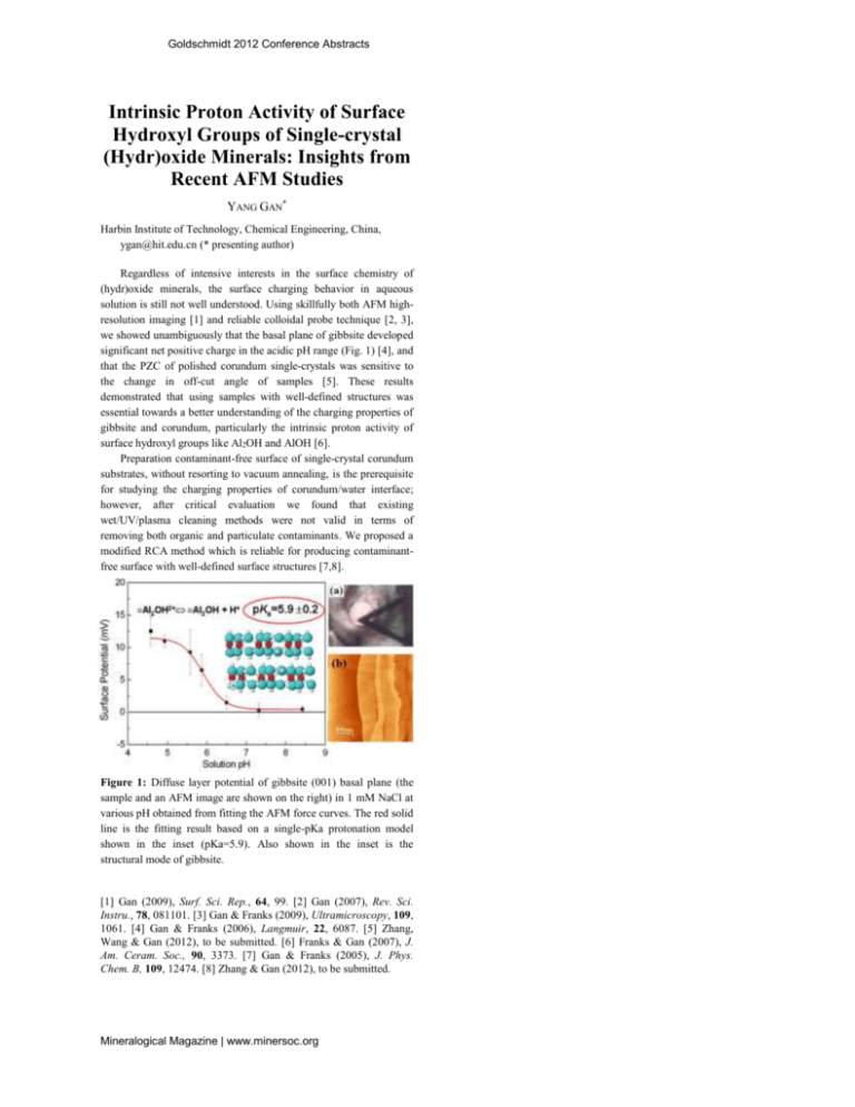
Goldschmidt 2012 Conference Abstracts Intrinsic Proton Activity of Surface Hydroxyl Groups of Single-crystal (Hydr)oxide Minerals: Insights from Recent AFM Studies YANG GAN* Harbin Institute of Technology, Chemical Engineering, China, ygan@hit.edu.cn (* presenting author) Regardless of intensive interests in the surface chemistry of (hydr)oxide minerals, the surface charging behavior in aqueous solution is still not well understood. Using skillfully both AFM highresolution imaging [1] and reliable colloidal probe technique [2, 3], we showed unambiguously that the basal plane of gibbsite developed significant net positive charge in the acidic pH range (Fig. 1) [4], and that the PZC of polished corundum single-crystals was sensitive to the change in off-cut angle of samples [5]. These results demonstrated that using samples with well-defined structures was essential towards a better understanding of the charging properties of gibbsite and corundum, particularly the intrinsic proton activity of surface hydroxyl groups like Al2OH and AlOH [6]. Preparation contaminant-free surface of single-crystal corundum substrates, without resorting to vacuum annealing, is the prerequisite for studying the charging properties of corundum/water interface; however, after critical evaluation we found that existing wet/UV/plasma cleaning methods were not valid in terms of removing both organic and particulate contaminants. We proposed a modified RCA method which is reliable for producing contaminantfree surface with well-defined surface structures [7,8]. Figure 1: Diffuse layer potential of gibbsite (001) basal plane (the sample and an AFM image are shown on the right) in 1 mM NaCl at various pH obtained from fitting the AFM force curves. The red solid line is the fitting result based on a single-pKa protonation model shown in the inset (pKa=5.9). Also shown in the inset is the structural mode of gibbsite. [1] Gan (2009), Surf. Sci. Rep., 64, 99. [2] Gan (2007), Rev. Sci. Instru., 78, 081101. [3] Gan & Franks (2009), Ultramicroscopy, 109, 1061. [4] Gan & Franks (2006), Langmuir, 22, 6087. [5] Zhang, Wang & Gan (2012), to be submitted. [6] Franks & Gan (2007), J. Am. Ceram. Soc., 90, 3373. [7] Gan & Franks (2005), J. Phys. Chem. B, 109, 12474. [8] Zhang & Gan (2012), to be submitted. Mineralogical Magazine | www.minersoc.org
![Structural and electronic properties of GaN [001] nanowires by using](http://s3.studylib.net/store/data/007592263_2-097e6f635887ae5b303613d8f900ab21-300x300.png)

