ENT ITE Review EAR
advertisement
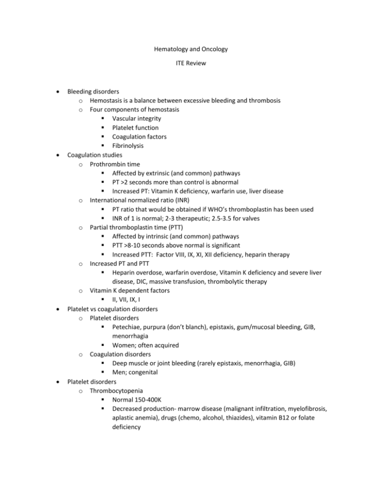
Hematology and Oncology ITE Review Bleeding disorders o Hemostasis is a balance between excessive bleeding and thrombosis o Four components of hemostasis Vascular integrity Platelet function Coagulation factors Fibrinolysis Coagulation studies o Prothrombin time Affected by extrinsic (and common) pathways PT >2 seconds more than control is abnormal Increased PT: Vitamin K deficiency, warfarin use, liver disease o International normalized ratio (INR) PT ratio that would be obtained if WHO’s thromboplastin has been used INR of 1 is normal; 2-3 therapeutic; 2.5-3.5 for valves o Partial thromboplastin time (PTT) Affected by intrinsic (and common) pathways PTT >8-10 seconds above normal is significant Increased PTT: Factor VIII, IX, XI, XII deficiency, heparin therapy o Increased PT and PTT Heparin overdose, warfarin overdose, Vitamin K deficiency and severe liver disease, DIC, massive transfusion, thrombolytic therapy o Vitamin K dependent factors II, VII, IX, I Platelet vs coagulation disorders o Platelet disorders Petechiae, purpura (don’t blanch), epistaxis, gum/mucosal bleeding, GIB, menorrhagia Women; often acquired o Coagulation disorders Deep muscle or joint bleeding (rarely epistaxis, menorrhagia, GIB) Men; congenital Platelet disorders o Thrombocytopenia Normal 150-400K Decreased production- marrow disease (malignant infiltration, myelofibrosis, aplastic anemia), drugs (chemo, alcohol, thiazides), vitamin B12 or folate deficiency o o o o Increased destruction- ITP, TTP, HUS, HELLP, sepsis, viral (HIV, mumps, varicella, EBV), SLE, drugs (PCN, sulfa, quinine, Lasix, heparin, ASA) Splenic sequestration- hypersplenism, cirrhosis, heme malignancy Dilution- massive transfusion Platelet transfusion Six pack increases platelet count approx. 50K Indications- platelet count <10-20K, life threatening bleeding Idiopathic thrombocytopenic purpura (ITP) Acute ITP Antiplatelet antibody IgG (both acute and chronic) Kids 2-6 yrs Follows viral infection Self limited; resolves spontaneously Tx supportive unless severe bleeding (plt <50K) or plt <20K Prednisone 1 mg/kg IVIG Platelet transfusion only for life-threatening hemorrhage Chronic ITP Women 20-50 yrs Remission rare Underlying autoimmune, collagen vascular, or malignant disease Steroids IVIG, immunosuppressives, splenectomy Platelet transfusion only for life-threatening hemorrhage Thrombotic thrombocytopenic purpura (TTP) Subset of thrombotic microangiopathies Subendothelial and intraluminal deposits of fibrin and platelet aggregation Females; 80% mortality if untreated Infection, autoimmune disease, immunosuppressives, chemo Treatment- steroids, plasmapheresis, dialysis, FFP Avoid platelet transfusions FAT RN Fever Anemia- MAHA; schistocytes on peripheral smear Thrombocytopenia Renal Neurologic Hemolytic uremic syndrome (HUS) Thrombotic microangiopathy Thrombocytopenia, hemolytic anemia, fever, neurologic, renal Less mental status change and more renal dysfunction than TTP Often kids Bacterial gastroenteritis (E coli O157:H7, Shigella) o o o o ASA/NSAIDs block cyclooxygenase, which decreases thromboxane formation decreased platelet aggregation ASA effect irreversible NSAID effect reversible Disseminated intravascular coagulation (DIC) Platelet, coagulation, fibrinolytic disorder Diffuse bleeding from multiple sites Acrocyanosis, thrombosis, pregangrenous changes of fingers, toes, genitalia, nsoe (purpura fulminans) Trauma, burns, pregnancy, sepsis, transfusion, carcinoma, acute leukemia, liver disease, snake bites, heat stroke Treat underlying cause If hemorrhageblood products If thrombosis low dose heparin More commonly seen with Gram (-) sepsis in pregnancy, retained fetus, chronic DIC, purpura fulminans Findings Increased PT/INR Increased PTT Decreased platelets Decreased fibrinogen Increased FSP (fibrin split products) Increased D-dimer Schistocytes Heparin induced thrombocytopenia (HIT) Antibodies to heparin/platelet factor 4 complex platelet activation and clot formation Occurs 5-10 days after exposure Heparin > LMWH Thrombocytopenia <150K or drop of 50% from baseline Arterial or venous thrombosis Stop the heparin! Avoid platelet transfusions, lower limb dopplers (high incidence of DVT), test for HIT antibodies Von Willebrand’s disease Most common genetic bleeding disorder Von Willebrand factor functions Facilitates platelet adhesion Links platelet to endothelium Plasma carrier for factor VIII Autosomal dominant Deficiency or dysfunction of vWF and mild factor VIII defect Type I- decreased vWF (most common) Type II- abnormal or dysfunctional vWF Type III- no vWF Mucocutaneous bleeding (epistaxis, menorrhagia, GIB) Abnormal platelet function studies, increased bleeding time, normal platelet count, normal PT/PTT Treatment- DDAVP (desmopressin), factor VIII concentrates, cryoprecipitate Coagulation disorders o Hemophilias Delayed/protracted bleeding after mild trauma or dental extraction Spontaneous hematuria Hemarthrosis and muscle hematomas Most common in ankle in kids; knee in adults Intracranial hemorrhage Proloned PTT; normal PT and platelets Hemophilia A X-linked recessive deficiency of Factor VIII Mild (hematuria, deep lac)- 20 U/kg Moderate (oral lac, dental, minor surgery, late hemarthrosis)- 25 U/kg Severe (CNS/GI/abd, major trauma)- 50 U/kg 1 U/kg increases activity level by 2% Cryoprecipitate, DDAVP, FFP Hemophilia B Aka Christmas Disease X-linked recessive deficiency of Factor IX Factor IX concentrate Prothrombin Complex Concentrates (PCC), FFP, NOT Cryo o Hypercoagulability Factor V Leiden (most common inherited hyercoagulability disorder) Prothrombin mutation Hyperhomocysteinemia Protein C deficiency Protein S deficiency Red blood cell disorders o RBC incides Mean corpuscular hemoglobin (MCH)- measure of hemoglobin content within RBC Mean corpuscular hemoglobin concentration (MCHC)- measure of concentration of hemoglobin within RBC Mean corpuscular volume (MCV)- measure of RBC size RBC distribution width- measure of deviation in volume of RBCs o Microcytic anemias Iron deficiency anemia o o o Hypochromic (decreased MCHC), decreased retics, decreased iron, increased TIBC, increased RDW Lead poisoning Hypochromic, basophilic stippling Lead lines- hyperdense lines at metaphyses (knee and wrist) Thalassemia Mediterranean, African, Asian Hypochromic Spherocytosis Hyperchromic (increased MCHC) Macrocytic anemias Vitamin B12 deficiency (pernicious anemia), folate deficiency (alcoholism)large, oval RBCs, hypersegmented neutrophils Liver disease, hypothyroidism Hemolytic anemias Findings in hemolysis- decreased hemoglobin, decreased haptoglobin, increased LDH, increased unconjugated bilirubin, schistocytes on peripheral smear G6PD deficiency X-linked nonimmune hemolytic anemia Coombs test negative Protects against malaria Stressors- infection, fava beans, medications (sulfa, nitrofurantoin, pyridium, antimalarials, dapsone) Heinz bodies on peripheral smear Sickle cell disease Autosomal recessive abnormal hemoglobin resulting from single amino acid substitution Deoxygenated HbS results in sickle-shaped, nondeformable rbc that cannot traverse small capillaries Sickle cell trait Spontaneous bleeding (hematuria, hyphema) Hyposthenuria (impaired ability to concentrate urine) Vaso-occlusive crises (rare, only extreme hypoxia) Presentation in kids Unexplained gnawing bone pain LUQ pain (splenic infarction) RUQ pain (gallstones) Severe hemolytic anemia Jaundice Painful swelling of hands and/or feet (dactylitis) Increased susceptibility to infection o Encapsulated organisms (S pneumoniae, H influenzae) o Salmonella osteomyelitis Stressors Global/local hypoxia High altitude Low temperature Acidosis Radiographic contrast dye Diagnostic studies Decreased hemoglobin, increased WBC, increased platelets Sickled RBCs on peripheral smear Increased retic count Increased alkaline phosphatase, increased bilirubin, increased LDH Vaso-occlusive (thrombotic) crisis Most common CBC to exclude aplastic/sequestration; retic count to exclude aplastic crisis Treatment- analgesics, hydration, supportive Hemolytic crisis Usually from infection or drugs Jaundice, pallor, increased retic count, decreased hemoglobin Sequestration crisis Kids 6 mo-6 yrs Abdominal pain, distention, splenomegaly, shock, pallor, hemoglobin very low, high retic count Treatment includes splenectomy Aplastic crisis Precipitated by infection (parvovirus B19) Hemoglobin very low, retic count very low (pathognomonic) Complications Infections o Due to functional asplenia, poorly migrating neutrophils o Encapsulated organisms o Broad spectrum antibiotics if suspected Acute chest syndrome o Pulmonary infiltrate on CXR with fever, chest pain, cough, wheezing, tachypnea o Can be from infection, fat embolism, rib infarction, thromboemboli, reactive airway disease, fluid overload, atelectasis o Broad spectrum antibiotics, ICU, exchange transfusion Priapism- 10-40% Cerebrovascular accident- thrombotic or hemorrhagic o Immediate exchange transfusion Renal papillary necrosis Congestive heart failure Pulmonary hypertension Leg ulcers Retinal infarction/detachment Avascular necrosis (digits in children, femoral head in young adults) Pigmented gallstones Proliferative disorders o Polycythemia vera Markedly increased hemoglobin Abnormal proliferation of all 3 cell lines JAK2 mutation Hypertension, plethora, headache, hepatosplenomegaly, epistaxis, engorged retinal veins, erythromelalgia Sludging, thrombosis, infarction in peripheral circulation Itching after hot showers- histamine release from increased basophil and mast cell production Treatment- phlebotomy o Hyperviscosity syndrome Blurred vision, headache, fatigue, somnolence, stroke, mesenteric ischemia Causes Waldenstrom macroglobulinemia (IgM)- most common Multiple myeloma (IgG or IgA) Leukemia with blast formation Polycythemia vera Management Phlebotomy for polycythemia Plasmapheresis for dysproteinemias Leukapheresis for blast transformations Multiple myeloma Proliferation of plasma cells causing a monoclonal immunoglobulin Extensive skeletal destruction and osteolytic lesions on imaging “punched out” Bone pain, anemia, hypercalcemia, renal insufficiency, hyperviscosity Rouleaux on peripheral smear Bence Jones protein on urine protein electrophoresis M spike on serum protein electrophoresis Transfusion reactions o o o TRALI (transfusion related acute lung injury) Presents acutely within 4-6 hours Noncardiogenic pulmonary edema Hypotension Looks like ARDS Stop transfusion immediately TACO (transfusion associated circulatory overload) Presents within several hours of transfusion Cardiogenic pulmonary edema Hypertension Massive transfusion Transfusion of volume of blood equivalent to patient’s entire blood volume within 24 hour period OR Transfusion of the equivalent of one-half of patient’s blood volume at one time Coagulopathy- dilution of clotting factors, platelet destruction, DIC Hypothermia Microembolization- degeneration products of platelets, leukocytes, fibrinARDS Micropore filtration decreases Hypocalcemia- citrate toxicity Cyanosis and methemoglobinemia o Cyanosis >5 g/dL reduced unsaturated hgb, 0.5 g/dL sulfhemoglobin, 1.5 g/dL methemoglobin Central- decreased arterial oxygen saturation Decreased atmospheric pressure (high altitude) Impaired pulmonary function- hypoventilation, V/Q mismatch, impaired O2 diffusion Anatomic shunts- congenital heart disease, pulmonary AV fistulas, multiple small intrapulmonary shunts Hgb abnormalities- methemoglobinemia, sulfhemoglobinemia Peripheral- normally saturated arterial blood with increased O2 extraction CHF, cold exposure, shock states, peripheral vascular disease Methemoglobinemia Cyanosis when concentration >15% Pulse ox 85% regardless of O2 sat Blood is dark brown-purple Precipitated by meds- nipride, nitroglycerin, local anesthetics (lidocaine, benzocaine), quinolones Treatment is methylene blue Oncologic emergencies o Cardiac tamponade Carcinoma of lung and breast, lymphoma (Hodgkin’s and NHL), leukemia, malignant melanoma o SVC syndrome Carcinoma of lung, lymphoma Slow, progressive tumor development Symptoms occur in early morning Edema/venous distention of face and upper extremities, SOB, headache, feeling of fullness in head, facial plethora, telangiestasia, papilledema Treatment- radiation, chemo, elevate HOB; steroids or diuresis if laryngeal or cerebral edema o Infections Neutropenia- absolute neutrophil count (ANC) <500 Nadir occurs 5-10 days after chemo Bacterial, viral, or fungal Most common Gram negative in neutropenic fever- Pseudomonas Isolate, broad spectrum antibiotics o Spinal cord compression Lung (most common), breast, prostate, multiple myeloma, lymphoma Back pain, motor loss, paresthesias, incontinence Treatment- steroids, radiation, surgery o Hypercalcemia Stones- kidney stones Bones- pain, pathological fx, osteoporosis, osteomalacia, arthritis Groans- constipation, indigestion, N/V, PUD, pancreatitis Moans- lethargy, fatigue, depression, memory loss, psychosis, delirium, coma o Carcinoma of lung, breast prostate; multiple myeloma; parathyroid hormone like substance (SCC lung); osteoclast activating factor (NHL, Adult T-cell lymphoma-leukemia) Check ionized calcium level Shortened QT interval Treatment- IV hydration, Lasix diuresis, bisphosphonates Acute tumor lysis syndrome 1-5 days after chemo for hematologic malignancy (leukemia, lymphoma) Also with SCLC, high tumor burden, highly chemosensitive tumors More likely if underlying renal dysfunction Hyperuricemia (DNA breakdown) Hyperkalemia (cytosol breakdown) Hyperphosphatemia (protein breakdown) Hypocalcemia (secondary to hyperphosphatemia) AKI, dysrhythmias (K, Ca), neuromuscular instability (Ca) Treatment- Hydration, allopurinol Dialysis if K >6 Uric acid >10 Cr >10 Phos >10 Volume overload Symptomatic hypocalcemia ENT ITE Review EAR Sudden hearing loss Conductive loss: lesions of external auditory canal, TM, middle ear, ossicles o Cerumen impaction (most common) o Canal obstruction (FB) o Otitis externa o Middle ear effusion o TM perforation o Sclerosis of TM or ossicles Sensorineural loss: lesions of cochlea, auditory nerve, brainstem auditory pathways o Bilateral ototoxic drugs (antibiotics-AG, emycin, vanco, antimalarials, NSAIDs, loops, antineoplastics-cisplatin, nitrogen mustard) exposure to loud noise o Unilateral viral neuritis (cochlear branch of CN VIII) Acoustic neuroma Meniere’s disease Temporal bone fracture Hearing loss tests o Rinne test (rinne rings right next to the ear) Tuning fork on mastoid, then next to ear Normal: air conduction better than bone (still hear vibrations next to ear) Conductive loss: bone > air conduction (vibration not heard next to ear) Sensorineural loss: test is normal o Weber (Weber wrinkles forehead) Tuning fork on forehead (normally sounds equally loud in both ears) Conductive loss: heard better in affected ear Sensorineural loss: heard better in unaffected ear o With bilateral sensorineural hearing loss, both tests are normal but there is decrease in hearing acuity Vertigo A sensation of movement of oneself (subjective vertigo) or the environment (objective vertigo) Peripheral (85-90%) vs central (10-15%) vertigo PERIPHERAL CENTRAL Onset Severity Pattern Worse on movement Nausea/sweating Nystagmus Fatigues Hearing loss/tinnitus Abnormal TM CNS symptoms Sudden Intense spinning Intermittent Yes Frequent Horizontal, rotatory (NEVER vertical Yes May occur May occur Absent Slow Less intense, ill-defined Constant No Infrequent Multi-directional, horizontal, rotatory, or vertical No No No Usually present (headache, diplopia, dysarthria, dysphagia, ataxia, facial numbness, hemiparesis) Peripheral vertigo causes o Benign paroxysmal positional vertigo (BPPV): most common cause Caused by canalolithiasis; delayed unilateral activation of posterior semicircular canal because of impaired endolymph flow caused by clumped otoliths Dix-Hallpike to help dx No hearing problems or tinnitus Tx: particle repositioning, sedative, antihistamines o Vestibular neuronitis Acute onset, viral etiology??; lasts days to weeks Symptoms limited to vestibular system (balance) o Laybrinthitis Infection of labyrinth (concurrent/recent URI) or result of ototoxic drugs Usually viral, rarely bacterial; look for OM/mastoiditis as cause Vestibular and hearing symptoms o Meniere’s disease Unilateral or bilateral excess production of endolymph Triad: tinnitus, vertigo, sensorineural hearing loss; also N/V Ages 40-60; spells last hours but can have long symptom free intervals b/t attacks o Ototoxicity AG, emycin, minocycline, quinolones, NSAIDs (ASA), loops, cytostatic drugs, antimalarials Vertigo is uncommon with these because the damage they cause is bilateral and vertigo requires an imbalance of sensory input between the vestibular mechanisms o VIII nerve lesions Involving VIII directly: meningioma, acoustic schwannoma; gradual onset of mild vertigo and unsteadiness Tumors of cerebellopontine angle: neuromas, meningiomas, dermoids; deafness, ataxia, ipsilateral facial weakness, cerebellar signs Herpes zoster oticus (Ramsay Hunt syndrome): deafness, vertigo, facial palsy; grouped vesicles on erythematous base inside the ear canal Central vertigo causes o Cerebellar or brainstem hemorrhage, infarction or tumor o Vertebrobasilar insufficiency o Multiple sclerosis: due to demyelination; look for other signs of MS o Migraine related dizziness and vertigo: basilar migraine aura—vertigo, decreased hearing, visual disturbances, dysarthria, diplopia, decreased LOC Otitis externa Pruritis, pain, tenderness of external canal, sense of ear fullness, white/cheesy/green discharge, pulling on ear or pressing tragus causes pain, erythema/edema of external canal Precipitated by excessive moisture (swimmer’s ear) or trauma Causes: Pseudomonas aeruginosa, Staph aureus (also Proteus, Enterobacteriaceae, strep) Treatment o Mild, nonpurulent: 2% acetic acid solution with hydrocortisone 1% o Edema and discharge: polymyxin B, neomycin, hydrocortisone (Cortisporin) suspension or solution; use suspension if TM perforation present b/c less toxic to middle ear o Avoid water for 2-3 weeks Malignant otitis externa o Extension of otitis externa into the mastoid or temporal bone o Seen in adult diabetics and debilitated and immunocompromised o Caused by Pseudomonas aeruginosa o Mortality up to 50% o Fever, excruciating pain, friable granulation tissue in external canal, edema/erythema of pinna and periauricular tissue; +/- CN palsies and trismus o Admit, IV abx, ENT consult Acute otitis media (AOM) Epidemiology: kids 6-36 months; winter and spring; often with viral URI; viral > bacterial Eustachian tube dysfunctionretention of secretionscolonization Pathogens: S. pneumo (most common), H. flu, M. catarrhalis, S. pyogenes; S. aureus, group B strep and gram-negative enterics may be seen in neonatal period Hx: irritability, poor feeding ear-pulling, otalgia hearing loss, URI sx Exam: TM red/opaque and may be bulging, otorrhea, loss of light reflex, decreased mobility of TM on pneumatic otoscopy (most reliable) Tx: WASP (Wait and see prescription): ask parents not to fill rx for 48 hours and only if kid is worse or no better o Amoxicillin (high dose): 80-90 mg/kg/day o Others: augmentin, clindamycin, cefuroxime, macrolides, erythromycin Beware of infants less than 2 months, especially if fever or toxic; need septic workup and broad spectrum coverage b/c more likely to be infected by coliforms, GBS, S. aureus Complications: TM perforation, hearing loss, mastoiditis, labyrinthitis, meningitis, brain abscess, cavernous sinus thrombosis, facial nerve palsy Bullous myringitis AOM with clear or hemorrhagic blisters within the layers of TM; ear pain and mild hearing loss Etiology: viral or bacterial (same organisms as AOM); not really Mycoplasma pneumonia as previously thought Mastoiditis Complication of untreated or inadequately treated AOM; also complication of leukemia, mono, sarcoma of temporal bone, Kawasaki disease S. pneumo (most frequent), H. flu, S. pyogenes, S. aureus Otalgia, otorrhea, fever, headache, hearing loss, outward and downward displacement of pinna, posterior auricular (mastoid) tenderness, abnormal TM Complications: osteitis, labyrinthitis, meningitis, encephalitis, brain abscess, damage to CN VII Dx with CT temporal bone Admit, IV abx (ceftriaxone, unasyn), ENT consult, +/- myringotomy with tubes or mastoidectomy TM perforation Penetrating object, loud noise, infection (AOM, myringitis), blast injury (explosion, slap, lightning), rapid pressure change (airplane, scuba), cerumen irrigation complication Sudden hearing loss, severe otalgia, vertigo If acute, see irregular borders with blood on edges or in canal; if chronic, smooth margins and no blood Most common area to perforate is pars tensa because most anterior and thinnest If complete hearing loss, nausea, vomiting, vertigo, facial palsyimmediate ENT because might have concurrent injury to ossicles, labyrinth or temporal bone Tx: no water in ear, reassurance, analgesia, ENT referral; consider abx if due to infection or forceful water entry (water skiing) or polluted water; if coexisting otitis externa, topical antibiotic suspension (not solution) should be used Ear FB: if live bug in ear, instill lidocaine to kill it prior to removal Auricular hematoma Must I&D and place protective pressure dressing to prevent formation of “cauliflower ear” Reassess in 24 hours for blood reaccumulation; may require repeat drainage NOSE Epistaxis Anterior (Kiesselbach’s plexus on anteroinferior nasal septum)-90%; posterior (sphenopalatine artery)-10% Posterior bleeds usually in elderly, hypertensive pts with atherosclerosis Causes o Trauma (look for septal hematoma) o Foreign body o Nose picking (most common) o Dry nasal mucosa (winter) o Allergies o Nasal irritants (cocaine, nasal sprays) o Anticoagulants o Pregnancy o Change in atmospheric pressure o Infection (rhinitis, sinusitis) o Osler-Weber-Rendu (telangiectasias, visceral lesions, family hx) Jury is out on whether hypertension causes nosebleeds Treatment o Direct pressure for 10 minutes o Blow nose to clear clots o Couple squirts of neosynephrine o Control focal anterior epistaxis with silver nitrate (only helpful when bleeding minimal; hold for 5-10 sec and on one side of septum-cauterizing both sides of septum can perforate) o Hemostatic materials-Surgicel, Gelfoam o Anterior nasal packing Leave in place 1-3 days Discharge on antistaph abx (Keflex) to prevent sinusitis, toxic shock syndrome o If posterior bleed, needs posterior packing, admission, ENT consult, supplemental O2 Complications o Rebleeding/severe bleeding (may require transfusion) o Sinusitis, otitis media-due to obstruction of sinus ostia and Eustachian tubes by packing o Toxic shock syndrome o Pressure necrosis of septum o Nasopulmonary reflex-with posterior packs; promotes bronchoconstriction and increases vascular resistancehypoxia, hypercarbia o Fatal airway obstruction with dislodgement of posterior packing o Bradycardia, dysrhythmias, coronary ischemia with posterior packing Nasal fractures Most commonly fractured facial bone Diagnose on exam; imaging not required unless other facial fractures suspected If no deformity, need only analgesia and nasal decongestant Refer to ENT in 2-7 days (when swelling has subsided) for reduction Gross angulation can be reduced in ED If fracture associated with laceration of nasal mucosa or skin, anti-staph abx needed Nasal septal hematoma Bluish-purple, grapelike swelling of nasal septum Need to vertically incise and drain, pack nasal cavity, anti-staph abx, ENT in 24-48 hrs Failure to drainavascular necrosis of nasal septum-“saddle nose” deformity CSF rhinorrhea Fracture of cribriform plate of ethmoid bone May not develop for days to weeks Clear nasal discharge following trauma; may have hyposmia/anosmia and headache Usually unilateral and increased by leaning forward or compression of jugular vein Dx: CT (most reliable); ring sign (filter paper on bed sheet—2 rings=CSF); dipstick CSF glucose >30 mg/dl Tx: place patient in upright position, neurosurgery consult, avoid coughing/sneezing/blowing/nasal packing Nasal FB Most common in kids 2-3 years May present only with unilateral foul smelling nasal discharge, persistent unilateral epistaxis, foul body odor Removal: topical vasoconstrictor and anesthetic facilitates exam and therapy; positive pressure techniques (have parent blow puff of air into child’s mouth while occluding uninvolved nostril), suction catheter, forceps, etc FACE Sinusitis Infection of paranasal sinuses (ethmoid, maxillary, frontal, sphenoid); maxillary most common Results from occlusion of sinus ostia, most commonly caused by local mucosal swelling secondary to viral URI (also allergic rhinitis, trauma, mechanical obstruction from tumors/FB/abnormal anatomy) Less than 3 weeks-acute; greater than 3 months-chronic Purulent nasal discharge, upper tooth/facial pain, maxillary sinus tenderness, headache, percussion tenderness, swollen nasal mucosa, opacification on transillumination, nasal congestion Acute: same bugs as AOM (S. pneumo, H. flu, S. pyogenes, M. catarrhalis, S. aureus) Chronic: anaerobes Usually doesn’t require imaging for initial dx o Water’s view—sinus opacification, air-fluid levels, >6mm mucosal thickening o CT—most sensitive/gold standard—sinus opacification, air-fluid levels, >4mm mucosal thickening, sinus wall displacement o CT not specific—40% asx and 87% pts with recent URI have abnormal findings Tx: most resolve spontaneously o Decongestants –topical and oral o Consider abx if sx >7 days—amoxicillin, Bactrim, augmentin, doxy, azithromycin for 1014 days Complications o Orbital cellulitis (ethmoid) o Skull osteomyelitis-Pott’s puffy tumor (frontal)—doughy-feeling tender mass o Meningitis, epidural abscess, subdural empyema, brain abscess (frontal) o Cavernous sinus thrombosis (sphenoid or ethmoid) Also caused by central face infection Veins of face, oral cavity, middle ear, mastoid drain to cavernous sinus High fever, toxic, eyelid edema, proptosis, chemosis, facial edema, altered mental status, headache, cranial nerve palsies (III, IV, V1, V2, VI); VI most commonly affected-lateral gaze palsy Parotitis/sialolithiasis Paramyxovirus (mumps) o Kids 5-15; winter o Tender bilateral parotid swelling, low grade fever, headache, malaise, clear saliva from Stensen’s duct o Can affect gonads (epididymitis, orchitis), meninges (meningoencephalitis), pancreas (pancreatitis) o Other complications—transverse myelitis, Guillain-Barre, myocarditis, deafness Suppurative o Elderly, debilitated, postop, decreased salivary flow (dehydration, drugs, irradiation) o S. aureus o Unilateral parotid swelling, trismus, purulent discharge from Stensen’s duct, fever o Tx: heat, massage, abx, sialogogues Sialolithiasis o Usually submandibular o Dry mouth, pain, worse at meal times o 90% seen on xrays o Can get secondary staph infections o Tx-massage, analgesics, sialogogues, warm compresses, milking, sometimes surgery Parotid duct/facial nerve proximity o A vertically oriented laceration posterior to the corner of the eye and bisecting a line drawn from the tragus of the ear to the center of the upper lip and involve both the facial nerve and the parotid ductENT should repair these Facial fractures Le Fort fractures o Rarely occur in pure form; usually in combination o Beware of airway and concurrent C-spine injury!!! o Avoid NG tube to avoid intracranial passage o Check for CSF rhinorrhea o Le Fort I: horizontal fracture of maxilla at level of nasal floor Upper dental arch mobile o Le Fort II: fractures through maxilla, nasal bones, and infraorbital rim Upper dental arch and nose mobile Look for injury to infraorbital nerve o Le Fort III: fractures through zygomaticofrontal suture or zygoma and frontal bone above nose Entire face is mobile; “dish pan” face Basilar skull fracture o Skull base involves floor or anterior/middle/posterior cranial fossa o Battle’s sign, raccoon eyes, hemotympanum, CSF rhinorrhea o May take hours to develop signs Mandible fracture o Second most commonly fractured facial bone o Ring structure—look for second fracture (>50%) o Jaw deviates TO the side of fracture, difficulty with mouth opening, decreased ROM, malocclusion (most sensitive)—tongue blade test o o o Badly fx mandible can result in airway compromise (tongue support is compromised) Dx with panorex, CT Any fracture in tooth-bearing region considered open because periodontal ligament communicates with oral cavity—antibiotics o Teeth that are angulated and sometimes avulsed—alveolar fractures o Lateral crossbite—unilateral condylar fractures o Displacement of lower incisors, interruption of arch continuity—symphysis fractures o Ecchymosis or hematoma of floor of mouth—suspicious for mandibular fx o Anesthesia of lower lip—injury of inferior alveolar or mental nerve secondary to fx o Admit for airway compromise, excessive bleeding, severely displaced, grossly infected, comorbid disease Mandible dislocation o Can result from trauma, yawning, laughing o Jaw locked open (condyle locked anterior to articular eminence), difficulty talking/swallowing o Bilateral—anterior open bite o Unilateral—jaw displaced AWAY from dislocation o Manual reduction with downward pressure applied to posterior teeth to dislodge condyle; chin then pressed posteriorly so condyle returns to fossa. Protect your thumbs!! Tripod fractures (zygomatic-maxillary complex) o Blow to cheek results in fx of zygomatic arch, zygomaticofrontal suture, infraorbital foramen. Also, fx of lateral wall of maxillary sinus and orbital floor o Flattening of cheek, periorbital swelling/ecchymosis, diplopia, step-off deformity of inferior orbital rim, anesthesia of cheek/upper teeth/lip/gum Orbital floor fractures (“blowout” fx) o Orbital fat, bone, extraocular muscles may protrude into maxillary sinus and become entrapped o Diplopia, enophthalmos, upward gaze palsy, hypesthesia of infraorbital nerve o CT, abx if sinus involvement, surgery if persistent enophthalmos, visual changes, muscle entrapment MOUTH Numbering the teeth o Adults: start from upper right third molar (#1) and around to upper left third molar (#16). Then down to lower left third molar (#17) and around to lower right third molar (#32) o Kids: lettered starting on upper right second molar (A) and around to upper left second molar (J). Then down to lower left second molar (K) and around to lower right second molar (T) Nerve blocks o Supraorbital nerve block: forehead/scalp o Posterior superior alveolar nerve block: maxillary molars (except portion of first molar) o Infraorbital nerve block: midface, maxillary incisors, premolars, lower eyelid, upper lip, side of nose, portion of first molar o Inferior alveolar nerve block: mandibular teeth, lower lip, chin Tooth emergencies o Tooth fractures Ellis I: fracture of enamel only; no hot/cold sensitivity; tx elective Ellis II: fracture of enamel and dentin; hot/cold sensitivity; see yellow dentin on exam; tx with calcium hydroxide paste and see a dentist w/in 24 hrs Ellis III: fracture of enamel, dentin, pulp; severe pain; see pink dot on exam; moist cotton or calcium hydroxide paste and see dentist ASAP o Alveolar osteitis “dry socket” Loss of clot with localized osteomyelitis 2-5 days postextraction (most often 3rd molar) Tx: pain meds not that great, nerve block, irrigate socket, pack with iodoform gauze dampened with eugenol, antibiotics, dentist ASAP o Dental pain Reversible pulpitis with caries: sharp intermittent tooth pain subsides quickly, worse with cold temps; tx with filling Irreversible pulpitis with caries: dull, continuous pain persists minutes to hours, worse with hot temps; tx with PCN, pain meds, root canal Pericoronitis: gum inflammation due to food impaction around crowded, malerupted or impacted third molars; tx with irrigation and abx if surrounding cellulitis Periapical abscess: most common cause of severe tooth pain. Inflammation, infection, necrosis of apical portion of tooth; can erode into abscess through cortical bone. Suspect if tooth severely painful on percussion. Parulis: abscess draining externally on gums. Tx=abx, extraction Periodontal abscess: gum disease is MCC of tooth loss; gum inflammation, calculus, infection, abscess; tx with I&D, clinda, flagyl o Avulsed teeth Do not reimplant primary teeth—risk of alveolar ankylosis Reimplant quickly--1% loss of survival per minute Rinse gently with saline; do not brush or will remove periodontal ligament Transport medium: saliva, milk, Hank’s solution (best) Prophylactic PCN, soft diet, Td Electrical burns to lip o Worry about delayed hemorrhage from labial artery 3-14 days later Acute necrotizing ulcerative gingivostomatitis (“trench mouth”) o Infection of gingiva precipitated by psychological stress, smoking, poor oral hygiene o Only periodontal lesion in which bacterial actually invade nonnecrotic tissue o Fusobacterium, spirochetes o Pt c/o pain, metallic taste, foul breath, fever, malaise, lymphadenopathy o Gingivae swollen, fiery red; interdental papillae swollen, ulcerated, “punched out” and covered with grayish pseudomembrane o Warm saline irrigation, hexadine rinse, abx (PCN, clinda, flagyl) o Other conditions with gingival hyperplasia: phenytoin, diabetes, nifedipine, acute leukemia Aphthous ulcer vs herpetic lesions o Aphthous ulcers: single circular ulcer <1cm with central yellow area surrounded by prominent band of erythema. Can occur anywhere in oral cavity EXCEPT lips, hard palate, attached gingiva. Tx with abx, topical steroids, anesthetic rinses o Herpetic lesions: clusters of small vesicles that coalesce. Occurs exclusively on the lips, hard palate, attached gingiva. Tx with topical acyclovir Candidiasis vs hairy leukoplakia o Candidiasis: painless, white, curd-like plaques on erythematous base that SCRAPE off with tongue blade. Risk: extremes of age, abx, dentures, diabetes, steroids, HIV, chemotherapy o Hairy leukoplakia: asymptomatic white patches with hair-like projections, often on lateral tongue, that CANNOT be scraped off with tongue blade. Caused by EBV; 80% develop AIDS within 3 years Ludwig’s angina o Progressive cellulitis of floor of mouth; submandibular, sublingual, submaxillary spaces involved bilaterally o Airway obstruction occurs in 33% o Precipitated by abscess to posterior mandibular molars (most commonly 2nd) o Anaerobes (Bacteroides) and aerobes (staph, strep) o Dysphagia, odynophagia, dysphonia, trismus, drooling, neck/sublingual pain, massive brawny edema of floor of mouth/anterior neck, fever, elevated tongue o Tx—keep sitting up, airway/airway/airway, ENT, abx (PCN+flagyl, cefoxitin, clinda, unasyn), ICU o Complications—Airway compromise is #1; extension to deeper layers of neck or chestmediastinitis, mediastinal abscess Masticator space infection o Bounded by muscles of mastication (masseter and internal pterygoid muscles) o From extension of anterior space infection (buccal, sublingual, submandibular space) or infection of third molar o Strep and anaerobes o Lateral facial swelling, pain, fever, trismus o Tx—abx (PCN, clinda), ENT, admission NECK/THROAT Croup (laryngotracheobronchitis) o Most common cause of upper respiratory obstruction in childhood o Usually 6 mo-3yrs, male>female; caused by virus (parainfluenza); fall and winter o Affects glottis and sublottic tissues o Preceding viral URI for 2-3 days, gradually increasing cough; insidious onset, nontoxic appearing o Barking cough (worse at night), hoarse voice, respiratory distress (tachypnea, dyspnea, retractions, stridor), nasal discharge, low-grade fever o CXR—subglottic narrowing of tracheal air column-“steeple sign” o Tx--cool mist, O2, hydration, racemic epi if stridor at rest (observe for rebound), steroids (decadron 0.15-0.6 mg/kg IM), no antibiotics o Admit—persistent stridor at rest, unable to tolerate po, unreliable social, incomplete response to racemic epi, multiple doses of racemic epi, severe presentation Epiglottitis o Adults >children since Hib vaccine; peak incidence 20-40 yrs; nonseasonal o S. pneumo (also GABHS, toxic fumes, superheated steam, gasoline ingestion, angioedema) o Affects supraglottic tissues (epiglottis, aryepiglottic folds, arytenoids in kids; can extend to prevertebral soft tissues, valleculae, base of tongue, soft palate in adults) o Usually no prodome in kids; adults have 1-2 day URI; progression is rapid (adults can be insidious) o Toxic appearance, tripod position, “hot potato” voice, sore throat, dysphagia, drooling, respiratory distress (tachypnea, dyspnea, inspiratory stridor), restlessness, tachycardia out of proportion to fever; can be less impressive in adults o Unremarkable oropharynx exam (if it is done) o Diagnosis ideally in OR—cherry red epiglottis; do not disturb a child for exam and radiographs; notify ENT, anesthesia o If less severe, lateral xrayenlarged “thumbprint” epiglottis o Tx—ceftriaxone, ENT, ICU Bacterial tracheitis (membranous laryngotracheobronchitis) o Bacterial infection of subglottic region with copious tracheal secretions; superimposed upon viral URI o Has features of both croup and epiglottitis o Ages 3 mo -10 yrs (usually <3 yrs); nonseasonal o S. aureus (also H. flu, S. pyogenes, M. catarrhalis) o Prodrome of URI or croup then rapid progression, toxic appearance, barking cough, respiratory distress (stridor and retractions), high fever o Direct visualization confirms dx; should be done by ENT in OR; see pseudomembranes and purulent secretions o Tx—O2, ENT/anesthesia, abx (-cillin plus ceftriaxone), ICU Parapharyngeal abscess o Space lateral to pharynx and medial to masticator space; extends from base of skull to hyoid bone o Precipitated by dental, pharyngeal, tonsillar infection o Anaerobes and aerobes o Neck pain, sore throat, dysphagia, odynophagia, unilateral swelling of neck/angle of mandible, restricted neck movement, torticollis, pharyngitis, bulging of pharyngeal wall, drooling, cervical adenopathy, fever o Tx—airway, IV abx, ENT, steroids, ICU o Complications—airway obstruction, spread (CN IX-XII neuropathies, carotid artery extension, septic thrombosis of internal jugular vein) Peritonsillar abscess o Most common deep head/neck infection o Between tonsillar capsule and superior constrictor muscle; complication of untreated or partially treated suppurative tonsillitis, also mucosal trauma, odontogenic spread o Teenagers/young adults; rare in kids <12; males>females o Can occur after tonsillectomy o Polymicrobial (GABHS, strep, H. flu, staph, bacteroides, fusobacterium) o Unilateral sore throat, dysphagia, odynophagia, drooling, “hot potato” voice, inferior and medial displacement of tonsil, deviation of uvula to opposite side, ear pain, trismus, fever, tender cervical lymphadenopathy, foul breath o Abx (PCN, unasyn, clinda, cefoxitin, emycin), decadron, needle aspiration (not deeper than 1 cm and stay medial to avoid laterally located carotid artery) Retropharyngeal abscess o Anterior to prevertebral fascia and posterior to pharynx o o o Kids 6 mo-3yrs (b/c large retropharyngeal nodes prone to infection; these involute w age) S. aureus, GABHS, anaerobes Usually follows URI, pharyngitis, OM, wound infection s/p penetrating injury to posterior pharynx (popsicle) o Sore throat, dysphagia, labored respirations, stridor, muffled voice, fever, unilateral bulging of posterior pharyngeal wall, tender cervical adenopathy, toxic, sit with neck in extension o Soft tissue neck xray done in sniffing position—neck in extension and during inspiration normal retropharyngeal space <1/2 width of adjacent vertebral body; RPA will be thickened o Tx—airway, abx (-cillin + flagyl, clinda, cefoxitin, unasyn), ENT, ICU o Complications—airway obstruction, aspiration, spread (mediastinum, vessels), sepsis Prevertebral infection o Space between prevertebral fascia and cervical spine o Usually from cervical osteomyelitis (staph, TB) o Bilateral bulging of pharynx, tenderness of C-spine o Xray—retropharyngeal swelling or osteo of spine o Tx—abx, neurosurgery Infectious mononucleosis o Caused by Epstein-Barr virus (human herpes virus 4) o 10-25 yrs o Sore throat, fever, malaise, fatigue, exudative pharyngitis, tender POSTERIOR cervical adenopathy, splenomegaly, Kehr’s sign (left shoulder pain), atypical lymphocytosis, elevated transaminases o Monospot helpful if positive o If given ampicillin, 95% get EBV-induced antibodies to it and a rash o Care regarding potential splenic rupture is appropriate o Steroids if: airway obstruction, severe hemolytic anemia, thrombocytopenia, neurologic (encephalitis, GBS) Diphtheria o Secondary to noncompliance with DPT immunization; spread by contact with respiratory secretions; incubation one week o Corynebacterium diphtheria=club-shaped Gram + bacillus o Infectious invasiontissue necrosis produces pseudomembrane in posterior pharynx; can lead to airway obstruction o ToxinCV (myocarditis/AV block/endocarditis); nephritis; hepatitis; neuro (eyes-strabismus, ptosis; palate-first muscles affected; limb paralysis; loss of DTRs) o Sore throat, fever, malaise, toxic, tachycardia, hoarse/muffled/absent voice, exudative pharyngitis, white/gray adherent pseudomembrane, marked cervical adenopathy (“bull neck”), fetid breath (“dirty mouse”) o Culture on Loeffler’s or tellurite media o Tx—Abx (PCN, emycin), diphtheria antitoxin o Asymptomatic, immunized contacts—Td booster o Asymptomatic, partially immunized or unimmunized—one dose IM PCN and begin immunization series Group A beta-hemolytic strep (GABHS) o Late winter, crowded conditions; <20 yrs; rare in <3 yrs o Centor criteria: fever, tender cervical adenopathy, exudative tonsillitis, no cough o Abdominal pain, vomiting, headache common in kids o o Strep screen helpful if positive Tx—10 days PCN or single IM benzathine PCN; cephalosporins and azithro usually reserved for recurrent; steroids o Complications Suppurative: PTA, OM, sinusitis, necrotizing, fasciitis, bacteremia, meningitis, brain abscess; abx decrease incidence Nonsuppurative Strep toxic shock syndrome Glomerulonephritis-abx do not decrease incidence Rheumatic fever-abx within 9 days prevent this o Jones criteria for diagnosis (need 2 major or 1 major and 2 minor PLUS evidence of recent strep infection o Major criteria J (Joints): migratory polyarthritis of large joints, usually starting in legs and migrating up O-imagine a heart (carditis): CHF, pericarditis, new murmur (mitral valve damage) N (nodules): subcutaneous-painless, firm collections on back of wrist, outside elbow, front of knees E (erythema marginatum): begins on trunk or arms as macules and spreads outward to form snakelike ring while clearing in middle. Never starts on face; worse with heat S (Syndenham’s chorea): St. Vitus’ dance; rapid movements of face and arms o Minor criteria Fever Arthralgias Labs-ESR, CRP, leukocytosis EKG-prolonged PR interval Previous rheumatic fever or rheumatic heart disease o Evidence of Group A Strep infection Positive throat culture, elevated or rising ASO or DNAase titer, recent scarlet fever Airway obstruction o Labored respirations: tachypnea, retractions, nasal flaring o Stridor Inspiratory: (supraglottic or glottic) indicates obstruction above or at the larynx, ex. epiglottis Biphasic: (subglottic) indicates obstruction below the larynx, ex. croup Expiratory: indicates bronchial or lower tracheal obstruction o Hoarseness, dysphagia, coughing, cyanosis Foreign body aspiration o Kids 1-4 yrs; males>females o Peanuts most common agent; hot dogs most common cause of fatal aspiration o Narrowest part of airway is where FB get lodged Adults—vocal cords Kids—cricoid cartilage o Presenting signs o o Aphonia: complete upper airway obstruction Stridor: incomplete upper airway obstruction Wheezing: incomplete lower airway obstruction Coughing: incomplete obstruction at larynx or distal airways Is the coin in the esophagus or airway??? Esophageal FB: lies in frontal/coronal plane and will be round in PA view Tracheal FB: lies in sagittal plane and will be round in lateral view (on edge in PA view) Partially obstructing FB Seen best on expiratory films “ball valve” effect: hyperinflation of obstructed lung due to air trapping, shift of mediastinum away from affected side
