Infectious Diseases
Infectious Disease Diagnosis and Management
Diagnostic Testing
The dermatologic diagnostic minimum database includes skin scrapes, otic swabs, and cutaneous cytology. The goal should be to identify all secondary infections (e.g., pyoderma, demodicosis, dermatophytosis, otitis, Malassezia dermatitis, infectious pododermatitis), then formulate a diagnostic plan for identifying and controlling the underlying/primary disease (i.e., allergies, endocrinopathies, keratinization defects, and autoimmune skin diseases).
Ask yourself, “What are the infections” for every dermatitis cases every time you evaluate the patient.
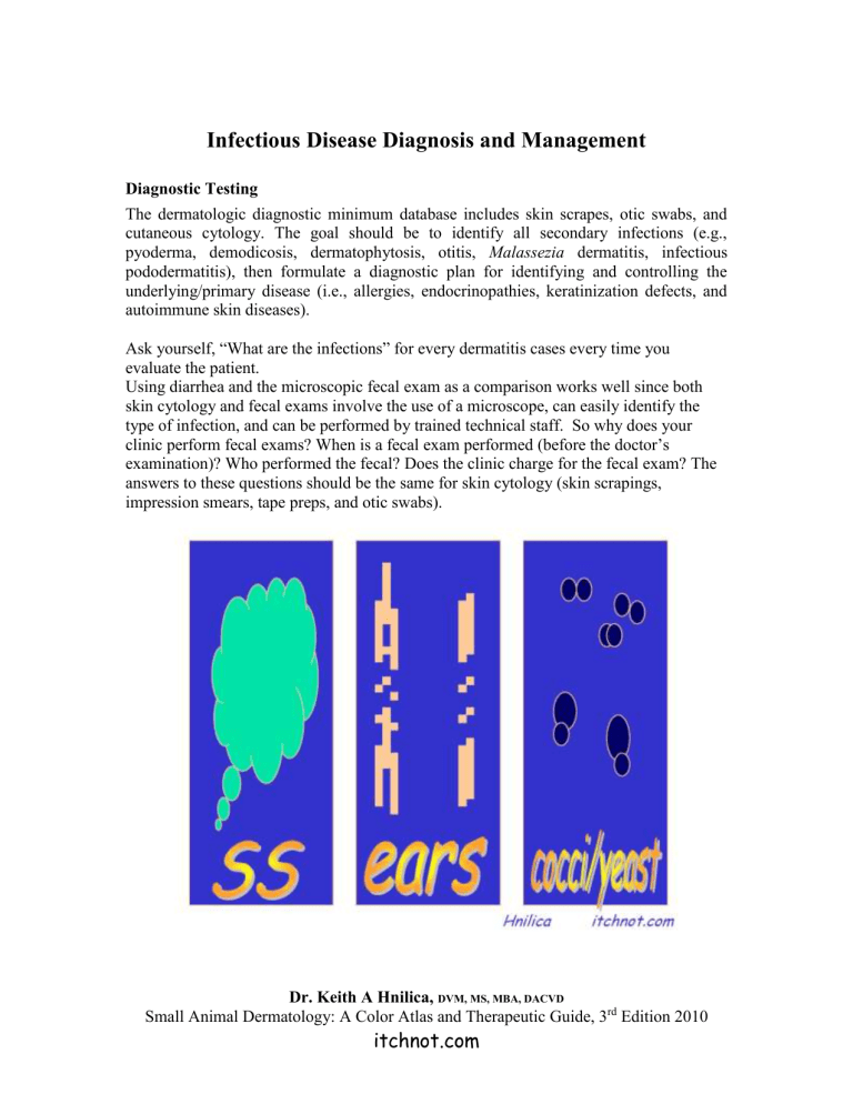
Using diarrhea and the microscopic fecal exam as a comparison works well since both skin cytology and fecal exams involve the use of a microscope, can easily identify the type of infection, and can be performed by trained technical staff. So why does your clinic perform fecal exams? When is a fecal exam performed (before the doctor’s examination)? Who performed the fecal? Does the clinic charge for the fecal exam? The answers to these questions should be the same for skin cytology (skin scrapings, impression smears, tape preps, and otic swabs).
Dr. Keith A Hnilica,
DVM, MS, MBA, DACVD
Small Animal Dermatology: A Color Atlas and Therapeutic Guide, 3 rd
Edition 2010 itchnot.com
The practical solution and the best method to answer the question, “What are the infections?” is to implement a minimum data base “infection screening” procedure performed by the technician before the veterinarian examines the patient. Every dermatology patient should have an otic cytology, skin cytology (either an impression smear or tape prep), and a skin scrape performed every time the patient is examined
(initially and at every recheck visit). This 3 Slide Technique TM can easily be performed and interpreted by a technician prior to the doctor’s evaluation; exactly how diarrhea cases and fecal exams are handled in most clinics.
Skin Scrapes (Slide #1 in the 3 Slide Technique)
Skin scrapes are the most common dermatologic diagnostic tests. These relatively simple and quick tests can be used to identify many types of parasitic infections. Although they are not always diagnostic, their relative ease and low cost make them essential tests in a dermatologic diagnostic minimum database.
Many practitioners reuse scalpel blades when performing skin scrapes; however, this practice should be stopped because of increased awareness of transmittable diseases (e.g.,
Bartonella , Rickettsia , feline leukemia virus [FeLV], feline immunodeficiency virus
[FIV], herpes, papillomavirus).
Procedure
Deep Skin Scrapes (for Demodex spp except D. gatoi ).
A dulled scalpel blade is held perpendicular to the skin and is used with moderate pressure to scrape in the direction of hair growth. If the area is covered with hair (usually, alopecic areas caused by folliculitis are selected), it may be necessary to clip a small window to access the skin. After several scrapes, the skin should become pink, with the capillaries becoming visible and oozing blood. This ensures that the material collected comes from deep enough within the skin to allow the collection of follicular Demodex mites. Most people also squeeze (pinch) the skin to express the mites from deep within the follicles into a more superficial area, so that they may be more easily collected. If the scraping fails to provide a small amount of blood, then the mites may have been left in the follicle, resulting in a false-negative finding. In some situations (with Shar peis or deep inflammation with scarring), it may be impossible to scrape deeply enough to harvest Demodex mites. These cases are few in number but require biopsy for identification of the mites within the hair follicles. Hairplucks from an area of lesional skin may be used to help find mites, but the accuracy of this technique compared with skin scrapes is unknown.
Regardless of the collection technique used, the entire slide should be searched for mites with the use of low power (usually a 10X objective). A search of the entire slide ensures that if only one or two mites are present (as is typical of scabies infection), the user will likely find them. It may be helpful to lower the microscope condenser; this provides greater contrast to the mites, thereby enhancing their visibility. (One must be sure to raise the condenser before looking for cells or bacteria on stained slides.)
Dr. Keith A Hnilica,
DVM, MS, MBA, DACVD
Small Animal Dermatology: A Color Atlas and Therapeutic Guide, 3 rd
Edition 2010 itchnot.com
There is no excuse for mistreating a patient who has demodicosis. Lesions caused by demodicosis can look identical to folliculitis lesion caused by bacterial pyoderma and dermatophytosis. Clinical appearance is not an acceptable method to rule-in or rule-out demodicosis. By having the technicians perform a skin scrape as part of the infection screen, 3 Slide Technique TM , demodicosis can easily and accurately be identified and treated.
Cutaneous Cytology (Slide #2 in the 3 Slide Technique)
Cutaneous cytology is the second most frequently employed dermatologic diagnostic technique. Its purpose is to help the practitioner to identify bacterial or fungal organisms
(yeast) and assess the infiltrating cell types, neoplastic cells, or acantholytic cells (typical of pemphigus complex).
The infections are always secondary to a primary disease; however, all too often, the patient is not evaluated or treated for the primary disease. This is due to 3 predominant factors: treating only the secondary infections over and over, the confusing nature of allergy, and access to cheap steroids which have delayed repercussions.
Superficial Pyoderma
(superficial bacterial folliculitis)
Features
Superficial pyoderma is a superficial bacterial infection involving hair follicles and the adjacent epidermis. The infection is almost alwayss secondary to an underlying cause; allergies and endocrine disease are the most common causes (Box 3-3). Superficial pyoderma is one of the most common skin diseases in dogs but rare in cats.
Superficial pyoderma is characterized by focal, multifocal, or generalized areas of papules, pustules, crusts, and scales, epidermal collarettes, or circumscribed areas of erythema and alopecia that may have hyperpigmented centers. Short-coated dogs often present with a “motheaten” patchy alopecia, small tufts of hair that stand up, or reddish brown discoloration of white hairs. In long-coated dogs, symptoms can be insidious and may include a dull, lusterless hair coat, scales, and excessive shedding. In both short- and long-coated breeds, primary skin lesions are often obscured by remaining hairs but can be readily appreciated if an affected area is clipped. Pruritus is variable, ranging from none to intense levels. Bacterial infections secondary to endocrine disease may cause pruritus, thereby mimicking allergic skin disease.
Staphylococcus pseudintermedius (previously Staphylococcus intermedius)is the most common bacterium isolated from canine pyoderma and is usually limited to dogs. Staphylococcus schleiferi is a bacterial species in dogs and humans that is emerging as a common canine isolate in patients with chronic infections and previous antibiotic exposure. Both Staphylococcus pseudintermedius and Staphylococcus schleiferi may develop methacillin-resistence especially if subtherapeutic doses of antibiotics or flouroquinilone antibiotics have been previously used in the patient.
Additionally, methicillin-resistant Staphylococcus aureus (human MRSA) is becoming more common among veterinary species. All three Staphylococcus may be zoonotic, moving from humans to canines or from canine to human; immunosuppressed individuals are at most risk.
Dr. Keith A Hnilica,
DVM, MS, MBA, DACVD
Small Animal Dermatology: A Color Atlas and Therapeutic Guide, 3 rd
Edition 2010 itchnot.com
Causes of Secondary Superficial and Deep Pyoderma
Demodicosis, scabies, Pelodera
Hypersensitivity (e.g., atopy, food, flea bite)
Endocrinopathy (e.g., hypothyroidism, hyperadrenocorticism, sex hormone imbalance, alopecia X)
Immunosuppressive therapy (e.g., glucocorticoids, progestational compounds, cytotoxic drugs)
Autoimmune and immune-mediated disorders
Trauma or bite wound
Foreign body
Poor nutrition
Treatment and Prognosis
1. The underlying cause must be identified and controlled.
2.
3.
4.
Systemic antibiotics (minimum 3-4 weeks) should be administered and continued 1 week beyond complete clinical and cytological resolution (see Box 3-1).
Concurrent bathing every 2 to 7 days with an antibacterial shampoo that contains chlorhexidine or benzoyl peroxide is helpful.
If lesions recur within 7 days of antibiotic discontinuation, the duration of therapy was
5. inadequate and antibiotics should be reinstituted for a longer time period and better attempts to identify and control the underlying disease should occur.
If lesions do not completely resolve during antibiotic therapy or if there is no response to the antibiotics, antibiotic resistance should be assumes and a bacterial culture and sensitivity
7. submitted.
If antibiotics resistance is suspected or confirmed, frequent bathing (up to daily) and the frequent application of topical chlorhexidine solutions combined with the simultaneous administration of two different class antibiotics at high doses seem to produce the best results.
Monitoring the infection with cytology and cultures with antibiotic sensitivities is important to determine when the treatments can be stopped. Premature discontinuation of therapy, not completely controlling the primary disease, and the use of fluoroquinilone antibiotics will likely perpetuate the resistant infection.
The prognosis is good if the underlying cause can be identified and corrected or controlled. 8.
Author's Note:
** Superficial Pyoderma is one of the most common skin diseases in dogs and almost always has an underlying cause (allergies or endocrine disease).
** Cefpodoxime, Ormetoprim/sulfadimethoxine (Primor), and Convenia provide the most consistent compliance which seem to help reduce the development of resistance when used at high doses.
** MRSA, MRSS, MRSI, and MRSP are becoming an emerging problem in some regions of the
US.
>>The most likely risk factors include previous exposure to fluoroquinilone antibiotics, sub-therapeutic antibiotic dosing, and concurrent steroid therapy.
>> Daily baths and topical treatments can be very beneficial in the resolution of the infection.
Dr. Keith A Hnilica,
DVM, MS, MBA, DACVD
Small Animal Dermatology: A Color Atlas and Therapeutic Guide, 3 rd
Edition 2010 itchnot.com
>> Maximize the dose of antibiotics and consider using two antibiotics simultaneously to protect additional resistance from developing.
>> Practice good hygiene (HAND WASHING) to prevent zoonosis.
** Consider screening dogs who visit the elderly or sick to prevent zoonosis. Cultures from the nose, lips, ears, axilla, and perianal areas are best for screening patients for MRS.
Malasseziasis
(Malassezia dermatitis)
Features
Malassezia pachydermatis is a yeast that is normally found in low numbers in the external ear canals, in perioral areas, in perianal regions, and in moist skin folds. Skin disease occurs in dogs when a hypersensitivity reaction to the organisms develops, or when there is cutaneous overgrowth. In dogs, Malassezia overgrowth is almost always associated with an underlying cause, such as atopy, food allergy, endocrinopathy, keratinization disorder, metabolic disease, or prolonged therapy with corticosteroids. In cats, skin disease is caused by Malassezia overgrowth that may occur secondary to an underlying disease (e.g., feline immunodeficiency virus, diabetes mellitus or an internal malignancy). In particular, generalized Malassezia dermatitis may occur in cats with thymoma-associated dermatosis or paraneoplastic alopecia. Malasseziasis is common in dogs, especially among West Highland White terriers, Dachshunds, English setters, Basset hounds, American cocker spaniels, Shih tzus, Springer spaniels, and German shepherds. These breeds may be predisposed. Malasseziasis is rare in cats.
Moderate to severe pruritus is seen, with regional or generalized alopecia, excoriations, erythema, and seborrhea. With chronicity, affected skin may become lichenified, hyperpigmented, and hyperkeratotic (leathery or elephant-like skin). An unpleasant body odor is usually present. Lesions may involve the interdigital spaces, ventral neck, axillae, perineal region, or leg folds. Paronychia with dark brown nail bed discharge may be present. Concurrent yeast otitis externa is common.
Diagnosis
1.
2.
Rule out other differentials
Cytology (tape preparation, impression smear): yeast overgrowth is confirmed by the finding round-toorganisms may be difficult to find
Treatment and Prognosis
1.
2.
Any underlying cause (allergies, endocrinopathy, keratinization defect) must be identified and corrected.
For mild cases, topical therapy alone is often effective. The patient should be bathed every 2 to
3 days with shampoo that contains 2% ketoconazole, 1% ketoconazole/2% chlorhexidine, 2% miconazole, 2% to 4% chlorhexidine, or 1% selenium sulfide (dogs only). Shampoos that have
Dr. Keith A Hnilica,
DVM, MS, MBA, DACVD
Small Animal Dermatology: A Color Atlas and Therapeutic Guide, 3 rd
Edition 2010 itchnot.com
3. two active ingredients provide better efficacy. Treatment should be continued until the lesions resolve and follow-up skin cytology reveals no organisms (approximately 4 weeks).
The treatment of choice for moderate to severe cases is ketoconazole (dogs) or fluconazole
4.
10mg/kg PO with food every 24 hours, Treatment should be continued until lesions resolve and follow-up skin cytology reveals no organisms (approximately 4 weeks).
Alternatively, treatment with terbinafine 5-40mg/kg PO every 24 hours or itraconazole
(Sporonox) 5-10mg/kg every 24 hours for 4 weeks may be effective.
5. Pulse therapy protocols have been published using several drugs and a variety of schedules; however, these often take longer to resolve the active infection.
5. The prognosis is good if the underlying cause can be identified and corrected. Otherwise, regular once- or twice-weekly antiyeast shampoo baths may be needed to prevent relapse.
This disease is not considered contagious to other animals or to humans, except for immunocompromised individuals.
Authors’s Note:
** Yeast dermatitis is currently the most commonly missed diagnosis in US general practices. Any patient with leathery, elephant-skin like lesions on the ventrum should be suspected of having
Malassezia dermatitis.
** Cutaneous cytology is not always successful for finding Malassezia organisms requiring the clinician to rely on clinical lesion patterns to make a tentative diagnosis.
** Yeast dermatitis is severely pruritic with owners reporting an itch level of 10 on a 0-10 visual analog scale.
Canine Generalized Demodicosis
Features
Canine generalized demodicosis may appear as a generalized skin disease that may have genetic tendencies and can be caused by three different species of demodectic mites: D. canis, D. injai, and an unnamed short-bodied Demodex mite. D. canis, a normal resident of the canine pilosebaceous unit (hair follicle, sebaceous duct, and sebaceous gland), is primarily transmitted from the mother to neonates during the first 2 to 3 days of nursing, but adult-to-adult transmission may rarely occur. D. injai, a recently described, large, long-bodied Demodex mite, is also found in the pilosebaceous unit, but its mode of transmission is unknown. Mode of transmission is also unknown for the short-bodied unnamed Demodex mite, which, unlike the other two species, lives in the stratum corneum. Depending on the dog’s age at onset, generalized demodicosis is classified as juvenile-onset or adult-onset. Both forms are common in dogs. Juvenile-onset generalized demodicosis may be caused by D. canis and the short-bodied unnamed Demodex mite. It occurs in young dogs, usually between 3 and 18 months of age, with highest incidence in medium-sized and large purebred dogs. Adult-onset generalized demodicosis can be caused by all three mite species and occurs in dogs older than 18 months of age, with highest incidence in middle-aged to older dogs that are immunocompromised because of an underlying condition such as endogenous or iatrogenic hyperadrenocorticism, hypothyroidism, immunosuppressive drug therapy, diabetes mellitus, or neoplasia. To date, only adult-onset disease has been reported with D. injai, with highest incidence noted in terriers.
Dr. Keith A Hnilica,
DVM, MS, MBA, DACVD
Small Animal Dermatology: A Color Atlas and Therapeutic Guide, 3 rd
Edition 2010 itchnot.com
Clinical signs of infestation with either D. canis or the unnamed Demodex mite are variable.
Generalized demodicosis is defined as five or more focal lesions, or two or more body regions affected. Usually, patchy, regional, multifocal, or diffuse alopecia is observed with variable erythema, silvery grayish scaling, papules, or pruritus. Affected skin may become lichenified, hyperpigmented, pustular, eroded, crusted, or ulcerated from secondary superficial or deep pyoderma. Lesions can be anywhere on the body, including the feet. Pododemodicosis is characterized by any combination of interdigital pruritus, pain, erythema, alopecia, hyperpigmentation, lichenification, scaling, swelling, crusts, pustules, bullae, and draining tracts.
Peripheral lymphadenomegaly is common. Systemic signs (e.g., fever, depression, anorexia) may be seen if secondary bacterial sepsis develops.
D. injai infestations are typically characterized by a focal areas of greasy seborrhea (seborrhea oleosa), especially over the dorsum of the trunk. Other skin lesions may include alopecia, erythema, hyperpigmentation, and comedones. Small breeds and terriers seem to predisoosed to
Demodex injai infections.
Diagnosis
1. Microscopy (deep skin scrapes): many demodectic adults, nymphs, larvae, and ova are typically found with D. canis and the short-bodied, unnamed demodectic mite, although D.
canis may be difficult to find in fibrotic lesions and in feet. With D. injai, mites may be low in number and difficult to find requiring skin biopsies.
Treatment and Prognosis
1.
2.
3.
If adult-onset, any underlying conditions should be identified and corrected. All steroid containing therapies should be discontinued as steroid administration is the mostcommon cause of adult onset demidicosis.
Intact dogs, especially females, should be neutered. Estrus or pregnancy may trigger relapse.
Any secondary pyoderma should be treated with appropriate long-term (minimum 3-4 weeks) systemic antibiotics that are continued at least 1 week beyond clinical resolution of the pyoderma.
4. Topical shampoo therapy using a 1-3% benzoyl peroxide shampoo every 3-7 days will help speed resolution and enhance the mitacidal treatments.
5.
Effective Mitacidal therapies include the following:
*I vermectin 0.2-0.6mg/kg PO every 24 hours is often effective against generalized demodicosis. Initially, ivermectin 0.1mg/kg PO is administered on day 1, then 0.2mg/kg PO is administered on day 2, with oral daily increments of 0.1mg/kg until 0.2-0.6mg/kg/day is being administered, assuming that no signs of toxicity develop. The cure rate for
0.4mg/kg/day ivermectin is 85% to 90%.
*M ilbemycin oxime, 0.5 to 2mg/kg PO every 24 hours. The cure rate is 85% to 90%.
* Doramectin is also reported to be effective against canine demodicosis at a dose of 0.6mg/kg
SC once weekly. The cure rate is approximately 85%. Adverse effects are uncommon but include, as for ivermectin, dilated pupils, lethargy, blindness, and coma.
* amitraz collars alone may be as effective as ivermectin (0.6mg/kg/day PO).
Dr. Keith A Hnilica,
DVM, MS, MBA, DACVD
Small Animal Dermatology: A Color Atlas and Therapeutic Guide, 3 rd
Edition 2010 itchnot.com
* Topical application of Promeris (topical metaflumizone and amitraz solution) (topical metaflumizone and amitraz solution) every two weeks has demonstrated good efficacy.
* Moxidectin has demonstrated variable efficacy when applied every 2-4 weeks.
Historical Treatment Include:
Traditional miticidal treatment entails the following:
Total body hair coat clip if dog is medium- to long-haired
Weekly bath with 2.5% to 3% benzoyl peroxide shampoo, followed by a total body application of 0.03% to 0.05% amitraz solution. The cure rate ranges from 50% to 86%.
* For demodectic pododermatitis, in addition to weekly amitraz dips, foot soaks in 0.125%
6. amitraz solution should be performed every 1 to 3 days.
Regardless of the miticidal treatment chosen, therapy is administered over the long term
7.
(weeks to months). Treatments should be continued for at least 1 month beyond the time when the first follow-up skin scrapings becomes negative for mites (total of two negative skin scrapings).
The prognosis is good to fair. Relapses may occur, requiring periodic or lifelong treatment in some dogs. The use of glucocorticosteroids in any dog that has been diagnosed with demodicosis should be avoided. Because of its hereditary predisposition, neither female nor male dogs with juvenile-onset generalized demodicosis should be bred. D canis is not considered contagious to cats or to humans. It is transmitted from bitch to newborn puppies during the first 2 to 3 days of nursing, and possibly between adult dogs that are close cohabitants. The mode of transmission for D injai and the unnamed short-bodied Demodex mite is unknown.
Author’s Note:
Steroids are the most common cause of adult onset Demodicosis.
Products containing amitraz tend to be the most toxic usually due to the product vehicle.
Aggressive treatment should be tried for up to six months before giving up.
One of the most common causes of treatment failure is that the patient will look greatly improved before negative skin scrapes are achieved. Many owners will discontinue treatment prematurely resulting in relapse.
The average time to achieve clinical improvement is 4-6 weeks; the first negative skin scrape usually occurs around 6-8 weeks; and most patients need approximately 3 months of treatment to resolve the infection based on two negative skin scrapes at least 3 weeks apart.
Dr. Keith A Hnilica,
DVM, MS, MBA, DACVD
Small Animal Dermatology: A Color Atlas and Therapeutic Guide, 3 rd
Edition 2010 itchnot.com
