TECHNICAL CORNER: LOW-PASS (HIGH FREQUENCY) FILTERS
advertisement

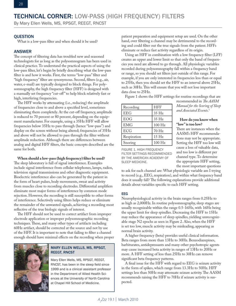
28 TECHNICAL CORNER: low-pass (high frequency) filters By Mary Ellen Wells, MS, RPSGT, REEGT, RNCST QUESTION What is a low-pass filter and when should it be used? ANSWER The concept of filtering data has troubled new and seasoned technologists for as long as the polysomnogram has been used in clinical practice. To understand the practical aspects of using the low-pass filter, let’s begin by briefly describing what the low-pass filter is and how it works. First, the terms “low-pass” filter and “high frequency” filter are synonymous. Second, filters (e.g., air, water, e-mail) are typically designed to block things. For polysomnography, the high frequency filter (HFF) is designed with a manually set frequency “cut-off ” to help block relatively fast or high, interfering frequencies. The HFF works by attenuating (i.e., reducing) the amplitude of frequencies close to and above a specified level, sometimes eliminating them completely. At the cut-off frequency, amplitude is reduced to 70 percent or 80 percent, depending on the equipment manufacturer. For example, using a 35Hz HFF will allow frequencies below 35Hz to pass through (hence “low-pass”) and display on the screen without being altered; frequencies of 35Hz and above will not be allowed to pass through the filter without amplitude reduction. Although there are differences between analog and digital HFF filters, the basic concepts described are the same for both. When should a low-pass (high frequency) filter be used? The sleep laboratory is full of signal interference. Examples include signal interference from cellular telephones, lamps, radios, television signal transmissions and other diagnostic equipment. Bioelectric interference also can be generated by the patient in the form of heart pulses, body movements, sweat and activity from muscles close to recording electrodes. Differential amplifiers eliminate most major forms of interference by common mode rejection. However, the recording is still susceptible to other forms of interference. Selectively using filters helps reduce or eliminate the remainder of the unwanted signals, achieving a recording most reflective of the true biologic signals of interest. The HFF should not be used to correct artifact from improper electrode application or improper polysomnographic recording techniques. These, and many other types of artifacts including 60Hz artifact, should be corrected at the source and not by use of the HFF. It is important to note that failing to filter a channel enough should have minimal effects on the recording when proper Mary Ellen Wells, MS, RPSGT, REEGT, RNCST Mary Ellen Wells, MS, RPSGT, REEGT, RNCST, has been in the sleep field since 1999 and is a clinical assistant professor in the Department of Allied Health Sciences at the University of North Carolina at Chapel Hill School of Medicine. patient preparation and equipment setup are used. On the other hand, over filtering a channel may be detrimental to the recording and could filter out the true signals from the patient. HFFs eliminate or reduce fast activity regardless of its origin. Using an HFF in combination with a low frequency filter (LFF) creates an upper and lower limit so that only the band of frequencies you need are allowed to go through. All physiologic variables recorded during polysomnography fall within a frequency band or range, so you should set filters just outside of this range. For example, if you are only interested in frequencies less than or equal to 25Hz, then you should set the HFF to an interval above 25Hz, such as 30Hz. This will ensure that you will not lose important data close to 25Hz. Figure 1 shows the HFF settings for routine recordings that are recommended in The AASM Manual for the Scoring of Sleep Recording HFF and Associated Events.1 EEG 35 Hz EOG 35 Hz How do you know when “low” is too low? There are instances when the ECG 70 Hz AASM’s HFF recommendaRespiration 15 Hz tions may not be appropriate. Snoring 100 Hz Setting the HFF too low will cause a loss of valuable data, FIGURE 1. High frequency filter settings recommended and too low is different per channel type. To determine by the American Academy of the appropriate HFF setting, Sleep Medicine. the most important questions to ask for each channel are: What physiologic variable am I trying to record (e.g., EEG, respiration), and within what frequency band does it usually fall? The following explanations provide additional details about variables specific to each HFF setting. EMG 100 Hz EEG Neurophysiological activity in the brain ranges from 0.25Hz to as high as 2,000Hz. In routine polysomnography, sleep stages are usually recognizable within the range 0.5-16Hz, with 16Hz being the upper limit for sleep spindles. Decreasing the HFF to 15Hz may reduce the appearance of sleep spindles, yielding unrecognizable stage N2 epochs as seen in Figure 2. Also, when the HFF is set too low, muscle activity may be misleading, appearing as normal brain activity. A higher frequency (beta) provides useful clinical information. Beta ranges from more than 13Hz to 30Hz. Benzodiazepines, barbiturates, antidepressants and many other psychotropic agents may cause increased beta activity in ranges of 13Hz to 20Hz or more. A HFF setting of less than 25Hz to 30Hz can remove significant beta frequency patterns. A final issue for the HFF with regard to EEG is seizure activity in the form of spikes, which range from 13.3Hz to 50Hz. HFF settings less than 50Hz may attenuate seizure activity. The AASM recommends raising the HFF to 70Hz if seizure activity is suspected. A2Zzz 19.1 | March 2010 29 FIGURE 2. The top image demonstrates typical stage N2 sleep. The bottom image demonstrates how decreasing the HFF attenuates the spindles seen in the top image, making it difficult to identify the sleep stage. EOG The fastest eye movements occur during rapid eye movement (REM) sleep and have a frequency of more than 1Hz. The sharpest REM slopes last 50 to 200 milliseconds. Muscle artifact is of concern in the EOG due to the close proximity of active muscles; therefore, a HFF setting of 30Hz to 35Hz will reduce muscle artifact while recording the fastest eye movements. EmG Muscle activity is very fast and produces high frequency waveforms, even above 1,000Hz. The HFF should be set as high as possible (usually 70Hz to 100Hz) or turned off to provide a faithful representation of EMG potentials. ECG In adults most of the diagnostic information in the ECG is below 100Hz. The fastest component of the ECG may be as fast as 0.04 seconds or about 25Hz. A HFF setting of 30 to 35Hz produces a stable, relatively artifact-free ECG. However, this setting may be too low for diagnostic purposes. A HFF setting of 70Hz is recommended to produce a wider bandwidth for a more detailed ECG analysis. Respiration For respiratory channels, very slow frequencies are of primary interest. Therefore, the HFF can be fairly low (about 15Hz). However, if snoring is recorded within the primary respiratory channels, the HFF should be raised to allow for faster frequencies up to 100Hz. Snoring Snoring produces very fast frequencies (like EMG). Using HFF settings similar to the EMG settings (i.e., 70Hz to 100Hz, or turned off ) should adequately represent snoring. REFERENCE 1. American Academy of Sleep Medicine. The AASM manual for the scoring of sleep and associated events: rules, terminology and technical specifications. Westchester, Ill: American Academy of Sleep Medicine;2007. p. 19. A2Zzz 19.1 | March 2010


