2011 Elsevier Ltd All rights reserved DOI 10.1016/j.cub.2011.02.002
Please cite this article in press as: Meyer et al., Interkinetic Nuclear Migration Is a Broadly Conserved Feature of Cell Division in Pseudostratified Epithelia, Current Biology (2011), doi:10.1016/j.cub.2011.02.002
Current Biology 21 , 1–7, March 22, 2011
ª
2011 Elsevier Ltd All rights reserved DOI 10.1016/j.cub.2011.02.002
Report
Interkinetic Nuclear Migration
Is a Broadly Conserved Feature of Cell
Division in Pseudostratified Epithelia
Summary
Aissam Ikmi,
1 Stowers Institute for Medical Research, 1000 East 50th Street,
Kansas City, MO 64110, USA
Animal development requires tight integration between the processes of proliferative growth and epithelial morphogenesis, both of which play out at the level of individual cells. In this respect, not only must polarized epithelial cells assume complex morphologies, these distinct forms must be radically and repeatedly transformed to permit mitosis. A dramatic illustration of this integration between epithelial morphogenesis and cell proliferation is interkinetic nuclear migration (IKNM), wherein the nuclei of pseudostratified epithelial cells translocate to the apical epithelial surface to execute cell division [
Results and Discussion
1, 2 ]. IKNM is widely considered a
hallmark of pseudostratified vertebrate neuroepithelia, and prior investigations have proposed both actomyosin- and microtubule-dependent mechanisms for apical localization of the mitotic nucleus [
3–8 ]. Here, using comparative
functional analysis in arthropod and cnidarian systems
(
Drosophila melanogaster and
Nematostella vectensis
), we show that actomyosin-dependent IKNM is likely to be a general feature of mitosis in pseudostratified epithelia throughout Eumetazoa. Furthermore, our studies suggest a mechanistic link between IKNM and the fundamental process of mitotic cell rounding.
A conserved feature of metazoan cell division is a rounding-up phase prior to spindle assembly, which occurs in systems as diverse as cultured potoroo kidney cells and the pseudostratified epithelia of Drosophila
imaginal discs [ 9–11 ]. Cytoskeletal
mechanisms of cell rounding have been probed with in vitro experiments, which have defined essential functions for
RhoA and actin dynamics at the cell cortex [ 9, 12, 13
]. Recent studies have also explored the contribution of membrane recycling to surface area changes during cell rounding in
vertebrate tissue culture [ 14
], as well as critical roles for the
ERM-domain protein moesin in controlling rounding and cortical stiffness in Drosophila
]. Still, despite these advances, precisely how mitotic cell rounding is coordinated with the maintenance of epithelial architecture remains poorly understood.
Perhaps the most conspicuous example of the morphological transformations associated with epithelial cell mitosis is interkinetic nuclear migration (IKNM), often considered a hall-
mark of pseudostratified vertebrate neuroepithelia [ 5–7, 17,
18 ]. First described in 1935 by F.C. Sauer in the pig neural
tube, IKNM is the process by which mitotic nuclei move to
the apical epithelial surface to execute cell division [ 1
]. Sauer
*Correspondence: mg2@stowers.org
2 These authors contributed equally to this work envisioned IKNM as monotonic, cyclical oscillations of the nucleus between apical positions in mitosis and basal positions in interphase, and studies since have pointed to a key role for microtubule-based force generation in this process
[
4, 7, 8 ]. Recently, through time-lapse analyses and pharma-
cological inhibitor experiments, actomyosin contractility was shown to be the primary motile force in IKNM in retinal epithelia of the zebrafish Danio rerio [
favor the interpretation of IKNM as a stochastic process, challenging Sauer’s original conception of monotonic nuclear movements. In the present analysis, we address two main questions. First, if IKNM is a hallmark of vertebrate neuroepithelia, how is the division of pseudostratified epithelial cells controlled in nonneural and nonvertebrate systems? Second, is there a mechanistic link between mitotic rounding and
IKNM, which also relies on actin dynamics at the cell cortex?
A Septate Junction-Delimited Mitotic Zone in
Drosophila
Wing Discs
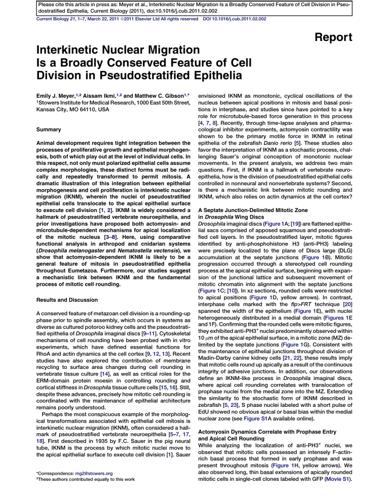
Drosophila imaginal discs (
]) are flattened epithelial sacs comprised of apposed squamous and pseudostratified cell layers. In the pseudostratified layer, mitotic figures identified by anti-phosphohistone H3 (anti-PH3) labeling were precisely localized to the plane of Discs large (DLG)
accumulation at the septate junctions ( Figure 1
B). Mitotic progression occurred through a stereotyped cell rounding process at the apical epithelial surface, beginning with expansion of the junctional lattice and subsequent movement of mitotic chromatin into alignment with the septate junctions
(
]). In xz sections, rounded cells were restricted
to apical positions ( Figure 1
D, yellow arrows). In contrast, interphase cells marked with the flp>FRT
] spanned the width of the epithelium (
Figure 1 E), with nuclei heterogeneously distributed in a medial domain (
zebrafish [ 5, 23 ], S phase nuclei labeled with a short pulse of
EdU showed no obvious apical or basal bias within the medial nuclear zone (see
A available online).
and 1F). Confirming that the rounded cells were mitotic figures, they exhibited anti-PH3
+ nuclei predominantly observed within
10 m m of the apical epithelial surface, in a mitotic zone (MZ) delimited by the septate junctions (
G). Consistent with the maintenance of epithelial junctions throughout division of
Madin-Darby canine kidney cells [
], these results imply that mitotic cells round up apically as a result of the continuous integrity of adhesive junctions. In addition, our observations define an IKNM-like process in Drosophila imaginal discs, where apical cell rounding correlates with translocation of prophase nuclei from the medial zone into the MZ. Extending the similarity to the stochastic form of IKNM described in
Actomyosin Dynamics Correlate with Prophase Entry and Apical Cell Rounding
While analyzing the localization of anti-PH3
+ nuclei, we observed that mitotic cells possessed an intensely F-actinrich basal process that formed in early prophase and was present throughout mitosis (
Figure 1 H, yellow arrows). We
also observed long, thin basal extensions of apically rounded mitotic cells in single-cell clones labeled with GFP ( Movie S1 ).
Please cite this article in press as: Meyer et al., Interkinetic Nuclear Migration Is a Broadly Conserved Feature of Cell Division in Pseudostratified Epithelia, Current Biology (2011), doi:10.1016/j.cub.2011.02.002
Current Biology Vol 21 No 6
2
Figure 1. Interkinetic Nuclear Migration-like Mitotic Cell Behavior in the Drosophila Wing Disc
(A) neuroglian-GFP;histone H2-RFP third-instar wing imaginal disc.
(B) Anti-phosphohistone H3 (PH3)
+ mitotic figures (blue) colocalize with the plane of the septate junctions as labeled by Discs large (DLG, green). Phalloidin staining of F-actin (ACT) is red. Terminal stages of cytokinetic furrow contraction are visible as F-actin-rich foci, also confined to the plane of the septate junctions (white arrows).
(C) Stages of mitosis ( i nterphase, p rophase, m etaphase, a naphase, t elophase, and c ytokinesis) from fixed samples labeled for DLG (green), mitotic chromatin (blue), and F-actin (red). Note the continuous localization of mitotic figures to the plane of the septate junctions.
(D) Phalloidin-stained cross-section through a wing imaginal disc. Yellow arrows indicate rounded and presumably mitotic cells at the apical epithelial surface.
(E) Stochastically labeled GFP
+ clones (green) illustrate the variability in nuclear positioning (white asterisks) in interphase cells.
(F) Schematic representation of interphase nuclei relative to the polarized cell-cell junctions (adherens junctions, red; septate junctions, green; nuclei, blue;
Ap, apical; Ba, basal).
(G) Whereas interphase nuclei occupy medial positions within the w
50 m m-thick epithelium (white asterisks), rounded cells and anti-PH3
+
(blue) are restricted to the DLG-delimited mitotic zone (MZ, green).
mitotic figures
(H) In fixed discs stained as above, intense basal F-actin accumulation is first observed in prophase cells (leftmost panel, yellow arrow) and persists throughout mitosis.
(I) In discs stained for anti-DLG (blue) and F-actin (red), anti-p-MRLC (green) accumulates at the cortex of early prophase figures and persists throughout mitotic rounding. Notably, p-MRLC is not detected in the F-actin-rich basal process (white arrows). Uniform cortical p-MRLC is lost or redistributed during formation and progression of the contractile ring (green arrowheads).
These findings indicate that, during mitotic rounding, imaginal disc cells can maintain some connectivity with the basal side of the epithelium despite the dramatic apical translocation of both the nucleus and bulk cytoplasm.
We next measured myosin II activity using a phospho-myosin regulatory light chain 2 antibody (anti-p-MRLC). Most interphase cells in the wing imaginal disc exhibited little or no cortical p-
MRLC enrichment (
C and S1D). By contrast, strong p-MRLC staining was observed at the cell cortex concomitant
with the earliest signs of mitotic rounding ( Figure 1 I). Uniform
cortical localization was maintained through cell rounding until metaphase and then became concentrated in the contractile ring during cytokinesis (green arrowheads in
I and
E and S1F). In mitotic cells, p-MRLC was excluded from the basal process (white arrows in
I and
indicating that the cortex of the rounded cell body has distinct molecular properties from the basal extension. Although the contribution of actomyosin contractility to cytokinesis is well established, our observations suggest an additional, earlier function in mitotic rounding of polarized epithelial cells.
Cell Rounding and Apical Translocation of the Mitotic
Nucleus Require Cortical Contractility and Rho Kinase
Activity
Previous studies in vertebrate neuroepithelia suggest that
IKNM could be driven by either actomyosin contractility
[
5, 24 ] or microtubule dynamics [ 7, 8, 25
]. We therefore used an ex vivo pharmacological assay to determine the requirement for different cytoskeletal systems in the apical movement of prophase nuclei into the MZ of the Drosophila wing disc
Please cite this article in press as: Meyer et al., Interkinetic Nuclear Migration Is a Broadly Conserved Feature of Cell Division in Pseudostratified Epithelia, Current Biology (2011), doi:10.1016/j.cub.2011.02.002
Cell Proliferation in Pseudostratified Epithelia
3
Figure 2. Rho Kinase and Cortical Contractility Are Required for Nuclear Translocation to the Mitotic Zone
(A) Experimental design for cytoskeletal inhibitor studies.
(B) Latrunculin A (LatA) treatment caused anti-PH3
+ nuclei to accumulate basal to the MZ (yellow arrows).
(C) Distance of 200 anti-PH3
+ and 500 m M LatA.
nuclei from the MZ for control discs (uncultured and 30 min of culture) compared with discs treated for 30 min with 100, 250,
(D) Percentages of anti-PH3
+ nuclei outside the MZ following LatA treatment (n = 300 nuclei per condition).
(E and F) CytoD treatment (at 5, 50, and 100 m M) disrupted apical translocation of mitotic nuclei in a manner similar to LatA.
(G) Percentage of PH3
+ nuclei outside the MZ for CytoD treatments (n = 300 nuclei per condition).
(H and I) Effects of Y-27632 (at 1, 2.5, and 5 mM) on the positions of 200 anti-PH3
+ nuclei.
(J) Percentage of PH3
+ nuclei outside the MZ following treatment with Y-27632 (n = 300 nuclei per condition). Statistical analyses were performed with the
Mantel-Haenszel test using controls cultured for 30 min.
(K–N) Third-instar wing imaginal discs stained for phalloidin (F-actin), anti-p-MRLC, and anti-PH3.
(K) UAS-dicer-2;nubbin-Gal4 control disc showing rounded mitotic cells specifically labeled with anti-p-MRLC. Box in main image indicates position of inset.
(L) nubbin-Gal4 > rok
RNAi wing discs display a severe reduction of anti-p-MRLC staining as well as a reduction in the number of apically rounded cells within the nubbin-Gal4 expression domain (dotted line). Note that mitotic cells outside the nubbin-Gal4 domain exhibit normal anti-p-MRLC staining (white arrows).
(M) xz section of a control disc showing cortical anti-p-MRLC staining in PH3
+
(N) In nubbin-Gal4>rok
RNAi discs, cortical anti-p-MRLC is eliminated in PH3
+ cells.
cells (white arrows).
(O) UAS-dicer-2;nubbin-Gal4 control disc.
(P) nubbin-Gal4 > rok
RNAi
(Q) Percentage of PH3
+ disc showing numerous anti-PH3
+ nuclei (blue) basal to DLG accumulation at the septate junction (green).
nuclei out of the MZ in nubbin-Gal4 > rok
RNAi discs (n = 450 nuclei per condition). Statistical analysis was performed using
Student’s t test.
*p < 0.0001; **p < 0.00001. Error bars indicate standard deviation.
( Figure 2 A). Uncultured control discs exhibited approximately
90% of anti-PH3
+ nuclei within the MZ, specifically defined as from the cell apex to 4 m m basal to the DLG domain (n = 300 nuclei). Similarly, 91% of anti-PH3
+ nuclei were within the MZ in control discs cultured for 30 min (n = 300 nuclei). Generally, anti-PH3 + nuclei basal to the septate junctions were in prophase, whereas metaphase and anaphase figures were restricted to the plane of the MZ.
To test the requirements for actin polymerization, we introduced latrunculin A (LatA) at three concentrations during a
30 min culture. Disrupting actin polymerization significantly increased the percentage of anti-PH3 + nuclei at abnormally
Please cite this article in press as: Meyer et al., Interkinetic Nuclear Migration Is a Broadly Conserved Feature of Cell Division in Pseudostratified Epithelia, Current Biology (2011), doi:10.1016/j.cub.2011.02.002
Current Biology Vol 21 No 6
4 basal positions with the epithelium, suggesting that the cells entered prophase but that the apical translocation was either blocked or delayed (
Figures 2 B–2D). LatA treatment also
increased the average distance of these basal anti-PH3
+ nuclei
). The majority of these basal nuclei exhibited multiple foci of intense anti-PH3 staining, contrasting with the more diffuse nuclear signal in controls.
Furthermore, only prophase figures were detected basal to the
MZ. Similar results were obtained in discs treated with cytochalasin D (CytoD;
E–2G;
effects in these assays were partially masked by the large percentage of anti-PH3 + nuclei already in the MZ at the time of drug application; among these, we observed a significant mitotic arrest (
Table S2 ). Nevertheless, disruption of actin dynamics
with either LatA or CytoD caused a significant increase in the number of anti-PH3 +
nuclei basal to the MZ ( Figures 2
D and 2G).
Actomyosin contractility is a key force-generating mechanism in eukaryotic cells. In epithelia, Rho GTPases regulate myosin II activity via Rho kinase-dependent phosphorylation
of the MRLC and inactivation of myosin phosphatase [ 26, 27 ].
RhoA has been implicated in the mitotic rounding of HeLa cells
], which has also been shown to phosphorylate myosin during thrombin-induced rounding of human tissue culture cells [
28 ]. We therefore tested the effect of the Rho kinase inhibitor Y-27632 [ 29, 30
] on cell rounding and nuclear movement during ex vivo culture. Treatment of imaginal discs with Y-27632 blocked p-MRLC accumulation in mitotic cells
A–S2D and S2G–S2J) and caused a more severe disruption of apically directed prophase movements than either
CytoD or LatA (
H–2J). We again analyzed the position of anti-PH3 + nuclei and found 44% basal to the MZ, compared with 9% for controls (at 5 mM Y-27632;
J). Similar to both LatA- and CytoD-treated discs, these basal anti-PH3
+ nuclei tended to be further from the MZ than those observed in controls (
I;
Table S1 ). Suggesting that the basal
anti-PH3
+ nuclei were in prophase, they were not associated with g
-tubulin-positive centrosomes ( Figures S3 A and S3B).
Together, the experiments above indicate that actin dynamics and Rho kinase play a critical function in the apical translocation of mitotic nuclei during wing disc IKNM. To confirm this genetically, we expressed a Rho kinase RNAi construct ( rok RNAi ) under the control of a Gal4 driver specific to the wing blade territory ( UAS-dicer-2;nubbin-Gal4 ). Wing discs expressing rok
RNAi exhibited mild morphological defects, and the anti-p-MRLC staining normally associated with mitotic cells was severely and specifically reduced within the nubbin-
Gal4
expression domain ( Figures 2 K and 2L). Consistent with
the Y-27632 experiments, anti-PH3 + nuclei accumulated at medial positions in the rok
RNAi expression domain (
2 M and 2N). In these discs, more than 60% of anti-PH3
+ nuclei were basal to the MZ, compared to 15% for controls (
O–2Q). In addition, apical cell rounding was strongly affected
( Figures S3 C–S3F). Together, these results are most consistent
with the view that Rho kinase activates cortical contractility at the onset of prophase, resulting in cell rounding and the apical translocation of the mitotic nucleus. One important consideration is that Rho kinase could utilize multiple effectors in addition to myosin. Moesin, for example, which is regulated by
RhoA/Rho kinase [ rounding [
31–33 ] and is implicated in mitotic cell
], is also likely to play an important role.
Microtubule Dynamics during Wing Disc IKNM
Microtubules play a central role in nuclear positioning in a wide variety of eukaryotic cells [
Drosophila eye disc,
[ microtubule motors control nuclear positioning in postmitotic
], and previous work on vertebrate neuroepithelia indicates a function for microtubules in regulating IKNM
]. Indeed, in the most thickened regions of the
Drosophila wing disc, interphase cells exhibit an intense apical accumulation of microtubules oriented parallel to the apico-
prophase entry (
]. In cross-sections at the apical epithelial surface, polymerized microtubules formed a meshwork filling
the polygonal profile of each cell ( Figure 3 A, asterisks), similar
to what has been described in pupal stages [
]. Intriguingly, this apical mesh was cell-autonomously disassembled at
A, arrow). As the prophase nucleus moved into the MZ, polymerized microtubules reappeared in association with the forming mitotic spindle (
head). During cytokinesis, midbody microtubules persisted in the narrowed bridge between the coequal daughters and progressively turned basally to become oriented along the apicobasal axis of the cell, perhaps reconstituting the inter-
phase arrays ( Figures S4 A–S4F). These events were more
clearly observed in xz sections through fixed wing discs, with loss of the apical microtubules in prophase (
Figures 3 B and 3C) followed by formation of the mitotic spindle ( Figures
D and 3E) and the appearance of apicobasally aligned mid-
F–3H).
To test the function of microtubule dynamics in mitotic cell rounding and IKNM, we used the same ex vivo culture approach for the application of three different concentrations
of paclitaxel ( Figures 3 I–3K) and colchicine ( Figures 3 L–3N).
Surprisingly, inhibiting microtubule dynamics had comparatively minor effects in our assay, even though both drugs
caused significant prophase arrest ( Table S3
) and colchicine visibly disrupted microtubule organization (
E, S2F,
S2K, and S2L). Interestingly, stabilizing microtubules with paclitaxel had a stronger effect than colchicine treatment, suggesting that disassembly of the interphase microtubules may be important for efficient apical translocation of the prophase nucleus. Still, given the limitations of pharmacological inhibitor treatments, further studies are needed to define the precise function of microtubule dynamics in Drosophila IKNM.
The Ancient Origin of IKNM: Beyond Bilateria
Our descriptive and functional studies in Drosophila indicate a prominent role for Rho kinase and cortical contractility in driving cell rounding and apical translocation of the mitotic nucleus. In this respect, IKNM in Drosophila imaginal discs is similar or identical to what is observed in some vertebrate neu-
]). To test whether IKNM is a widespread mechanism for pseudostratified epithelial cell division, we also analyzed cell proliferation during development of a cnidarian, the sea anemone Nematostella vectensis . Cnidaria and Bilateria diverged about 500 million years ago, prior to the emergence of centralized nervous systems (e.g., [
Nematostella larvae exhibit a pseudostratified ectodermal layer during the planula stage of development (
A and 4B). Using confocal microscopy, we observed rounded mitotic cells with cortical enrichment of F-actin at the apical epithelial surface (
C). In transverse sections, these mitotic figures closely resembled those observed in
Drosophila imaginal discs, including the presence of an
F-actin-rich basal process ( Figures 4 D–4F
0
). Also similar to
Drosophila , interphase nuclei occupied a densely packed
E and 4F) and anti-PH3
+ nuclei localized to an apical MZ (
G). We did not detect an obvious bias in the position of S phase nuclei at this developmental stage
Please cite this article in press as: Meyer et al., Interkinetic Nuclear Migration Is a Broadly Conserved Feature of Cell Division in Pseudostratified Epithelia, Current Biology (2011), doi:10.1016/j.cub.2011.02.002
Cell Proliferation in Pseudostratified Epithelia
5
Figure 3. Microtubule Dynamics during Mitosis in the Drosophila Wing Disc
(A) Apices of fixed wing disc cells exhibit a diffuse microtubule web during interphase (white asterisks). As cells enter prophase and begin to round up, the apical microtubule web disappears (white arrow). The mitotic spindle begins to form in subsequent stages (white arrowhead).
(B–H) In xz sections through fixed discs, apical microtubules orient along the apicobasal axis. Apical anti-tubulin staining (green) diminishes in early prophase (B) and vanishes altogether as the anti-PH3
+ nucleus (blue) reaches the MZ (C). After formation of the mitotic spindle (D and E) and cytokinesis, the midbody microtubules turn basally, ultimately becoming oriented along the apicobasal axis of the daughter cells (F–H).
(I–K) Paclitaxel treatments were sufficient to cause a mitotic arrest (
Table S3 ) but did not strongly affect the apicobasal positions of mitotic nuclei (yellow
arrows in I) or mitotic cell rounding.
(J) Plotted positions of 200 nuclei relative to the MZ for 50, 200, and 500 m M paclitaxel.
(K) Percentages of anti-PH3
+ nuclei basal to the MZ following paclitaxel treatments (n = 300 nuclei per condition). Statistical analyses in (K) and (N) were performed with the Mantel-Haenszel test using controls cultured for 30 min. **p < 0.0001. Error bars indicate standard deviation.
(L–N) Colchicine treatment (at 1, 50, and 500 m M) caused a mitotic arrest (
Table S3 ) but did not strongly affect the apicobasal positions of mitotic nuclei or
mitotic cell rounding (yellow arrows in L).
Please cite this article in press as: Meyer et al., Interkinetic Nuclear Migration Is a Broadly Conserved Feature of Cell Division in Pseudostratified Epithelia, Current Biology (2011), doi:10.1016/j.cub.2011.02.002
Current Biology Vol 21 No 6
6
Figure 4. Interkinetic Nuclear Migration in the Ectoderm of Nematostella vectensis
(A) Differential interference contrast image of a 4-day-old planula larvae showing ectoderm (ect), endoderm (end), blastopore/oral pole (asterisk), and pharynx (pha). Scale bars represent 50 m m in (A)–(C).
(B) Nematostella larvae stained with phalloidin to label F-actin (ACT, green) and propidium iodide to label nuclei (PI, red).
(C) As in Drosophila (e.g.,
(D) Transverse view showing apical rounding of cells (white arrowheads). Scale bars represent 25 m m in (D) and (E).
(E–F
0
) Detailed view of the ectoderm. Rounded cells in the MZ exhibit enriched cortical F-actin (white arrowheads).
(G) Apical localization of anti-PH3
+ nuclei (green) in the ectoderm of control larvae stained with antia -tub (red) and PI (blue). Scale bar represents 10 m m.
(H and I) CytoD-treated (H) and colchicine-treated (I) animals exhibit anti-PH3
+ nuclei in abnormal positions basal to the MZ.
(J) Effects of CytoD and colchicine on the position of anti-PH3
+ nuclei. Statistical analyses were performed with the Mantel-Haenszel test using controls cultured in dimethyl sulfoxide. *p < 0.0001; **p < 0.00001. Error bars indicate standard deviation.
( Figure S1 B), although a basal positional bias may exist at later
stages of development (data not shown).
To assess the contribution of different cytoskeletal elements to Nematostella IKNM, we treated 4-day-old planula larvae with CytoD, Y-27632, or colchicine and then analyzed the position of anti-PH3
+ nuclei with respect to the MZ. In controls, approximately 20% of anti-PH3 + nuclei were observed basal to the MZ (n = 354). Consistent with our results in Drosophila , disrupting actin polymerization with CytoD nearly doubled the percentage of anti-PH3 + nuclei in the medial zone (
4 H and 4J). An even stronger effect was observed when 2.5 mM
Y-27632 was used to inhibit Rho kinase activity ( Figures S3
G–
S3J). These animals also exhibited intrusion of interphase
nuclei into the MZ ( Figure S3
H), an effect we did not observe in Drosophila . Also contrasting with our results in Drosophila , disrupting microtubules with colchicine increased the number of anti-PH3 + nuclei at abnormally basal positions (
and 4J). This indicates a role for microtubules in apical translocation of the mitotic nucleus and suggests that both cortical contractility and microtubule dynamics contribute to IKNMlike movements in Nematostella .
Conclusions
The results presented here have two main implications. First, we describe IKNM-like processes in the pseudostratified epithelia of arthropods and cnidarians, indicating that this mode of cell division is widespread and not a unique feature of vertebrate neuroepithelia. Mechanistically, our findings are most consistent with the view that IKNM is a highly conserved process primarily driven by cortical contractility associated with mitotic cell rounding at prophase entry but that it has been specialized in some instances to require microtubuledependent nuclear movements ([
];
I and 4J).
Second, the deep evolutionary conservation of IKNM-like
Please cite this article in press as: Meyer et al., Interkinetic Nuclear Migration Is a Broadly Conserved Feature of Cell Division in Pseudostratified Epithelia, Current Biology (2011), doi:10.1016/j.cub.2011.02.002
Cell Proliferation in Pseudostratified Epithelia
7 mitotic behavior throughout Eumetazoa suggests the existence of strong constraints on the mechanism of cell division in pseudostratified epithelia. We favor a model wherein IKNM is required to restrict cell divisions to the plane of the apical junctions, thereby ensuring continuous monolayer integrity of proliferating epithelial sheets.
Supplemental Information
Supplemental Information includes four figures, three tables, Supplemental
Experimental Procedures, and one movie and can be found with this article online at doi:10.1016/j.cub.2011.02.002
.
Acknowledgments
This work was supported by a Burroughs Wellcome Fund Career Award in
Biomedical Sciences to M.C.G., as well as generous support from the Stowers Institute for Medical Research. We would like to thank Eugenia Park for helpful comments on an early draft of the manuscript; Trey Ammons, Jaime
High, Adam Petrie, and Diana Baumann for assistance with Nematostella husbandry; Yale Passamaneck and Mark Martindale for the Nematostella
EdU protocol; Hua Li for assistance with statistical analyses; and Lynnette
Gutchewsky for administrative support.
Received: August 4, 2010
Revised: December 7, 2010
Accepted: February 1, 2011
Published online: March 3, 2011
References
1. Sauer, F.C. (1935). Mitosis in the neural tube. J. Comp. Neurol.
62 ,
377–405.
2. Baye, L.M., and Link, B.A. (2008). Nuclear migration during retinal development. Brain Res.
1192 , 29–36.
3. Messier, P.E., and Auclair, C. (1974). Effect of cytochalasin B on interkinetic nuclear migration in the chick embryo. Dev. Biol.
36 , 218–223.
4. Messier, P.E. (1978). Microtubules, interkinetic nuclear migration and neurulation. Experientia 34 , 289–296.
5. Norden, C., Young, S., Link, B.A., and Harris, W.A. (2009). Actomyosin is the main driver of interkinetic nuclear migration in the retina. Cell 138 ,
1195–1208.
6. Schenk, J., Wilsch-Brauninger, M., Calegari, F., and Huttner, W.B.
(2009). Myosin II is required for interkinetic nuclear migration of neural progenitors. Proc. Natl. Acad. Sci. USA 106 , 16487–16492.
7. Del Bene, F., Wehman, A.M., Link, B.A., and Baier, H. (2008). Regulation of neurogenesis by interkinetic nuclear migration through an apicalbasal notch gradient. Cell 134 , 1055–1065.
8. Xie, Z., Moy, L.Y., Sanada, K., Zhou, Y., Buchman, J.J., and Tsai, L.H.
(2007). Cep120 and TACCs control interkinetic nuclear migration and the neural progenitor pool. Neuron 56 , 79–93.
9. Cramer, L.P., and Mitchison, T.J. (1997). Investigation of the mechanism of retraction of the cell margin and rearward flow of nodules during mitotic cell rounding. Mol. Biol. Cell 8 , 109–119.
10. Gibson, M.C., Patel, A.B., Nagpal, R., and Perrimon, N. (2006). The emergence of geometric order in proliferating metazoan epithelia. Nature
442 , 1038–1041.
11. The´ry, M., and Bornens, M. (2008). Get round and stiff for mitosis. HFSP
J 2 , 65–71.
12. Maddox, A.S., and Burridge, K. (2003). RhoA is required for cortical retraction and rigidity during mitotic cell rounding. J. Cell Biol.
160 , 255–265.
13. Cortese, J.D., Schwab, B., 3rd, Frieden, C., and Elson, E.L. (1989). Actin polymerization induces a shape change in actin-containing vesicles.
Proc. Natl. Acad. Sci. USA 86 , 5773–5777.
14. Boucrot, E., and Kirchhausen, T. (2007). Endosomal recycling controls plasma membrane area during mitosis. Proc. Natl. Acad. Sci. USA
104 , 7939–7944.
15. Kunda, P., Pelling, A.E., Liu, T., and Baum, B. (2008). Moesin controls cortical rigidity, cell rounding, and spindle morphogenesis during mitosis. Curr. Biol.
18 , 91–101.
16. Carreno, S., Kouranti, I., Glusman, E.S., Fuller, M.T., Echard, A., and
Payre, F. (2008). Moesin and its activating kinase Slik are required for cortical stability and microtubule organization in mitotic cells. J. Cell
Biol.
180 , 739–746.
17. Bort, R., Signore, M., Tremblay, K., Martinez Barbera, J.P., and Zaret,
K.S. (2006). Hex homeobox gene controls the transition of the endoderm to a pseudostratified, cell emergent epithelium for liver bud development. Dev. Biol.
290 , 44–56.
18. Gotz, M., and Huttner, W.B. (2005). The cell biology of neurogenesis.
Nat. Rev. Mol. Cell Biol.
6 , 777–788.
19. Cohen, S.M. (1993). Imaginal disc development. In The Development of
Drosophila melanogaster , Volume II , M. Bate and A. Martinez Arias, eds.
(Cold Spring Harbor, NY: Cold Spring Harbor Laboratory Press), pp. 747–841.
20. Pignoni, F., and Zipursky, S.L. (1997). Induction of Drosophila eye development by decapentaplegic. Development 124 , 271–278.
21. Baker, J., and Garrod, D. (1993). Epithelial cells retain junctions during mitosis. J. Cell Sci.
104 , 415–425.
22. Reinsch, S., and Karsenti, E. (1994). Orientation of spindle axis and distribution of plasma membrane proteins during cell division in polarized MDCKII cells. J. Cell Biol.
126 , 1509–1526.
23. Baye, L.M., and Link, B.A. (2007). Interkinetic nuclear migration and the selection of neurogenic cell divisions during vertebrate retinogenesis.
J. Neurosci.
27 , 10143–10152.
24. Webster, W., and Langman, J. (1978). The effect of cytochalasin B on the neuroepithelial cells of the mouse embryo. Am. J. Anat.
152 , 209–221.
25. Tsai, J.W., Chen, Y., Kriegstein, A.R., and Vallee, R.B. (2005). LIS1 RNA interference blocks neural stem cell division, morphogenesis, and motility at multiple stages. J. Cell Biol.
170 , 935–945.
26. Quintin, S., Gally, C., and Labouesse, M. (2008). Epithelial morphogenesis in embryos: Asymmetries, motors and brakes. Trends Genet.
24 , 221–230.
27. Kimura, K., Ito, M., Amano, M., Chihara, K., Fukata, Y., Nakafuku, M.,
Yamamori, B., Feng, J., Nakano, T., Okawa, K., et al. (1996).
Regulation of myosin phosphatase by Rho and Rho-associated kinase
(Rho-kinase). Science 273 , 245–248.
28. Sandquist, J.C., Swenson, K.I., Demali, K.A., Burridge, K., and Means,
A.R. (2006). Rho kinase differentially regulates phosphorylation of nonmuscle myosin II isoforms A and B during cell rounding and migration.
J. Biol. Chem.
281 , 35873–35883.
29. Uehata, M., Ishizaki, T., Satoh, H., Ono, T., Kawahara, T., Morishita, T.,
Tamakawa, H., Yamagami, K., Inui, J., Maekawa, M., et al. (1997).
Calcium sensitization of smooth muscle mediated by a Rho-associated protein kinase in hypertension. Nature 389 , 990–994.
30. Ishizaki, T., Uehata, M., Tamechika, I., Keel, J., Nonomura, K., Maekawa, M., and Narumiya, S. (2000). Pharmacological properties of Y-27632, a specific inhibitor of Rho-associated kinases. Mol. Pharmacol.
57 , 976–983.
31. Fehon, R.G., McClatchey, A.I., and Bretscher, A. (2010). Organizing the cell cortex: The role of ERM proteins. Nat. Rev. Mol. Cell Biol.
11 ,
276–287.
32. Matsui, T., Maeda, M., Doi, Y., Yonemura, S., Amano, M., Kaibuchi, K., and Tsukita, S. (1998). Rho-kinase phosphorylates COOH-terminal threonines of ezrin/radixin/moesin (ERM) proteins and regulates their head-to-tail association. J. Cell Biol.
140 , 647–657.
33. Shaw, R.J., Henry, M., Solomon, F., and Jacks, T. (1998). RhoA-dependent phosphorylation and relocalization of ERM proteins into apical membrane/actin protrusions in fibroblasts. Mol. Biol. Cell 9 , 403–419.
34. Morris, N.R. (2003). Nuclear positioning: The means is at the ends. Curr.
Opin. Cell Biol.
15 , 54–59.
35. Whited, J.L., Cassell, A., Brouillette, M., and Garrity, P.A. (2004).
Dynactin is required to maintain nuclear position within postmitotic
Drosophila photoreceptor neurons. Development 131 , 4677–4686.
36. Gibson, M.C., and Perrimon, N. (2005). Extrusion and death of DPP/
BMP-compromised epithelial cells in the developing Drosophila wing.
Science 307 , 1785–1789.
37. Shen, J., and Dahmann, C. (2005). Extrusion of cells with inappropriate Dpp signaling from Drosophila wing disc epithelia. Science 307 , 1789–1790.
38. Shimada, Y., Yonemura, S., Ohkura, H., Strutt, D., and Uemura, T. (2006).
Polarized transport of Frizzled along the planar microtubule arrays in
Drosophila wing epithelium. Dev. Cell 10 , 209–222.
39. Marlow, H.Q., Srivastava, M., Matus, D.Q., Rokhsar, D., and Martindale,
M.Q. (2009). Anatomy and development of the nervous system of
Nematostella vectensis, an anthozoan cnidarian. Dev. Neurobiol.
69 ,
235–254.
40. Tsai, J.W., Lian, W.N., Kemal, S., Kriegstein, A.R., and Vallee, R.B.
(2010). Kinesin 3 and cytoplasmic dynein mediate interkinetic nuclear migration in neural stem cells. Nat. Neurosci.
13 , 1463–1471.

